. 2014 Jul 1;127(Pt 13):2811-7.
doi: 10.1242/jcs.146167. Epub 2014 May 2.

Distinct functional roles for the two SLX4 ubiquitin-binding UBZ domains mutated in Fanconi anemia

Affiliations

Distinct functional roles for the two SLX4 ubiquitin-binding UBZ domains mutated in Fanconi anemia

Christophe Lachaud et al. J Cell Sci. .

Abstract

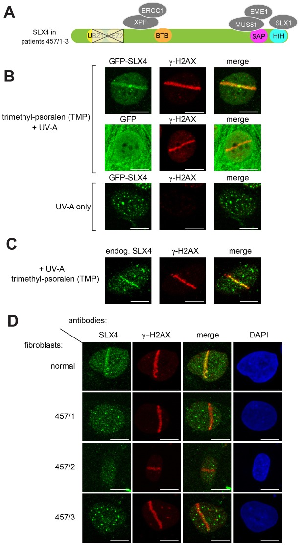
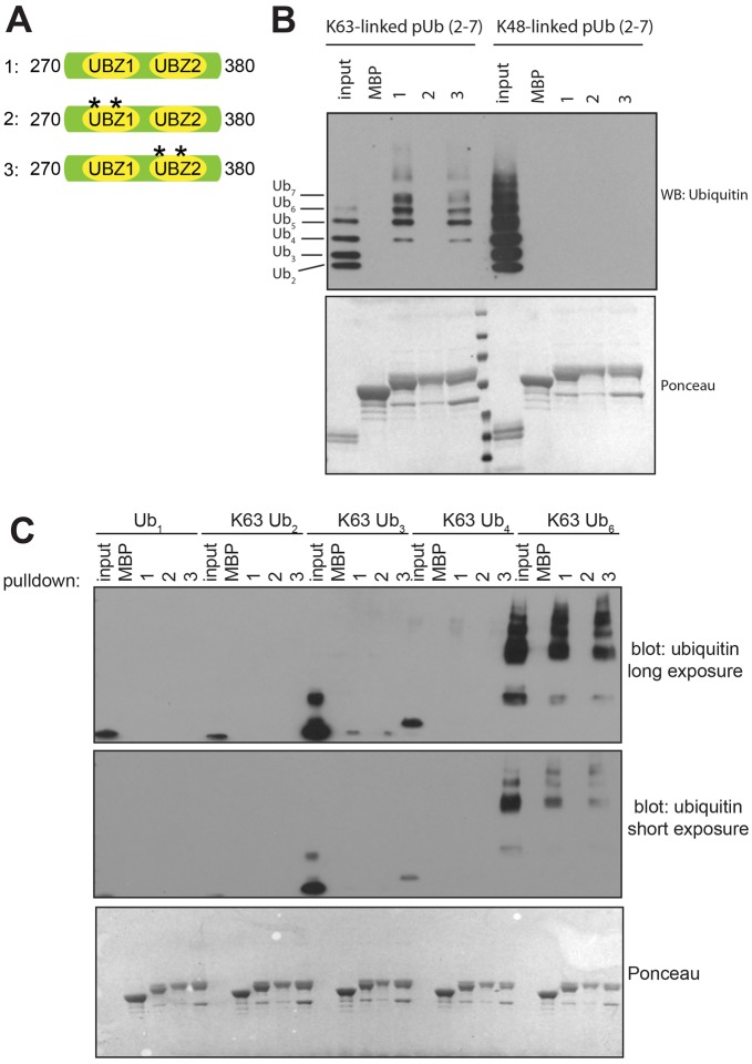
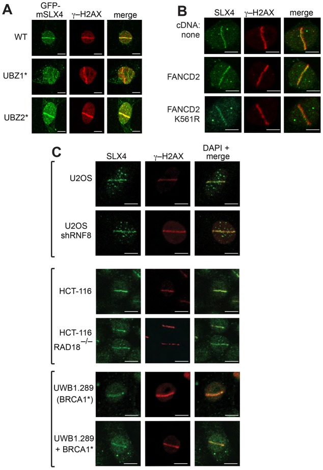
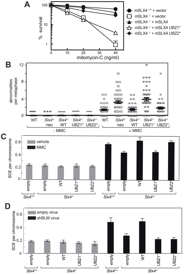
Defects in SLX4, a scaffold for DNA repair nucleases, result in Fanconi anemia (FA), due to the defective repair of inter-strand DNA crosslinks (ICLs). Some FA patients have an SLX4 deletion removing two tandem UBZ4-type ubiquitin-binding domains that are implicated in protein recruitment to sites of DNA damage. Here, we show that human SLX4 is recruited to sites of ICL induction but that the UBZ-deleted form of SLX4 in cells from FA patients is not. SLX4 recruitment does not require either the ubiquitylation of FANCD2 or the E3 ligases RNF8, RAD18 and BRCA1. We show that the first (UBZ-1) but not the second UBZ domain of SLX4 binds to ubiquitin polymers, with a preference for K63-linked chains. Furthermore, UBZ-1 is required for SLX4 recruitment to ICL sites and for efficient ICL repair in murine fibroblasts. The SLX4 UBZ-2 domain does not bind to ubiquitin in vitro or contribute to ICL repair, but it is required for the resolution of Holliday junctions in vivo. These data shed light on SLX4 recruitment, and they point to the existence of currently unidentified ubiquitylated ligands and E3 ligases that are crucial for ICL repair.

Keywords: FANCP; Fanconi anemia; ICL; SLX4; UBZ; Ubiquitin.


Figures

Fig. 1.
Fig. 1.
Recruitment of SLX4 to sites of localized DNA damage induced by PUVA. (A) Diagram showing SLX4 domain organization and associated nucleases. Siblings 457/1, 457/2 and 457/3 are FA patients with a deletion (outlined in black) removing all of the second of the tandem SLX4 UBZ domains and part of the first. BTB; Broad-complex, Tramtrack, Bric-a-brac domain: SAP; SAF-A/B, Acinus and PIAS motif: HtH; helix-turn-helix motif. (B) U2OS cells stably expressing GFP–SLX4 (upper and lower panels) or GFP only (middle panels) were incubated (or not) with trimethyl-psoralen (TMP; 20 µM, 60 min) and subjected to subnuclear micro-irradiation using a 355-nm UV-A laser. Cells were fixed and subjected to indirect immunofluorescence analysis with antibodies against GFP or γ-H2AX. (C) As for B except the localization of endogenous SLX4 (endog.) in U2OS cells was examined. (D) Cells from FA patients 457/1, 457/2 and 457/3 or normal human fibroblasts were treated as in B and endogenous SLX4 localization was analyzed by using indirect immunofluorescence. Scale bars: 10 µm.
Fig. 2.
Fig. 2.
Testing the ability of the SLX4 UBZ domains to bind to ubiquitin. (A) Diagram of the MBP-tagged SLX4 fragments used in ubiquitin-binding assays. Asterisks denote cysteine to alanine mutations at Cys 296 and Cys 299 in UBZ-1 (fragment 2) or Cys 336 and Cys 339 in UBZ-2 (fragment 3). (B) The fragments shown in A or MBP alone were immobilized on amylose–agarose and incubated with K48-linked or K63-linked poly-ubiquitin (pUb, 2–7) chains. Pulldowns were subjected to SDS-PAGE and immunoblotted (WB) with anti-ubiquitin antibodies. The bottom panel shows a Ponceau staining of the membrane performed prior to blotting. (C) Same as for B, except that mono-ubiquitin or K63-linked poly-ubiquitin chains of the indicated length were used with fragments 1–3.
Fig. 3.
Fig. 3.
Relative contributions of the SLX4 UBZ domains to DNA repair and recruitment to DNA damage sites. (A) Slx4−/− MEFs were infected with retroviruses expressing GFP-tagged mouse (m)SLX4 wild-type (WT), SLX4 UBZ-1* or SLX4 UBZ-2*. Cells were incubated with trimethyl-psoralen (TMP) and subjected to subnuclear micro-irradiation using a 355-nm UV-A laser. Cells were fixed and subjected to indirect immunofluorescence analysis with antibodies against GFP or γ-H2AX. (B) PD20 cells lacking FANCD2 (upper panels), or PD20 cells stably expressing FANCD2 (middle panels) or FANCD2 K561R (lower panels), were subjected to laser micro-irradiation as in A. Endogenous SLX4 and γ-H2AX were analyzed. (C) Same as for B, except that U2OS cells stably expressing RNF8 shRNA plus parental cells, HCT-116 cells lacking RAD18 plus parental cells or UWB1.289 cells lacking BRCA1 were used. UWB1.289 cells expressing BRCA1 were used as a control. For each population, ∼300 cells were counted, and representative images are shown. Scale bars: 10 µm.
Fig. 4.
Fig. 4.
Relative contributions of the SLX4 UBZ domains to the resolution of Holliday junctions. (A) Clonogenic survival analysis of Slx4−/− MEFs stably expressing untagged mouse (m)SLX4, SLX4 UBZ-1* or SLX4 UBZ-2*, exposed to the indicated doses of MMC. For each genotype, the viability of untreated cells was defined as 100%. Wild-type MEFs and Slx4−/− MEFs infected with empty virus (+vector) were used as controls. Data represent the mean±s.e.m., n = 3. (B) Metaphase spreads of MEFs treated with MMC (20 ng/ml) were stained with DAPI and analyzed for the presence of radial and broken chromosomes. Each cell line was either treated with MMC or left untreated, and 50 metaphase spreads were analyzed for each condition to determine the number of abnormalities per metaphase. WT, wild-type. Data show the mean±s.d. (C) Wild-type (Slx4+/+) MEFs, Slx4−/− MEFs and Slx4−/− MEFs infected with retroviruses expressing wild-type SLX4 (WT) or SLX4 bearing mutations in domains UBZ-1 (UBZ-1*) or UBZ-2 (UBZ-2*) were exposed to MMC (10 ng/ml) and the frequency of SCEs was measured. (D) Same as for C, expect that SCE frequencies in MEFs were measured after the depletion of BLM. In C and D, 400 chromosomes in three metaphase spreads (1200 in total) were analyzed. Data represent the mean±s.e.m.

References

    1. Aressy B., Greenberg R. A. (2012). DNA damage: placing BRCA1 in the proper context. Curr. Biol. 22, R806–R808 10.1016/j.cub.2012.07.046 - DOI - PMC - PubMed
    1. Castor D., Nair N., Declais A. C., Lachaud C., Toth R., Macartney T. J., Lilley D. M., Arthur J. S., Rouse J. (2013). Cooperative control of Holliday junction resolution and DNA repair by the SLX1 and MUS81-EME1 Nucleases. Mol. Cell 52, 221–233 10.1016/j.molcel.2013.08.036 - DOI - PMC - PubMed
    1. Fekairi S., Scaglione S., Chahwan C., Taylor E. R., Tissier A., Coulon S., Dong M. Q., Ruse C., Yates J. R., III, Russell P. et al.(2009). Human SLX4 is a Holliday junction resolvase subunit that binds multiple DNA repair/recombination endonucleases. Cell 138, 78–89 10.1016/j.cell.2009.06.029 - DOI - PMC - PubMed
    1. Garcia-Higuera I., Taniguchi T., Ganesan S., Meyn M. S., Timmers C., Hejna J., Grompe M., D'Andrea A. D. (2001). Interaction of the Fanconi anemia proteins and BRCA1 in a common pathway. Mol. Cell 7, 249–262 10.1016/S1097--2765(01)00173--3 - DOI - PubMed
    1. Geng L., Huntoon C. J., Karnitz L. M. (2010). RAD18-mediated ubiquitination of PCNA activates the Fanconi anemia DNA repair network. J. Cell Biol. 191, 249–257 10.1083/jcb.201005101 - DOI - PMC - PubMed

Publication types