Review
. 2014 Oct;71(19):3685-710.
doi: 10.1007/s00018-014-1633-0. Epub 2014 May 4.

Eph- and ephrin-dependent mechanisms in tumor and stem cell dynamics

Affiliations
Review

Eph- and ephrin-dependent mechanisms in tumor and stem cell dynamics

Erika Gucciardo et al. Cell Mol Life Sci. 2014 Oct.

Abstract

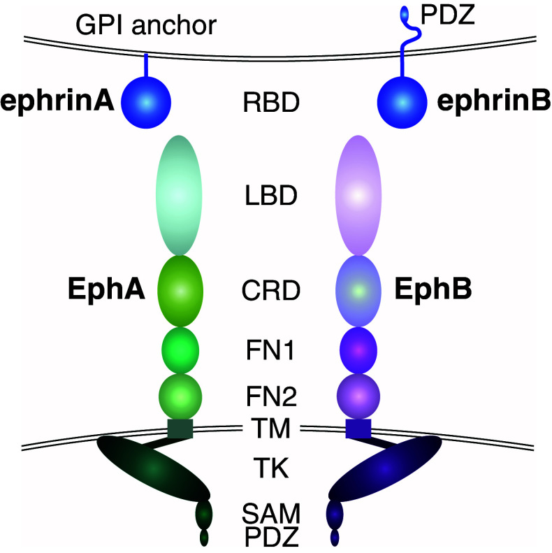
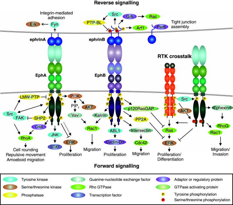
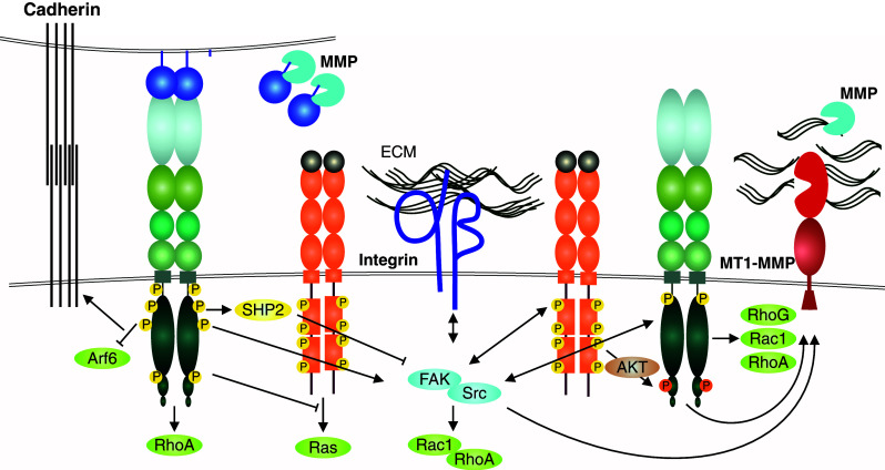
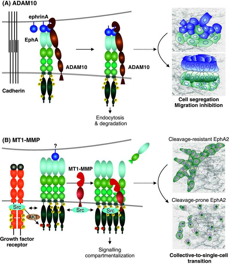
The erythropoietin-producing hepatocellular (Eph) receptors comprise the largest family of receptor tyrosine kinases (RTKs). Initially regarded as axon-guidance and tissue-patterning molecules, Eph receptors have now been attributed with various functions during development, tissue homeostasis, and disease pathogenesis. Their ligands, ephrins, are synthesized as membrane-associated molecules. At least two properties make this signaling system unique: (1) the signal can be simultaneously transduced in the receptor- and the ligand-expressing cell, (2) the signaling outcome through the same molecules can be opposite depending on cellular context. Moreover, shedding of Eph and ephrin ectodomains as well as ligand-dependent and -independent receptor crosstalk with other RTKs, proteases, and adhesion molecules broadens the repertoire of Eph/ephrin functions. These integrated pathways provide plasticity to cell-microenvironment communication in varying tissue contexts. The complex molecular networks and dynamic cellular outcomes connected to the Eph/ephrin signaling in tumor-host communication and stem cell niche are the main focus of this review.


Figures

Fig. 1
Fig. 1
Structure of Eph receptors and ephrin ligands. EphA and EphB receptors share a conserved multi-domain structure [24]. The extracellular domain contains an N-terminal ligand-binding domain (LBD), a cysteine-rich domain (CRD) followed by two fibronectin type-III repeats (FN1 and FN2). A single-pass transmembrane domain (TM) is followed by an intracellular region containing a juxtamembrane region, a tyrosine kinase domain (TK), a sterile α motif (SAM), and a postsynaptic density protein PSD95, Drosophila disc large tumor suppressor DlgA, and zonula occludens-1 protein ZO-1 (PDZ)-binding motif [3, 6]. The ephrin ligands contain a conserved extracellular N-terminal receptor binding domain (RBD). EphrinA ligands are attached to the cell membrane through a glycosylphosphatidylinositol (GPI)-anchor, whereas ephrinBs contain a transmembrane domain, and a C-terminal cytoplasmic tail including a PDZ-binding motif [29]
Fig. 2
Fig. 2
Eph/ephrin signaling. Eph forward signaling (bottom). Ligand binding at cell–cell contact (or involving soluble shed ligand) triggers receptor clustering and phosphorylation of tyrosine residues in juxtamembrane and tyrosine kinase domains [, , –36]. The ligand-induced Eph activation mediates downstream signaling pathways such as PI3K–Akt, Janus kinase (JNK)-STAT, as well as focal adhesion kinase (FAK) and Src kinase-mediated signals [, , –49]. Forward signaling can also induce transient ERK activation, although the overall effect of EphAs on ERK activity appears suppressive, as e.g., loss of EphA2 results in increased overall Ras/ERK pathway activation [12, 61, 65, 154, 214, 215]. Eph forward signaling also regulates Rho GTPase-mediated actin dynamics through interaction with guanine exchange factors (GEFs), e.g., Ephexin and Vav (EphA), as well as Kalirin and Intersectin (EphB) [, –86]. Low molecular weight protein tyrosine phosphatase (LMW-PTP) activated by Src regulates Eph signaling attenuation or termination by receptor dephosphorylation [82]. Ephrin reverse signaling (top). Eph-ephrin interaction triggers intracellular signaling into the ligand-expressing cell. Reverse signals through GPI-anchored ephrinAs rely on lipid raft-mediated clustering with proteins such as Src family kinase (Fyn) to induce e.g., ERK signaling [53, 87]. EphB/ephrinB interaction triggers reverse signals more directly via ephrinB cytoplasmic tail phosphorylation followed by recruitment of SH2-domain-containing proteins such as Src and Grb4, as well as Rac1 activation, which is attenuated by EphB dephosphorylation by PTP-Basophil-like (PTP-BL) phosphatase [90, 93]. EphrinBs also recruit PDZ-domain-containing proteins through their C-terminus [90], whereas non-phosphorylated ephrinB1 can interact with PAR6, promoting tight junctions [97]. Receptor tyrosine kinase crosstalk (middle right). EphA overexpression coupled with low expression of ephrinA ligand is associated with low tyrosine phosphorylation of the ligand-unbound receptor [32]. Akt downstream of growth factor receptors phosphorylates ligand-unbound EphA2 at serine residue (Ser897) [32, 101]. Src activation can regulate ligand-independent EphA signaling [22, 118]. RTK-EphA2 crosstalk promotes Rho-GTPase activities and EGF-induced Ras/ERK pathway activation in proliferating/differentiated cancer cells whereas suppression of the differentiation-promoting ERK activity in tumor-propagating cells (TPCs) in glioblastoma multiforme (GBM) is mediated by serine-phosphorylated EphA2 [10, 83, 86, 101]
Fig. 3
Fig. 3
Growth factor and adhesion receptor interactions with EphA2 in tumor cell invasion. The context-dependent, often tumor-suppressive, EphA/ephrinA forward signaling supports the maintenance of cadherin junctions abundant in noninvasive epithelial tumors, and involved also in collective cell invasion (left) [63, 157]. In invasive cancer cells, the ligand-independent EphA2 signaling involves physical interaction with EGFR and Akt-mediated EphA2 phosphorylation at Ser897 (right) [32, 100, 101, 103]. This RTK crosstalk between the Eph and growth factor RTKs is linked to the integrin-mediated cell–ECM communication through intracellular pathways via non-receptor tyrosine kinases Src and FAK [22, 171]. These kinases are common mediators of both upstream and downstream RTK and integrin signals regulating cell adhesive, mitogenic, and migratory responses, including Rho family GTPase-mediated cytoskeletal rearrangements (through e.g., RhoA, Rac1, and RhoG). The integrin-mediated adhesion and MT1-MMP-mediated degradation of the ECM, both required for efficient mesenchymal or collective tumor cell invasion across collagen-rich tissues, ephrin cleavages by MMPs as well as EphA2-dependent MT1-MMP induction are also depicted [13, 22]. RhoA-mediated repulsive responses upon EphA2 activation, limited ECM proteolysis, or impaired integrin-mediated adhesion can instead lead to more amoeboid-type single cell invasion by increased actomyosin contractility (see also Fig. 4b)
Fig. 4
Fig. 4
Metalloproteinase regulation of Eph signaling and cellular responses. a EphrinA-EphA binding upon cell–cell contact induces activation and conformational changes in EphA, leading to the recruitment of ADAM10. The Eph-interacting ADAM10 then cleaves receptor-bound ephrinA ligands in trans from an adjacent cell, followed by endocytosis of the activated receptor-ligand complexes, and cell–cell repulsion or contact inhibition of locomotion [21]. As typical for RTKs, the transiently activated signal can be attenuated by receptor degradation after endocytosis. This Eph forward signaling leads to cell segregation and migration inhibition. b In invasive carcinoma cells, EphA2 overexpression and ephrinA suppression are frequently coupled with the induction of MT1-MMP [22]. In these cells, growth factor receptors such as ErbB2/EGFR are also often induced or mutationally activated to mediate ligand-independent EphA2 crosstalk as well as migration signaling through Src. In this context, Src activities can also promote the transcriptional activation and phosphorylation of MT1-MMP [198]. EphA2 kinase activity-independent cleavage of the adjacent EphA2 receptor in cis by MT1-MMP triggers Src and EphA2 activity-dependent intracellular translocation of the EphA2 signaling complexes and subsequent RhoA GTPase activation [22]. When coupled with the Src-mediated migration signaling, these events and the intracellular signaling compartmentalization lead to cytoskeletal contractility, cell–cell repulsion, and a switch from collective to single-cell invasion [22]
Fig. 5
Fig. 5
Eph signaling in stem cell dynamics. a EphrinB–EphB signaling regulates both proliferation and migration in intestinal stem cell niche [159, 254]. Intestine stem cells (ISC) reside together with Paneth cells at the bottom of the crypt, where they divide and give rise to differentiated progenitor cells. As these cells differentiate, they migrate up the crypt and villus to sustain renewal of the intestinal epithelium. At the bottom, Paneth cells express EphA3, whereas ISC and progenitor cells express EphB2 in a gradient that decreases towards the lumen. EphrinB1 and ephrinB2 are expressed by the differentiated epithelial cells in a counter gradient [159]. EphB2/ephrinB1 bidirectional signaling mediate repulsive responses required for cell compartmentalization along the crypt-villus axis [159, 253]. In early stage colorectal cancer (adenoma), Wnt pathway enhances the expression of EphB2/EphB3, leading to epithelial evaginations and development of adenomatous polyps (right top) [159]. Along progression to colorectal cancer, the EphB expression is lost allowing spread of tumor cells into ephrinB-expressing areas as well as into surrounding stroma (right bottom) [223]. b EphrinB–EphB signaling regulates lineage plasticity of adult neural stem cell niche cells. The subventricular zone (SVZ) of the brain lateral ventricles contains a single layer of multiciliated ependymal cells lined by differentiated niche astrocytes and ventricle contacting self-renewing astrocytes. In physiological conditions, ependymal cells are involved in maintaining the SVZ stem cell niche, while self-renewing astrocytes are able to differentiate into neural stem cells (progenitors) and further to neuroblasts. Ependymal cells express EphB1/EphB2 receptors and ephrinB1/ephrinB2 ligands, while SVZ astrocytes express EphB1 and ephrinB2 [249]. EphB forward signaling downstream of Notch contributes to the maintenance of ependymal cell and astrocyte characteristics [250, 257]. Upon brain injury, the Notch–EphB signaling is disrupted by EphB downregulation, resulting in cell lineage interconversion between ependymal cells and astrocytes [249]. c In bone marrow, Ephrin–Eph signaling regulates osteoclast differentiation and osteoclast-osteoblast communication [267, 268]. Bone remodeling is sustained by a balance between new bone formation by osteoblasts and old mineralized bone resorption by osteoclasts. Within osteoclast precursors EphA4-dependent ephrinB2 reverse signaling limits, whereas ephrinA2–EphA2 signaling promotes osteoclast differentiation. Upon interaction with EphB4 expressing osteoblasts, EphB4 forward signaling mediates osteoblast differentiation and bone formation, whereas ephrinB2 reverse signaling inhibits bone resorption. d EphA2 and EphA3 signaling maintains cancer stem cell characteristics in human glioblastoma multiforme (GBM) [10, 11]. These tumors are composed of heterogeneous populations of differentiated dividing tumor cells and less differentiated tumor-propagating cells (TPCs). Ligand-independent EphA2 signaling coupled with Ser897 phosphorylation, and ligand-independent EphA3 signaling maintains TPCs in an undifferentiated state, further promoting their self-renewal and tumor-propagating abilities

Similar articles

Cited by

References

    1. Hirai H, Maru Y, Hagiwara K, Nishida J, Takaku F. A novel putative tyrosine kinase receptor encoded by the eph gene. Science. 1987;238:1717–1720. - PubMed
    1. Pasquale EB. Eph-ephrin bidirectional signaling in physiology and disease. Cell. 2008;133:38–52. - PubMed
    1. Pasquale EB. Eph receptors and ephrins in cancer: bidirectional signalling and beyond. Nat Rev Cancer. 2010;10:165–180. - PMC - PubMed
    1. Nievergall E, Lackmann M, Janes PW. Eph-dependent cell–cell adhesion and segregation in development and cancer. Cell Mol Life Sci. 2012;69:1813–1842. - PMC - PubMed
    1. Wilkinson DG. Multiple roles of EPH receptors and ephrins in neural development. Nat Rev Neurosci. 2001;2:155–164. - PubMed

LinkOut - more resources