. 2014 May 12;29(3):263-76.
doi: 10.1016/j.devcel.2014.04.001. Epub 2014 May 1.

Collier/OLF/EBF-dependent transcriptional dynamics control pharyngeal muscle specification from primed cardiopharyngeal progenitors

Affiliations

Collier/OLF/EBF-dependent transcriptional dynamics control pharyngeal muscle specification from primed cardiopharyngeal progenitors

Florian Razy-Krajka et al. Dev Cell. .

Abstract

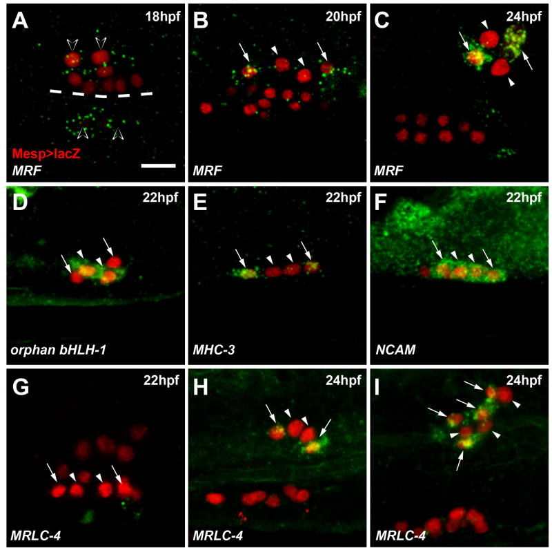
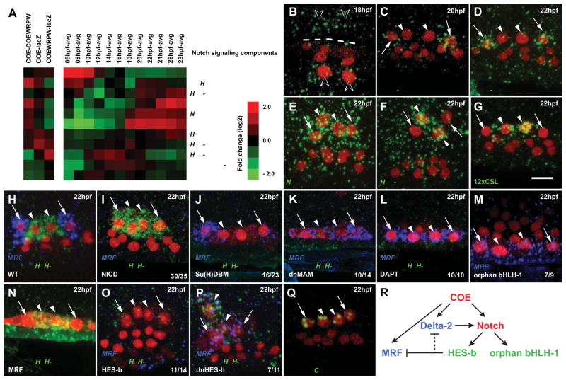
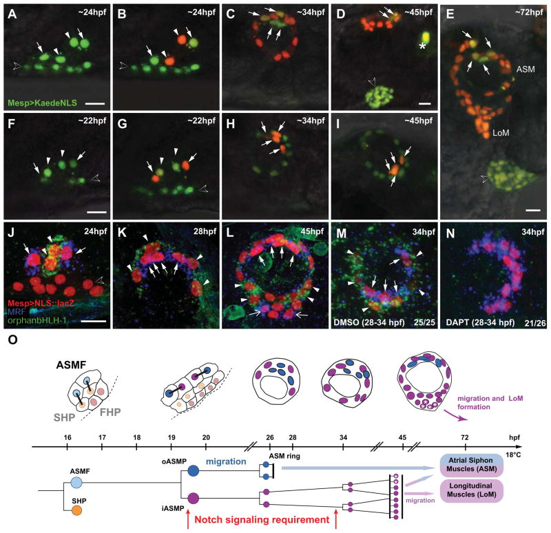
In vertebrates, pluripotent pharyngeal mesoderm progenitors produce the cardiac precursors of the second heart field as well as the branchiomeric head muscles and associated stem cells. However, the mechanisms underlying the transition from multipotent progenitors to distinct muscle precursors remain obscured by the complexity of vertebrate embryos. Using Ciona intestinalis as a simple chordate model, we show that bipotent cardiopharyngeal progenitors are primed to activate both heart and pharyngeal muscle transcriptional programs, which progressively become restricted to corresponding precursors. The transcription factor COE (Collier/OLF/EBF) orchestrates the transition to pharyngeal muscle fate both by promoting an MRF-associated myogenic program in myoblasts and by maintaining an undifferentiated state in their sister cells through Notch-mediated lateral inhibition. The latter are stem cell-like muscle precursors that form most of the juvenile pharyngeal muscles. We discuss the implications of our findings for the development and evolution of the chordate cardiopharyngeal mesoderm.


Figures

Figure 1
Figure 1. Schematic representation and microarray analysis of cardiopharyngeal development in Ciona intestinalis
(A) Schematic embryos, larvae and juveniles showing the divisions, migrations (green and blue arrows), and morphogenesis of the B7.5 lineage cells (higher magnifications): Trunk ventral cells (TVC, green), anterior tail muscles (ATM, grey), secondary TVC (2ary TVC, yellow), first heart precursors (FHP, red), atrial siphon muscle founder cells (ASMF, blue), second heart precursors (SHP, orange), inner and outer ASM precursors (ASMPs, blue), the ASM ring, heart (red/orange) and longitudinal muscles (LoM). Dashed line : midline. Oral Siphon Muscles (OSM) do not derive from the B7.5 lineage. The approximate time-line applies to the diagrams and lineage tree (hpf: hours post-fertilization). The B7.5 cardiopharyngeal lineage. Same colors as above, right side is shown (B7.5: left side), only one TVC lineage is shown. Mesp>dnFGFR and Mesp>ETS::VP16 perturbations alter ATM vs TVC specification. FoxF>COE and FoxF>COE::WRPW alter ASM vs Heart fates. See the text for details. (B) Schematic representation of published spatio-temporal expression patterns of the B7.5 lineage genes. (C)Transcription profiles of B7.5, TVC, heart (GATA-a) and ASM markers in perturbations conditions and control time series from B7.5-lineage sorted cells. Colors indicate fold change in pairwise comparisons, e.g. COE-COEW, means differential expression in FoxF>COE compared to FoxF>COE::WRPW samples sorted at 21 hpf. COEW, COE::WRPW; EtsVP, Ets::VP16; AVG, average over the whole time series; FC, Fold Change. 6hpf and “TVC/ATM perturbations” datasets were previously published (Christiaen et al., 2008). (D) Euler diagrams showing the logics and numbers of “heart”, “ASM”, “TVC” and “ATM” candidate transcripts. Green or red numbers indicate significantly enriched or depleted mutual overlaps, respectively. See Table S2 for detailed statistics. (E) Significant temporal profiles determined by STEM. See also Data S1.
Figure 2
Figure 2. Multilineage priming in cardiopharyngeal progenitors
(A) Transcription profiles of a subset of Heart candidates. Log2 fold-change scale and abbreviations as Figure 1C. TVC (cyan), ATM (magenta) and temporal profiles classifications are shown. (B) Mutual enrichments of heart and TVC candidate genes in “early” and “intermediate” temporal profiles. Green: numbers of transcripts significantly enriched in dual categories. (C–F) Expression of selected heart/TVC candidate genes. Left: chromogenic in situ hybridization on tailbud embryos. Black arrowheads: TVCs. Right: Close-ups on 18hpf TVCs expressing Mesp>NLS::lacZ (red). Asterisk in C′:TiKi expression in the endoderm. (D′) aPKC expression restricted to the heart precursors (white arrows, FHP; white arrowheads, SHP). (E′) 5HT-7r expression restricted to the ASMFs (open arrowheads). (F′) UNC-5 expression in the whole TVC progeny. (G) Lineage tree showing TVC genes’ segregation to the heart and ASM precursors between 14 and 18 hpf (BMP2/4 restricts to the heart precursors at 20 hpf). Dashed line: midline. Scale bars, 50 μm at 10 hpf, 10 μm for at 18 hpf. See also Data S1, Figure S2 and Table S1.
Figure 3
Figure 3. Hand-like/NoTrlc is restricted to the ASM founder cells and necessary for COE activation
(A–D) Hand-like/NoTrlc expression (green) in 14 to 20 hpf TVCs marked with Mesp>NLS:LacZ (red). (A) Hand-like is first restricted to the secondary TVCs (white arrowhead). (B) Expression persists in the 16hpf SHPs (white arrowheads) and ASMFs (open arrowheads). (C, D) Expression is restricted to the 18hpf ASMFs and vanishes in the 20 hpf ASMPs. (E–G) COE expression (green) in 18 hpf TVCs (red) electroporated with U6>shTyrA (control) or combined U6>shHand-like constructs. Transfected TVCs were visualized by Mesp>NLS::lacZ (red). (E) COE is expressed in control ASMFs. (F) Hand-like knock down disrupts COE expression in electroporated ASMFs (red), but not on the non-electroporated side. (G) Proportions of COE expression phenotypes in indicated conditions, n = number of electroporated larval halves. (H) Summary model of the Hand-like and COE expression dynamics in the TVC lineage. Dashed line: midline. Anterior to the left. Scale bar, 10 μm. See also Figure S2 and Figure S3.
Figure 4
Figure 4. COE activates a core muscle program among ASM-specific genes
(A) Experimental and temporal transcription profiles of 249 ASM candidates genes. TVC (cyan), ATM (magenta) and temporal profile classifications are shown. Selected “high-low-high” and “late” ASM candidates are shown. (B) Mutual enrichments of ASM candidate genes in “high-low-high” and “late” temporal profiles. Green: numbers of transcripts significantly enriched in dual categories. (C–E′) Expression of selected ASM candidates at 10 and 28 hpf. ASM genes are expressed in the atrial (white arrowheads) and oral (white arrows) siphon muscle rings at 28 hpf. NCAM is expressed in the endoderm and nervous system at 10 hpf and 28 hpf. MHC-3 is not expressed at 10 hpf. MRF is expressed in the ATMs (black arrows) and other tail muscles at 10 hpf and 28 hpf (open arrowhead). Anterior to the left. Scale bars, 50 μm. See also Data S1 and Tables S1–S6.
Figure 5
Figure 5. Restriction of muscle differentiation markers to a subset of ASMPs
(A–C) MRF transcripts (green) in the 18hpf ASMFs (open arrowheads), are restricted to the oASMPs (arrows). iASMPs (arrowheads) do not express MRF. (D) iASMP-specific expression of orphan bHLH-1. (E) oASMP-specific expression of MHC-3. (F) Pan-ASM expression of NCAM. (G–I) MRLC-4 expression starts in the oASMPs at ~24 hpf (H, arrows) and is maintained in their putative daughter cells (I, arrows). MRLC-4 is not expressed in the iASMPs (G–I, arrowheads). Mesp>NLS::lacZ (red) marks the TVC progeny. Dashed line: midline. Anterior to the left. Scale bars, 10μm. See also Data S1–5 and Figure S4.
Figure 6
Figure 6. HES-b-mediated Notch signaling restricts MRF expression to the differentiating outer ASM precursors
(A) Profiles of Notch signaling components: Delta-2, Notch, Serrate/Jagged and Su(H) show characteristics of ASM candidate genes (COE-COE::WRPW>0, increased expression after 16 hpf). HES-b expression is upregulated at ~20 hpf, when the iASMPs are born. Other components are constitutively expressed (e.g. MAM) or have different expression profiles in the B7.5 lineage (e.g. Delta). Abbreviations are as in Figure 1C. (B–D) Delta-2 expression in the ASMFs (open arrowheads, B), oASMPs (arrows, C and D) but not in the iASMPs (arrowheads, C and D). (E) Notch expression in all ASMPs. (F) iASMP-specific (arrowheads) expression of HES-b. (G) GFP mRNA (green) in larvae electroporated with 12xCSL:bpFOG>UNC-76::GFP reveal Notch signaling in the iASMPs. (H–P) Double FISH of MRF (blue) and orphan bHLH-1 (green) in control (WT) and indicated experimental conditions. Mesp>NLS::LacZ (red) marks TVC nuclei. (I) COE(-3299/-151):bpFOG-driven ASM-specific expression of NICD inhibits MRF and expands orphan bHLH-1 expression in all ASMPs. (J–L) ASM-specific expression of Su(H)DBM (J) and dnMAM (K), or incubation in DAPT (L) expands MRF and inhibits orphan bHLH-1. (M) orphan bHLH-1 misexpression expands MRF to all ASMPs. (N) MRF misexpression does not inhibit orphan bHLH-1 expression (N). (O) Islet(-3299/-151):bpFOG-driven ASM-specific expression of HES-b inhibits both MRF and orphan bHLH-1. (P) Dn-HES-b misexpression induces MRF expansion to all ASMPs without repressing orphan bHLH-1. The numbers of larvae with the indicated phenotype relative to the total are shown. Dashed line: midline. Anterior to the left. Scale bars, 10μm. (Q) Intron-specific probes show nascent COE transcripts in all ASMPs. (R) Summary model of Notch-mediated lateral inhibition of MRF through HES-b activation in the iASMPs. Red, expression in all ASMPs; green, expression in iASMPs; blue, expression in oASMPs. See also Figure S5.
Figure 7
Figure 7. Inner-ASMP derivatives either self-renew or re-activate MRF to form the majority of the B7.5 derived body-wall muscles
(A–I) Lineage tracing in individual larvae expressing Mesp>Kaede::NLS in the B7.5 lineage. (A, F) Before photoconversion: green-only Kaede::NLS fluorescence. (B) iASMPs-specific (arrowheads) green-to-red Kaede photoconversion.. Green-only fluorescence persists in the oASMPs nuclei (arrows). (F–I) oASMPs-specific Kaede photoconversion. (C, H) By 34 hpf, ASMPs form ~12-cell rings, oASMP and iASMPs have divided once and twice, respectively. (D, I) By ~45 hpf, iASMP derivatives have divided a third time and oASMP derivatives have stopped dividing. (E) iASMP-descendants (red nuclei from iASMPs in (B) form most ASMs and all nascent LoMs. Open arrowheads in D and E: heart primordium. LoM, Longitudinal Muscle. (J–N) Double in situ hybridization of MRF (blue) and orphan bHLH-1 (green) on 22 to 45 hpf Mesp>NLS::LacZ (red)-electroporated larvae. (JL) MRF and orphan bHLH-1 are expressed in the o- (arrows) and iASMP derivatives (arrowheads), respectively. (L) At ~45hpf, iASMP-derived LoM precursors either self-renew and continue to express orphan bHLH-1 (arrowheads), or shut it off and activate MRF (open arrows, I). (M, N) 28 to 34 hpf incubation in DAPT abolishes orphan bHLH-1 and expands MRF in all ASMP derivatives. (O) B7.5-derived pharyngeal muscle development. First heart precursors (FHPs, red), second heart precursors (SHPs, orange), ASM founder cells (ASMFs, light blue), inner ASM precursors and derivatives (iASMPs, violet), outer ASM precursors and derivatives (oASMPs, dark blue). Asterisks : MRF reactivation in some iASMP derivatives. Sustained Notch signaling is required for MRF repression in the iASMPs and for renewal of their orphan bHLH-1(+) derivatives. Dashed line : midline. Anterior to the left. Scale bars, 10μm. See also Figure S6.

Similar articles

Cited by

References

    1. Ando R, Hama H, Yamamoto-Hino M, Mizuno H, Miyawaki A. An optical marker based on the UV-induced green-to-red photoconversion of a fluorescent protein. Proceedings of the National Academy of Sciences of the United States of America. 2002;99:12651–12656. - PMC - PubMed
    1. Beh J, Shi W, Levine M, Davidson B, Christiaen L. FoxF is essential for FGF-induced migration of heart progenitor cells in the ascidian Ciona intestinalis. Development. 2007;134:3297–3305. - PubMed
    1. Bjornson CR, Cheung TH, Liu L, Tripathi PV, Steeper KM, Rando TA. Notch signaling is necessary to maintain quiescence in adult muscle stem cells. Stem cells. 2012;30:232–242. - PMC - PubMed
    1. Buckingham M, Vincent SD. Distinct and dynamic myogenic populations in the vertebrate embryo. Current opinion in genetics & development. 2009;19:444–453. - PubMed
    1. Chiba S, Sasaki A, Nakayama A, Takamura K, Satoh N. Development of Ciona intestinalis juveniles (through 2nd ascidian stage) Zoological science. 2004;21:285–298. - PubMed

Publication types

Associated data