. 2014 Jun 13;289(24):17163-73.
doi: 10.1074/jbc.M114.558288. Epub 2014 May 2.

Role of autophagy in cisplatin resistance in ovarian cancer cells

Affiliations

Role of autophagy in cisplatin resistance in ovarian cancer cells

Juan Wang et al. J Biol Chem. .

Abstract

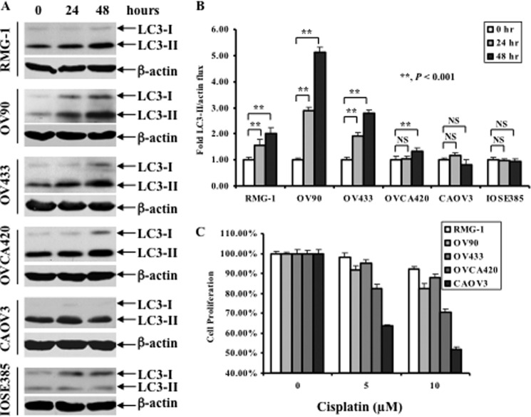
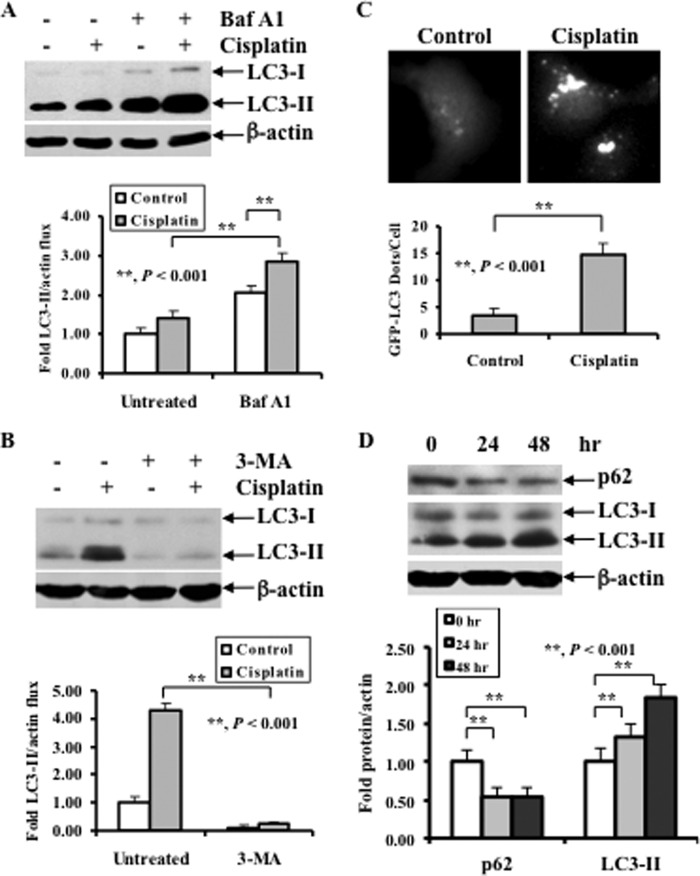
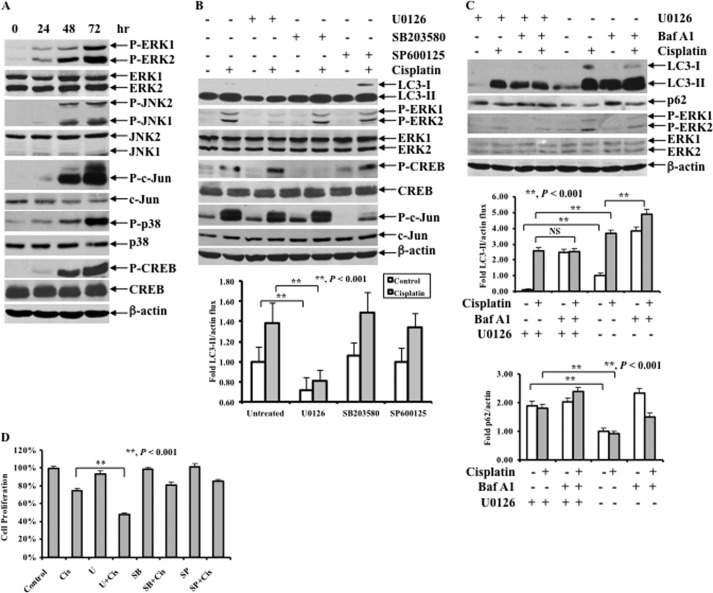
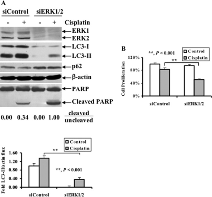
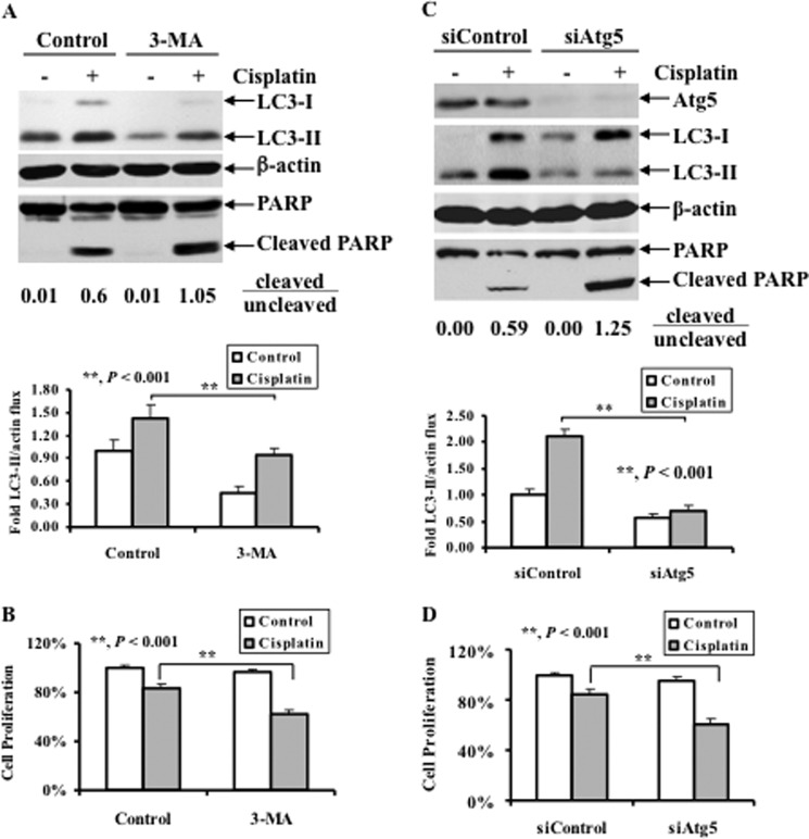
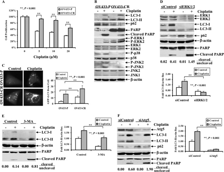
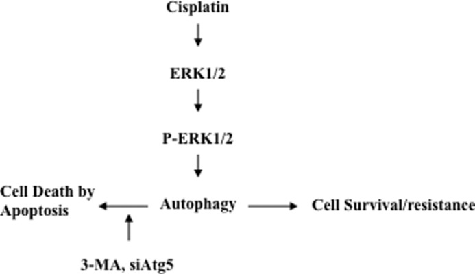
Cisplatin-based treatment is the first line chemotherapy for several cancers including ovarian cancer. The development of cisplatin resistance results in treatment failure, but the underlying mechanisms are not fully understood. Here we show that the induction of autophagy plays an important role in cisplatin resistance in ovarian cancer cells. Specifically, we show that cisplatin resistance is correlated with autophagy induction in a panel of ovarian cancer cells but not in immortalized human ovarian surface epithelial cells. Mechanistically, cisplatin treatment activates ERK and subsequently promotes autophagy. The inhibition of ERK activation with MEK inhibitors or knockdown of ERK expression with siRNA decreases cisplatin-induced autophagy and subsequently sensitizes ovarian cancer cells to cisplatin-induced apoptosis. In ovarian cancer cells that have developed acquired cisplatin resistance, both ERK activation and autophagy induction are increased. Importantly, knockdown of ERK or inhibition of autophagy promotes cisplatin-induced apoptosis in acquired cisplatin-resistant cells. Collectively, our data indicate that ERK-mediated autophagy can lead to cisplatin resistance and suggest that cisplatin resistance can be overcome by inhibition of autophagy in ovarian cancer cells.

Keywords: Autophagy; Cancer Therapy; Drug Resistance; Extracellular Signal-regulated Kinase (ERK); Ovarian Cancer.


Figures

FIGURE 1.
FIGURE 1.
Effect of cisplatin treatment on LC3 levels and growth inhibition in a panel of human ovarian cell lines. A, Western blot analyses of the levels of LC3. Ovarian cancer cell lines were left untreated or treated with 20 μm cisplatin (RMG-1, OV433, OV90, and OVCA420 cells) or 10 μm cisplatin (CAOV3 cells) for the indicated time periods. IOSE385 cells were treated with 3.33 μm cisplatin (at IC50) for the indicated time periods. β-Actin was used as a loading control. B, quantification of LC3-II. The level of LC3-II was quantified by densitometry. Data represent mean ± S.D. (error bars) of three independent experiments. **, p < 0.001, statistically significant; NS, not statistically significant. C, MTT assays of growth inhibition. Ovarian cancer cells in A were left untreated or treated with cisplatin with the indicated concentrations for 48 h.
FIGURE 2.
FIGURE 2.
Effect of Baf A1 and 3-MA on cisplatin-induced LC3-II, and the role for cisplatin treatment in p62 levels and LC3 punctate formation. A, Western blot analyses of the levels of LC3 in cells treated with Baf A1 (upper panel) and quantification of LC3-II (lower panel). OV433 cells were left untreated or treated with 20 μm cisplatin in the presence or absence of 2 nm Baf A1 for 24 h. B, Western blot analyses of LC3 in cells treated with 3-MA (upper panel) and quantification of LC3-II (lower panel). OV433 cells were left untreated or treated with 20 μm cisplatin in the presence or absence of 1 mm 3-MA for 24 h. C, representative fluorescent images of GFP-LC3 (upper panel) and quantification of GFP-LC3 dots (lower panel). OV433 cells transfected with GFP-LC3 were selected with G418 for stable GFP-LC3 and then subjected to cisplatin treatment (20 μm, 20 h) or left alone. Bright dots denote autophagosomes. Bars represent mean ± S.D. (error bars) of triplicate samples with >20 cells analyzed per sample. D, Western blot analyses of LC3 and p62 (upper panel) and quantification of LC3-II and p62 (lower panel). OV433 cells were left untreated or treated with 20 μm cisplatin for 24 h. In A, B, and D, data represent mean ± S.D. of three independent experiments. **, p < 0.001, statistically significant.
FIGURE 3.
FIGURE 3.
Effect of cisplatin treatment on MAPK pathways, LC3 levels, and cisplatin sensitivity following ERK inhibition. A, Western blot analyses of the activation of the MAPK pathways including P-ERK1/2, ERK1/2, P-JNK1/2, JNK1/2, P-c-Jun, c-Jun, P-p38, p38, P-CREB, and CREB. OV433 cells were left untreated or treated with 20 μm cisplatin for the indicated time periods. B, Western blot analyses of LC3, P-ERK1/2, ERK1/2, P-CREB, CREB, P-c-Jun, and c-Jun (upper panel) and quantification of LC3-II (lower panel). OV433 cells were left untreated or treated with 20 μm cisplatin in the presence or absence of 10 μm U0126 or 10 μm SB203580 or 10 μm SP600125 for 24 h. C, Western blot analyses of the levels of LC3 and p62 in untreated or 20 μm cisplatin-treated cells with or without 2 nm Baf A1 treatment in the presence or absence of 10 μm U0126 (upper panel) and quantification of LC3-II (middle panel) and p62 (lower panel) in OV433 cells. D, MTT analyses of growth inhibition. OV433 cells were left untreated or treated with 20 μm cisplatin (Cis) in the presence or absence of 10 μm MEK inhibitor U0126 (U), 10 μm p38 inhibitor SB203580 (SB), or 10 μm JNK inhibitor SP600125 (SP) for 24 h. Data represent mean ± S.D. (error bars) of three independent experiments. **, p < 0.001, statistically significant.
FIGURE 4.
FIGURE 4.
Effect of ERK1/2 knockdown on cisplatin-induced LC3 levels, PARP cleavage, and growth inhibition. A, Western blot analyses of ERK1/2, LC3, p62, and PARP cleavage (upper panel) and quantification of LC3-II (lower panel). OV433 cells transfected with either control nontarget siRNAs (siControl) or ERK1/2 siRNAs (siERK1/2) were left untreated or treated with 20 μm cisplatin for 24 h. Quantification of PARP cleavage detected by Western blots was performed using densitometry and ratios of cleaved to uncleaved PARP are indicated. Data represent mean ± S.D. (error bars) of three independent experiments. B, MTT analyses of growth inhibition. OV433 cells were treated as described in A. Data represent mean ± S.D. of three independent experiments. **, p < 0.001, statistically significant.
FIGURE 5.
FIGURE 5.
Effect of pharmacological inhibition of autophagy or knockdown of Atg5 on cisplatin-induced apoptosis and cisplatin sensitivity. A, Western blot analyses of LC3 and PARP cleavage (upper panel) and quantification of LC3-II (lower panel). OV433 cells were left untreated or treated with 20 μm cisplatin in the presence or absence of 1 mm 3-MA for 24 h. Quantification of PARP cleavage was described in Fig. 4A. B, MTT analyses of growth inhibition. OV433 cells were treated as in A. C, Western blot analyses of the levels of Atg5, LC3, and PARP cleavage (upper panel) and quantification of LC3-II (lower panel). OV433 cells transfected with either control nontarget siRNAs (siControl) or Atg5 siRNAs (siAtg5) were left untreated or treated with 20 μm cisplatin for 24 h. Quantification of PARP cleavage was described above. D, MTT analyses of growth inhibition. OV433 cells were treated as in C. Data represent mean ± S.D. (error bars) of three independent experiments. **, p < 0.001, statistically significant.
FIGURE 6.
FIGURE 6.
ERK activation, autophagy induction, and effect of autophagy inhibition on apoptosis in acquired cisplatin-resistant ovarian cancer cell line. A, MTT analyses of growth inhibition. OV433-P and OV433-CR cells were left untreated or treated with cisplatin at the indicated concentrations for 72 h. B, Western blot analyses of LC3, p62, P-ERK1/2, ERK1/2, P-p38, p38, p-JNK, JNK, and PARP. OV433-P and OV433-CR cells were left untreated or treated with 20 μm cisplatin for 24 h. C, representative fluorescent images of GFP-LC3 (left panel) and statistical analysis of GFP-LC3 dots (right panel). OV433-P and OV433-CR cells transfected with GFP-LC3 were selected with G418 for 3 weeks to isolate clones that stably express GFP-LC3. The resulting cells were left untreated or treated with cisplatin (20 μm, 20 h). Bright dots denote autophagosomes. Bars represent mean ± S.D. (error bars) of triplicate samples with >20 cells analyzed per sample. D, Western blot analyses of ERK1/2, LC3, p62, and PARP cleavage (upper panel) and quantification of LC3-II (lower panel). OV433-CR cells transfected with either control nontarget siRNAs (siControl) or ERK1/2 siRNAs (siERK1/2) were left untreated or treated with 20 μm cisplatin for 24 h. Quantification of PARP cleavage was described above. E, Western blot analyses of LC3, and PARP cleavage (left panel) and quantification of LC3-II (right panel). OV433-CR cells were left untreated or treated with 20 μm cisplatin in the presence or absence of 1 mm 3-MA for 24 h. Cleaved PARP was quantified as described above. F, Western blot analyses of Atg5, LC3, p62 and PARP (left panel) and quantification of LC3-II (right panel). OV433-CR cells transfected with siRNA against Atg5 (siAtg5) or control siRNA (siControl) were left untreated or treated with 20 μm cisplatin for 24 h. Cleaved PARP was quantified as described above. Data represent mean ± S.D. of three independent experiments. **, p < 0.001, statistically significant.
FIGURE 7.
FIGURE 7.
Proposed model for the mechanism by which cisplatin activates ERK1/2, which subsequently promotes an autophagic response, leading to cisplatin resistance in ovarian cancer cells.

References

    1. Cho K. R., Shih IeM. (2009) Ovarian cancer. Annu. Rev. Pathol. 4, 287–313 - PMC - PubMed
    1. Bryant C. S., Kumar S., Spannuth W., Shah J. P., Munkarah A. R., Deppe G., Alvarez R. D., Morris R. T. (2011) Feasibility of extension of platinum-free interval with weekly bolus topotecan and subsequent platinum retreatment outcomes in recurrent ovarian cancer. Arch. Gyn. Obstet. 283, 361–367 - PubMed
    1. Tummala M. K., McGuire W. P. (2005) Recurrent ovarian cancer. Clin. Adv. Hematol. Oncol. 3, 723–736 - PubMed
    1. Go R. S., Adjei A. A. (1999) Review of the comparative pharmacology and clinical activity of cisplatin and carboplatin. J. Clin. Oncol. 17, 409–422 - PubMed
    1. Kelland L. (2007) The resurgence of platinum-based cancer chemotherapy. Nat. Rev. Cancer 7, 573–584 - PubMed

Publication types