. 2014 Jun 20;289(25):17365-78.
doi: 10.1074/jbc.M114.567230. Epub 2014 May 2.

Contribution of pyruvate phosphate dikinase in the maintenance of the glycosomal ATP/ADP balance in the Trypanosoma brucei procyclic form

Affiliations

Contribution of pyruvate phosphate dikinase in the maintenance of the glycosomal ATP/ADP balance in the Trypanosoma brucei procyclic form

Kamel Deramchia et al. J Biol Chem. .

Abstract

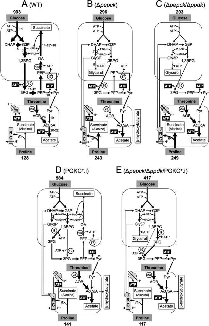
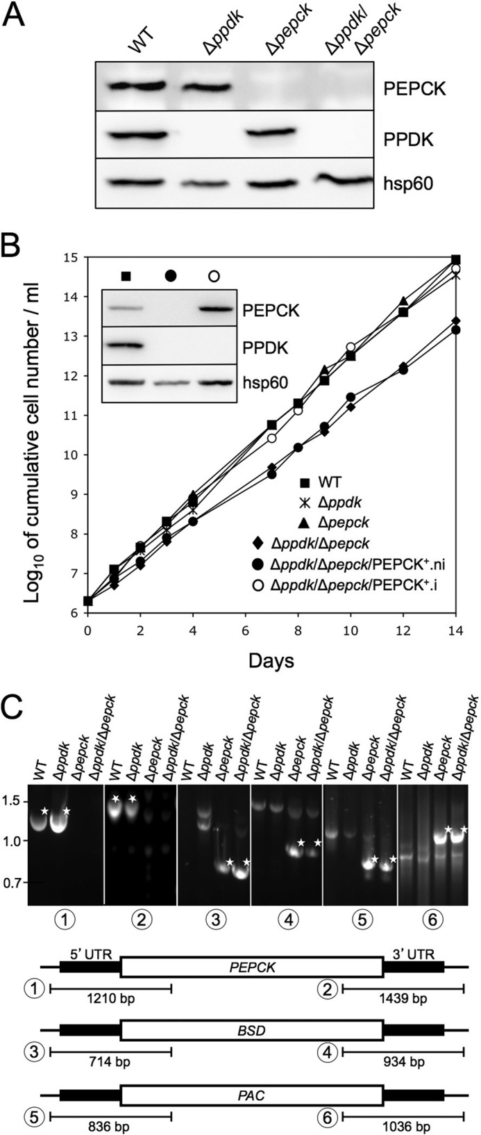
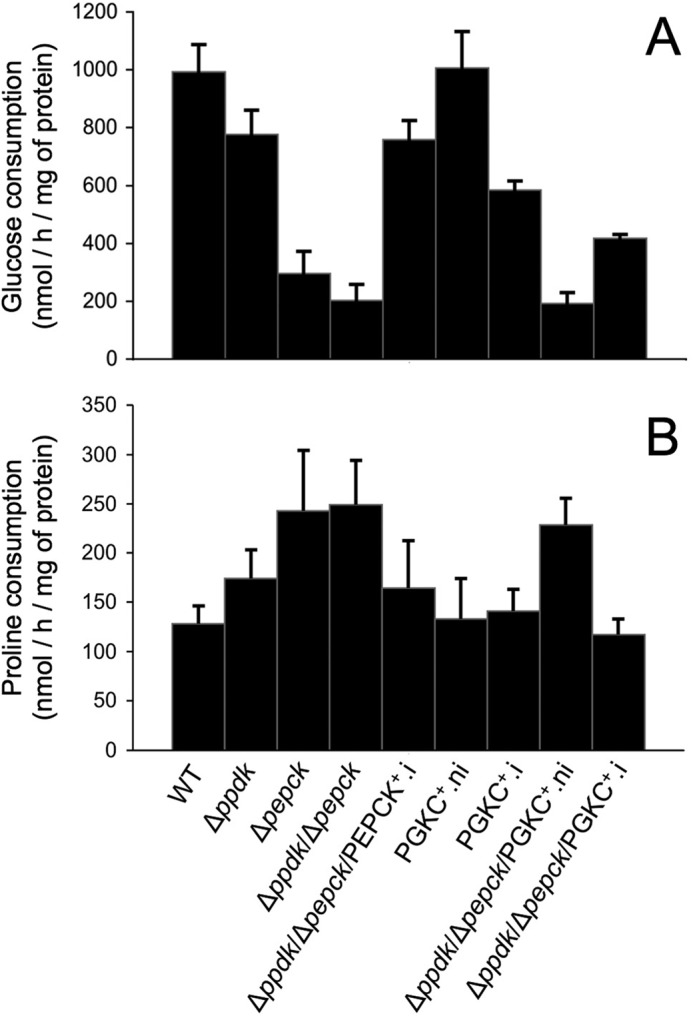
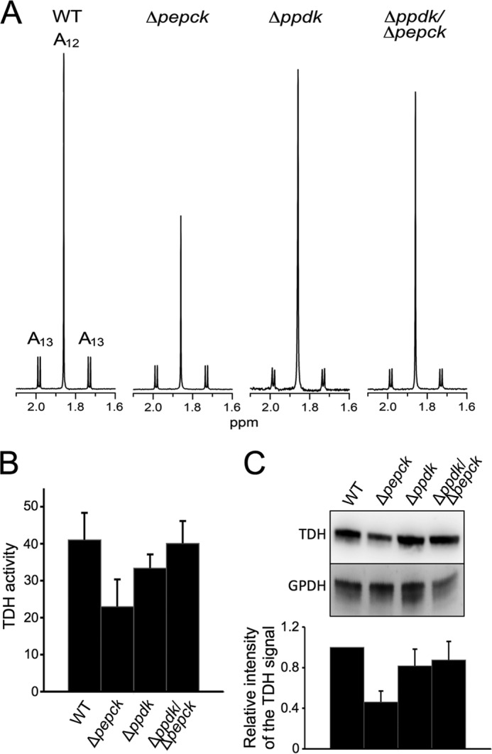
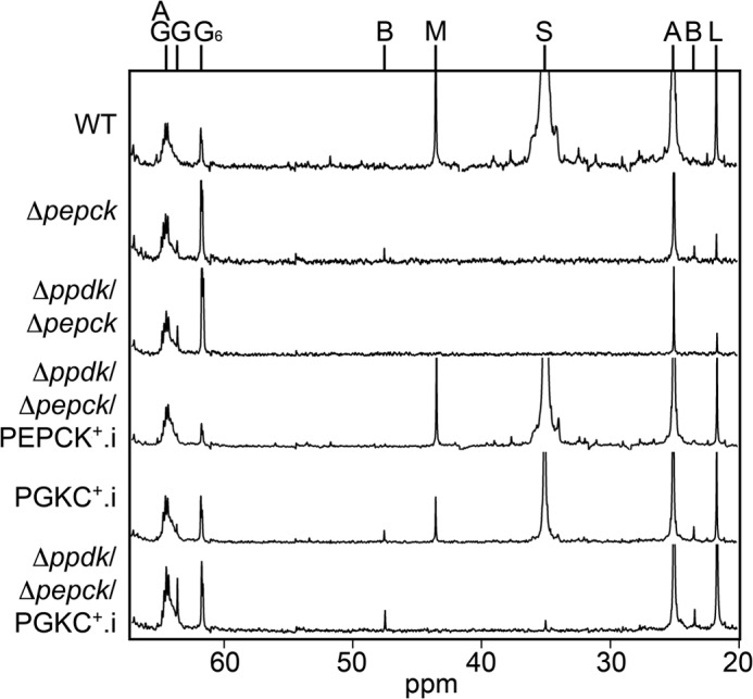
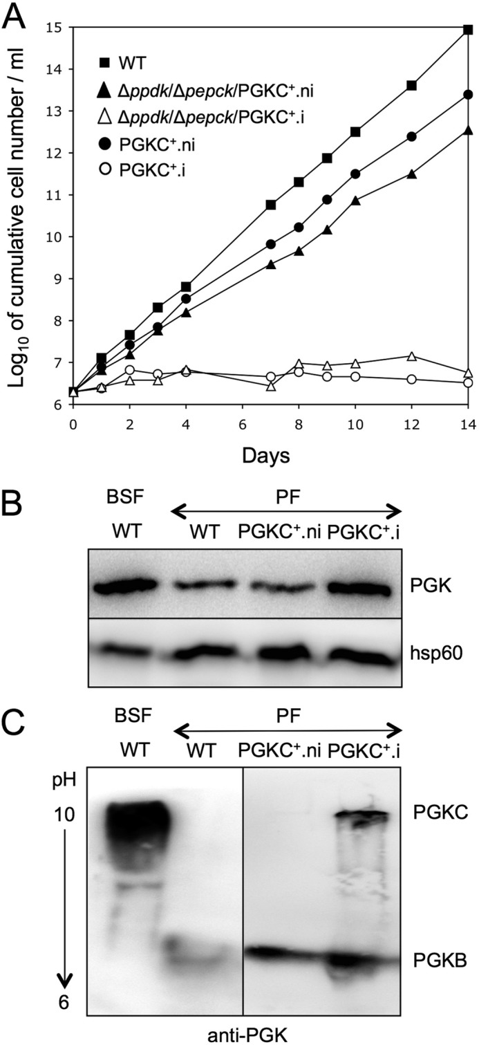
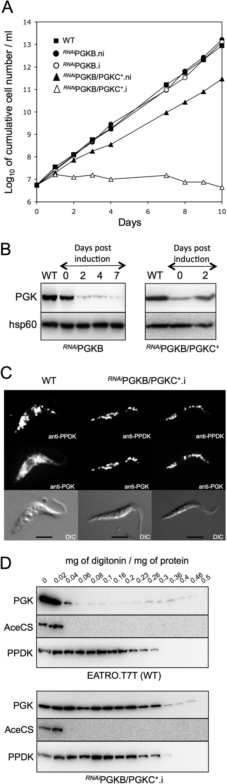
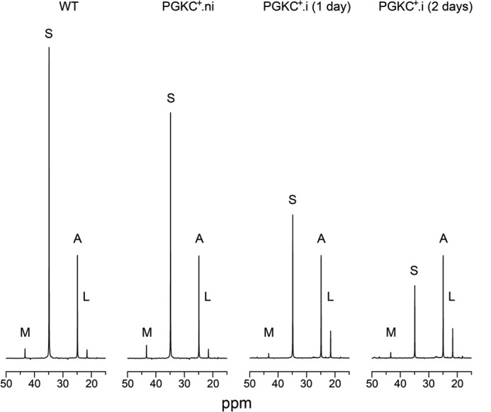
Trypanosoma brucei belongs to a group of protists that sequester the first six or seven glycolytic steps inside specialized peroxisomes, named glycosomes. Because of the glycosomal membrane impermeability to nucleotides, ATP molecules consumed by the first glycolytic steps need to be regenerated in the glycosomes by kinases, such as phosphoenolpyruvate carboxykinase (PEPCK). The glycosomal pyruvate phosphate dikinase (PPDK), which reversibly converts phosphoenolpyruvate into pyruvate, could also be involved in this process. To address this question, we analyzed the metabolism of the main carbon sources used by the procyclic trypanosomes (glucose, proline, and threonine) after deletion of the PPDK gene in the wild-type (Δppdk) and PEPCK null (Δppdk/Δpepck) backgrounds. The rate of acetate production from glucose is 30% reduced in the Δppdk mutant, whereas threonine-derived acetate production is not affected, showing that PPDK function in the glycolytic direction with production of ATP in the glycosomes. The Δppdk/Δpepck mutant incubated in glucose as the only carbon source showed a 3.8-fold reduction of the glycolytic rate compared with the Δpepck mutant, as a consequence of the imbalanced glycosomal ATP/ADP ratio. The role of PPDK in maintenance of the ATP/ADP balance was confirmed by expressing the glycosomal phosphoglycerate kinase (PGKC) in the Δppdk/Δpepck cell line, which restored the glycolytic flux. We also observed that expression of PGKC is lethal for procyclic trypanosomes, as a consequence of ATP depletion, due to glycosomal relocation of cytosolic ATP production. This illustrates the key roles played by glycosomal and cytosolic kinases, including PPDK, to maintain the cellular ATP/ADP homeostasis.

Keywords: Gene Knockout; Glucose Metabolism; Parasite Metabolism; Peroxisome; Trypanosoma brucei.


Figures

FIGURE 1.
FIGURE 1.
Schematic representation of the intermediary metabolism of procyclic wild-type and mutant cell lines. This figure highlights steps from the glucose metabolism involved in the maintenance of the glycosomal NAD+/NADH and ATP/ADP balances, as well as steps involved in ATP production in the cytosol and mitochondrion from the three main carbon sources used by the procyclic trypanosomes (glucose, proline, and threonine). For simplification, mitochondrial production of succinate from glucose is not shown, as well as the steps of proline degradation and the cytosolic localization of fumarase (step 15*). For enzymatic steps involved in net production of ATP molecules, ATP is represented by white characters on a black background, and the circled step numbers represent enzymes analyzed here. Excreted end products from degradation of glucose, proline and threonine are boxed (succinate and alanine are the main end products excreted from proline metabolism, in the presence or absence of glucose, respectively (14)). The arrow thickness is representative of the measured or estimated metabolic flux through the corresponding branches. The rate of glucose and proline consumption (nmol/h/mg of protein) is indicated above and below the carbon source name, respectively (values from Fig. 3). Abbreviations: AcCoA, acetyl-CoA; AOB, amino oxobutyrate; 1,3BPG, 1,3-biphosphoglycerate; DHAP, dihydroxyacetone phosphate; e, electrons; G3P, glyceraldehyde 3-phosphate; Gly3P, glycerol 3-phosphate; OA, oxaloacetate; 3PG, 3-phosphoglycerate; PEP, phosphoenolpyruvate; Pyr, pyruvate; RC, respiratory chain. Enzymes of glucose of threonine degradation are indicated in panel A, or when mentioned below, in panels B or D: 1, hexokinase; 2, glucose-6-phosphate isomerase; 3, phosphofructokinase; 4, aldolase; 5, triose-phosphate isomerase; 6, glyceraldehyde-3-phosphate dehydrogenase; 7, NADH-dependent glycerol-3-phosphate dehydrogenase (panel B); 8, glycerol kinase (panel B); 9, glycosomal phosphoglycerate kinase (PGKC) (panel D); 10, cytosolic phosphoglycerate kinase (PGKB); 11, phosphoglycerate mutase; 12, enolase; 13, PEPCK; 14, malate dehydrogenase; 15*, cytosolic fumarase (located in the glycosomes for simplification); 16, glycosomal NADH-dependent fumarate reductase; 17, pyruvate phosphate dikinase (PPDK); 18, pyruvate kinase; 19, pyruvate dehydrogenase complex; 20, acetate:succinate CoA-transferase; 21, succinyl-CoA synthetase; 22, acetyl-CoA thioesterase; 23, threonine dehydrogenase (TDH); 24, 2,2-amino-3-ketobutyrate coenzyme A ligase; 25, FAD-dependent glycerol-3-phosphate dehydrogenase (panel B); 26, F0F1-ATP synthase.
FIGURE 2.
FIGURE 2.
Production of Δppdk, Δppdkpepck, and rescue cell lines. Panel A shows a Western blot analysis of the parental procyclic (WT) and mutant cell lines with the immune sera indicated in the right margin. Panel B shows growth curves of the wild-type (WT), Δppdk, Δpepck, Δppdkpepck, and tetracycline-induced (.i) and uninduced (.ni) Δppdkpepck/PEPCK+ cell lines, as well as a Western blot analysis of the wild-type (■), Δppdkpepck/PEPCK+.ni (●), and Δppdkpepck/PEPCK+.i (○) cell lines with the immune sera indicated in the right margin of the inset. Panel C shows a PCR analysis of genomic DNA isolated from the parental wild-type and Δppdk, Δpepck, and ΔppdkΔpepck cell lines. Amplifications were performed with primers based on sequences that flank the 5′ UTR and 3′ UTR fragments used to target the PEPCK gene depletion (black boxes) and internal sequences of the blasticidin (BSD, PCR products 3 and 4), puromycin (PAC, PCR products 5 and 6) resistance genes and, as controls, the PEPCK gene (products 1 and 2). As expected, PCR amplification of the PEPCK gene was only observed in wild-type and Δppdk cell lines, whereas BSD and PAC PCR products were observed only in Δpepck and ΔppdkΔpepck cell lines. White stars indicate the expected PCR fragment.
FIGURE 3.
FIGURE 3.
Glucose and proline consumption by wild-type (WT) and mutant cell lines. The rate of glucose (panel A) and proline (panel B) consumed by the indicated cell lines incubated in SDM79 medium is expressed as nanomole consumed per h and per mg of protein. Mean of at least 3 biological replicates are presented.
FIGURE 4.
FIGURE 4.
TDH expression and activity are not reduced in the Δppdkpepck cell line. In panel A, contribution of threonine and glucose to acetate production was determined by 1H NMR analysis. Acetate excreted by the procyclic wild-type (WT), Δppdk, Δpepck, and Δppdkpepck cell lines from 4 mm d-[U-13C]glucose and 4 mm threonine was determined by 1H NMR. Each spectrum corresponds to one representative experiment from a set of at least 3. A part of each spectrum ranging from 1.6 to 2.1 ppm is shown. The resonances were assigned as indicated: A12, threonine-derived acetate; A13, 13C-enriched glucose-derived acetate. Panel B shows the TDH activity (milliunits/mg of protein), normalized with the pyruvate dehydrogenase activity measured in the same samples. In panel C, expression of TDH and glycerol-3-phosphate dehydrogenase (GPDH) was analyzed by Western blotting with specific immune sera. The ratio between the TDH and GPDH signals, indicated below the blot, represents a mean ± S.D. of 4 different experimental duplicates, with an arbitrary value of 1 for the parental cells (WT).
FIGURE 5.
FIGURE 5.
Analysis of β-hydroxybutyrate and glycerol production from glucose. Supernatant of wild-type (WT) and mutant cell lines incubated with 4 mm [1-13C]glucose were analyzed by 13C NMR. The resonances were assigned as follows: A, acetate; B, β-hydroxybutyrate; G, glycerol; G6, carbon C6 of glucose; L, lactate; M, malate; AG, acylglycerol; S, succinate.
FIGURE 6.
FIGURE 6.
Expression of PGKC in procyclic (PF) and bloodstream (BSF) forms of T. brucei. The growth curves of the parental (WT) and tetracycline-induced (.i) and uninduced (.ni) mutant procyclic cell lines expressing rPGKC in the wild-type or Δppdkpepck backgrounds is shown in panel A. Expression of the PGK isoforms was analyzed by Western blot on total cellular protein extracts separated by SDS-PAGE (panel B) or isoelectric focusing gel electrophoresis (panel C). The anti-hps60 immune serum was used as a loading control.
FIGURE 7.
FIGURE 7.
Subcellular localization of rPGKC. Panel A shows growth curves of the parental procyclic cells (WT) and tetracycline-induced (.i) and uninduced (.ni) RNAiPGKB and RNAiPGKB/PGKC+ cell lines. Expression of PGK isoforms upon induction in the RNAiPGKB and RNAiPGKB/PGKC+ mutants is shown by Western blotting analyses in panel B, with hsp60 immune serum serving as internal control. Panel C shows immunofluorescence analyses of procyclic cells stained with the rabbit anti-PGK (Alexa Fluor 488 channel green) and the monoclonal mouse anti-PPDK (Alexa Fluor 594 channel red) as glycosomal control. Differential interference contrast of cells is shown at the bottom of each panel. Scale bar, 5 μm. In panel D, the presence of the PGK isoforms, the cytosolic acetyl-CoA synthetase (AceCS, cytosolic marker), and PPDK (glycosomal marker), in the pellet fractions from wild-type (upper panel) and RNAiPGKB/PGKC+.i (lower panel) cells incubated with 0.02–0.5 mg of digitonin/mg of protein in STE buffer containing 150 mm NaCl was determined by Western blot analyses.
FIGURE 8.
FIGURE 8.
Carbon-13 NMR spectra of metabolic end products excreted by procyclic cell lines incubated with [1-13C]glucose. For these 13C NMR analyses, the parental EATRO1125.T7T (WT) and uninduced (.ni) and tetracycline-induced (.i, 1 and 2 days) PGKC+ mutant cell lines were incubated with 4 mm d-[1-13C]glucose in PBS/NaHCO3 buffer. The NMR spectra of the incubation medium were obtained after addition of 15 μl of dioxane, to quantify 13C-enriched excreted end products from glucose metabolism (Table 1). The size of the spectra is normalized on the acetate peaks. The resonances were assigned as follows: A, acetate; L, lactate; M, malate; S, succinate.

Similar articles

Cited by

References

    1. Marshall J. S., Ashton A. R., Govers F., Hardham A. R. (2001) Isolation and characterization of four genes encoding pyruvate, phosphate dikinase in the oomycete plant pathogen Phytophthora cinnamomi. Curr. Genet. 40, 73–81 - PubMed
    1. Nevalainen L., Hrdý I., Müller M. (1996) Sequence of a Giardia lamblia gene coding for the glycolytic enzyme, pyruvate, phosphate dikinase. Mol. Biochem. Parasitol. 77, 217–223 - PubMed
    1. Feng X. M., Cao L. J., Adam R. D., Zhang X. C., Lu S. Q. (2008) The catalyzing role of PPDK in Giardia lamblia. Biochem. Biophys. Res. Commun. 367, 394–398 - PubMed
    1. Saavedra E., Encalada R., Pineda E., Jasso-Chávez R., Moreno-Sánchez R. (2005) Glycolysis in Entamoeba histolytica. Biochemical characterization of recombinant glycolytic enzymes and flux control analysis. FEBS J. 272, 1767–1783 - PubMed
    1. Bringaud F., Baltz D., Baltz T. (1998) Functional and molecular characterization of a glycosomal PPi-dependent enzyme in trypanosomatids: pyruvate, phosphate dikinase. Proc. Natl. Acad. Sci. U.S.A. 95, 7963–7968 - PMC - PubMed

Publication types

MeSH terms

Substances

LinkOut - more resources