. 2014 May 2;3(6):409-17.
doi: 10.1242/bio.20147336.

Functional anatomy and ion regulatory mechanisms of the antennal gland in a semi-terrestrial crab, Ocypode stimpsoni

Affiliations

Functional anatomy and ion regulatory mechanisms of the antennal gland in a semi-terrestrial crab, Ocypode stimpsoni

Jyuan-Ru Tsai et al. Biol Open. .

Abstract

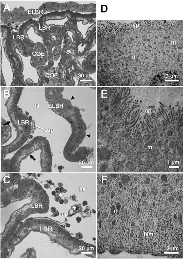
Brachyuran crabs from diverse habitats show great differences in their osmoregulatory processes, especially in terms of the structural and physiological characteristics of the osmoregulatory organs. In crustaceans, the antennal glands are known to be important in osmoregulation, and they play a functional role analogous to that of the vertebrate kidney. Nevertheless, the detailed structure and function of the antennal glands in different species have rarely been described. The aim of this study is to investigate the role of the antennal gland in ion regulation by examining the ultrastructure of the cells and the distribution of the ion regulatory proteins in each cell type in the antennal gland of a semi-terrestrial crab. The results showed that Na(+), K(+)-ATPase activity significantly increased in the antennal gland after a 4-day acclimation in dilute seawater and returned to its original (day 0) level after 7 days. Three major types of cells were identified in the antennal gland, including coelomic cells (COEs), labyrinthine cells (LBRs) and end-labyrinthine cells (ELBRs). The proximal tubular region (PT) and distal tubular region (DT) of the antennal gland consist of LBRs and COEs, whereas the end tubular region (ET) consists of all three types of cells, with fewer COEs and more ELBRs. We found a non-uniform distribution of NKA immunoreactivity, with increasing intensity from the proximal to the distal regions of the antennal gland. We summarise our study with a proposed model for the urine reprocessing pathway and the role of each cell type or segment of the antennal gland.

Keywords: Antennal gland; Coelomic cell; K+-ATPase; Labyrinthine cell; Na+; Osmoregulation.


Conflict of interest statement

Competing interests: The authors have no competing interests to declare.

Figures

Fig. 1.
Fig. 1.. The Na+, K+-ATPase (NKA) activity in the antennal gland of Ocypode stimpsoni in 0, 1, 4 and 7 days after crabs were transferred to 5‰ seawater.
Compared to the control group (0 day), the NKA activity increased and reached a significant level at the 4th day followed by a return to the normal level at the 7th day. These results are analyzed by one-way ANOVA (p<0.05) and grouping by Duncan's multiple range test. Different letters indicate significant differences (p<0.05).
Fig. 2.
Fig. 2.. The gross anatomy and the portions of the antennal gland.
(A) The antennal gland is located in the base of the eyestalk (as the dash circle shows). The arrowhead (◂) indicates the anterior portion of the antennal gland, and the arrow (←) indicates the posterior portion. (The dorsal view of the crab with the carapace removed. The upper side of this figure is the front of the crab.) (B,C) The removed antennal gland is coiled with the bladder (bd) and connective tissue (ct). (B) The dorsal view of the antennal gland has a smooth surface. (C) The ventral view of the antennal gland has an indentation (*) in the centre of the anterior portion. (D) The anterior portion of the antennal gland is composed of the proximal tubular region (PT) and the distal tubular region (DT). The posterior portion of the antennal gland is composed of the end tubular region (ET). Lines E and F represent the different sectioning levels of the antennal gland. In section (E), the sectioning level is crossing through the anterior part and includes both PT and DT. Tubular cells and large coelomosac (arrowheads) are found in the peripheral of the antennal gland defined as DT. A large haemolymph vessel (hv) is found in the centre of the antennal gland which is defined as PT. The haemocytes (*) are found in the haemolymph vessel (hv). In section (F), the sectioning level is on the peripheral region of the antennal gland and includes only DT. Numerous tubular cells and large coelomosac (arrowheads) filled up this region. Scale bars: 2 mm (A), 1 mm (B,C), 300 µm (E,F).
Fig. 3.
Fig. 3.. The cell arrangement and the two major cell types, COEs and LBRs, in PT and DT of the antennal gland.
(A,B) Haemolymph vessels (hv) are composed of endothelial cells (arrowheads) in between COEs and LBRs. The endothelial lining of the haemolymph vessel (hv) is connected to the COEs directly (arrow). Haemocytes (*) are only found in the haemolymph sinus (hs) or in the haemolymph vessels (hv). Lumen (lm) is always in the apical side of LBR. No haemocyte was found in the lumen (lm). (C,D) LBRs are cuboidal cells with apical brush-boarder microvilli (bbm), basolateral membrane folding and lots of mitochondria (m). Numerous vesicles (open triangles) are found in the sub-apical spaces. The length of the apical brush-boarder microvilli (bbm) of LBR increases from that in PT (C) to that in DT (D). (E) COE has irregular cell shape and has extended foot process (f) attached to the basal matrix (bm). Numerous vesicles (open triangle) are found in the COEs. Some of the COE contain a large endosome (en), or called vacuole, in the cytoplasm. (F) The enlarged picture of the foot process shows pedicels (black arrows) along the basal matrix (bm) and has very small vesicles (white arrows), or called the pinocytes, in the cell margin of COE. Scale bars: 20 µm (A,B), 2 µm (C–E), 500 nm (F).
Fig. 4.
Fig. 4.. The ultrastructure of the labyrinthine cells (LBRs) and end-labyrinthine cells (ELBRs) in the end tubular region (ET) of the antennal gland.
(A) ET is the end portion of the antennal gland and is composed of LBR, ELBR and COE. (B) ELBRs are always found in the peripheral side of ET. ELBRs have loose apical microvilli (arrowheads) while LBRs have the brush-boarder microvilli (black arrows). Capillaries (cap) are found between the basal side of the LBRs and ELBRs. (C) Haemocytes (*) are found in the capillary and the haemolymph space (hs). (D) The upper side of the ELBR shows irregular apical surface without a distinct sub-apical region. The mitochondria (m) are distributed from the base of the apical membrane to the top of the nucleus (located in the bottom-right of this photo). (E) The mitochondria (m) are distributed just below the apical surface. (F) The ultrastructure of the bottom side of ELBR. The mitochondria (m) are distributed in the whole cytoplasm and also twined with the basal membrane folding (blm). Scale bars: 30 µm (A), 20 µm (B,C), 3 µm (D), 1 µm (E), 2 µm (F).
Fig. 5.
Fig. 5.. The non uniform distribution of Na+, K+-ATPase (NKA) in the antennal gland.
(A,B) The continuous cross sections of the antennal gland. (A) A negative control slice which was only incubated with the secondary antibody and post stained with the hematoxylin. The blue color indicates the location of the nucleus. (B) The slice showed the localization of NKA in red color which mostly found in the DT and ET regions. (C) The enlarged picture from the PT and DT regions. The labyrinthine cell (LBR) in the PT (left side) shows little staining of NKA than that in the DT (right side). The coelomic cell (COE) shows little staining of NKA. (D) The enlarged picture from the DT and ET regions. The LBR shows a stronger staining pattern than that in the end-labyrinthine cell (ELBR). PT, proximal tubular region. DT, distal tubular region. ET, end tubular region. hc, haemocyte. Scale bars: 250 µm (A,B), 30 µm (C,D).
Fig. 6.
Fig. 6.. The locations of the ion-regulatory proteins in the proximal tubular region (PT) and the distal tubular region (DT) of the antennal gland.
(A) The NKA (green) is in the basolateral membrane and in the foot process (bottom) of the COE in the DT. VHA (red) showed little staining in the apical region of the LBR, with stronger signal aggregated in the centre of the COE. (B) NKA (green) was found in the basolateral membrane of the LBR. NHE (red) was found inside the COE and in the apical to sub-apical regions in the LBR. (C) NKCC (green) and NHE (red) were distributed in the centre of the COE. (D) NKCC (green) was found in the basolateral membrane while VHA (red) was in the apical membrane of the LBR. COE, coelomic cell. LBR, labyrinthine cell. hc, haemocyte. NKCC, Na+/K+/2Cl cotransporter. VHA, V-type H+-ATPase. NHE, Na+/H+ exchanger. NKA, Na+, K+-ATPase. Scale bars: 10 µm (A–C), 5 µm (D).
Fig. 7.
Fig. 7.. The localizations of Na+, K+-ATPase (NKA) and Na+/K+/2Cl cotransporter (NKCC) in the end-labyrinthine cell (ELBR).
The continuous sections (A–C) show different immuno-positive pattern of NKA and NKCC. (A) A negative control slice only incubated with the secondary antibody and post-stained with the hematoxylin. The blue color indicated the location of the nucleus of the ELBR. (B) The red color showed the localization of the NKA in the basolateral membrane (below the nucleus) of the ELBR. The upper side of this image is the apical side, while the bottom is basal side of the cell. (C) The red color indicated the localization of the NKCC-like protein in the apical and sub-apical region (above the nucleus) of the ELBR. The upper side of this image is the apical side, while the bottom is basal side of the cell. Scale bars: 30 µm.
Fig. 8.
Fig. 8.. Schematic drawing of the ion-regulatory models and the ultrastructure of the three cell types in the antennal gland.
(A) The labyrinthine cells (LBR) have apical brush-border microvilli, a sub-apical space with vesicles (open triangle) and basal folding with elongated mitochondria (m). On the outer side of the apical brush-border microvilli of the LBRs, numerous aposomes (black arrowheads) were frequently found. NKCC and NKA locate in the basolateral membrane. VHA and NHE locate in the apical membrane and in the sub-apical region. (B) The ELBRs have an irregular apical membrane and its mitochondria (m) are distributed in the whole cytoplasm. NKA locates in the basolateral membrane while NKCC in the apical membrane. VHA and NHE are in the apical membrane and in the sub-apical region. (C) The coelomic cells (COE) have small vesicles (open arrow), vesicles (open triangle) and a large endosome (en). NKA locates in the membrane of the foot process. The NKCC, NHE and VHA aggregate in the centre of the COE.
Fig. 9.
Fig. 9.. Schematic diagram of the proposed haemolymph/urine flow in the antennal gland.
The haemolymph/urine flow first enters into the antennal gland through COEs (dash arrow) into the lumen. The filtrate is transported to the apical side of LBRs and re-processed by the LBRs (gray arrows). COE, coelomic cell. LBR, labyrinthine cell. hs, haemolymph sinus. *haemocyte. Scale bar: 30 µm.

Similar articles

Cited by

References

    1. Ahearn G. A., Franco P. (1990). Sodium and calcium share the electrogenic 2 Na(+)-1 H+ antiporter in crustacean antennal glands. Am. J. Physiol. 259, F758–F767. - PubMed
    1. Brown D., Paunescu T. G., Breton S., Marshansky V. (2009). Regulation of the V-ATPase in kidney epithelial cells: dual role in acid-base homeostasis and vesicle trafficking. J. Exp. Biol. 212, 1762–1772 10.1242/jeb.028803 - DOI - PMC - PubMed
    1. Buranajitpirom D., Asuvapongpatana S., Weerachatyanukul W., Wongprasert K., Namwong W., Poltana P., Withyachumnarnkul B. (2010). Adaptation of the black tiger shrimp, Penaeus monodon, to different salinities through an excretory function of the antennal gland. Cell Tissue Res. 340, 481–489 10.1007/s00441-010-0971-y - DOI - PubMed
    1. Chung K. F., Lin H. C. (2006). Osmoregulation and Na,K-ATPase expression in osmoregulatory organs of Scylla paramamosain. Comp. Biochem. Physiol. 144A, 48–57 10.1016/j.cbpa.2006.02.003 - DOI - PubMed
    1. De Vries M. C., Wolcott D. L., Holliday C. W. (1994). High ammonia and low pH in the urine of the ghost crab, Ocypode quadrata. Biol. Bull. 186, 342–348 10.2307/1542280 - DOI - PubMed

LinkOut - more resources