. 2014 Jun;7(3):249-56.
doi: 10.1161/CIRCGENETICS.113.000480. Epub 2014 May 2.

A mutation causing Brugada syndrome identifies a mechanism for altered autonomic and oxidant regulation of cardiac sodium currents

Affiliations

A mutation causing Brugada syndrome identifies a mechanism for altered autonomic and oxidant regulation of cardiac sodium currents

Takeshi Aiba et al. Circ Cardiovasc Genet. 2014 Jun.

Abstract

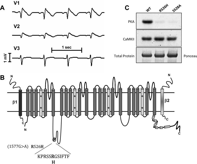
Background: The mechanisms of the electrocardiographic changes and arrhythmias in Brugada syndrome (BrS) remain controversial. Mutations in the sodium channel gene, SCN5A, and regulatory proteins that reduce or eliminate sodium current (INa) have been linked to BrS. We studied the properties of a BrS-associated SCN5A mutation in a protein kinase A (PKA) consensus phosphorylation site, R526H.

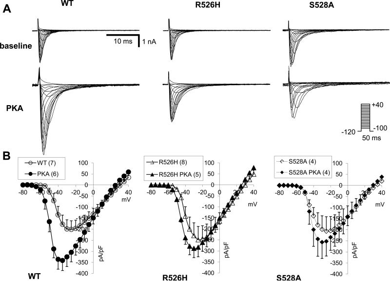
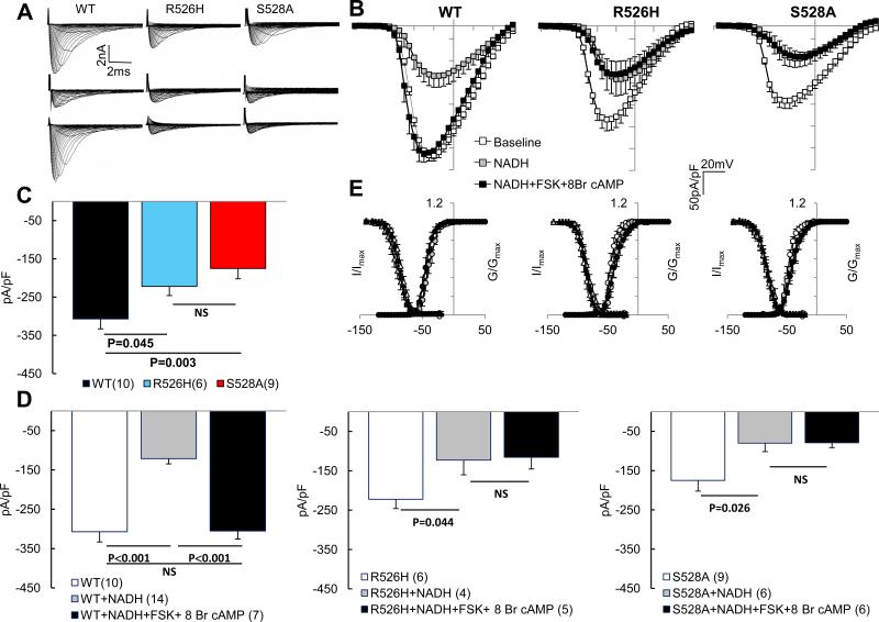
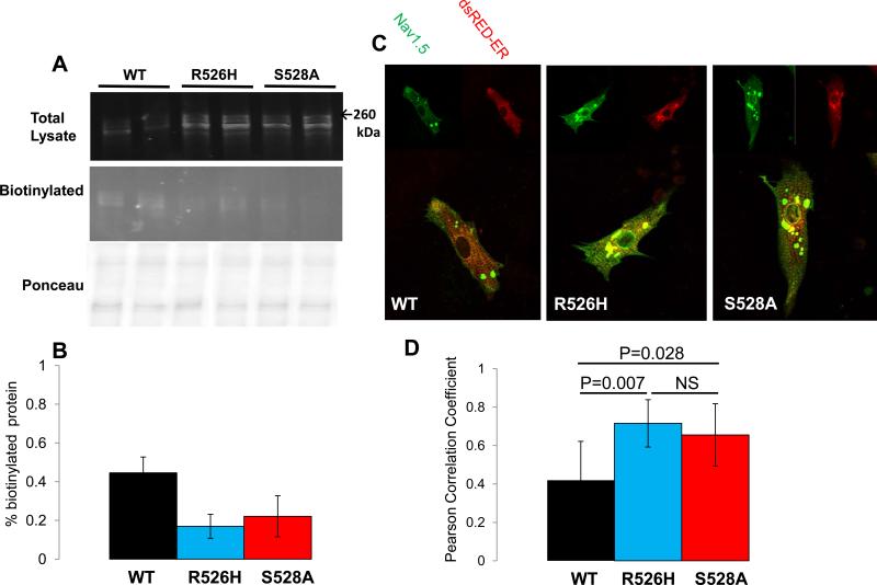
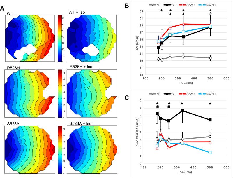
Methods and results: In vitro PKA phosphorylation was detected in the I-II linker peptide of wild-type (WT) channels but not R526H or S528A (phosphorylation site) mutants. Cell surface expression of R526H and S528A channels was reduced compared with WT. Whole-cell INa through all channel variants revealed no significant differences in the steady-state activation, inactivation, and recovery from inactivation. Peak current densities of the mutants were significantly reduced compared with WT. Infection of 2D cultures of neonatal rat ventricular myocytes with WT and mutant channels increased conduction velocity compared with noninfected cells. PKA stimulation significantly increased peak INa and conduction velocity of WT but not mutant channels. Oxidant stress inhibits cardiac INa; WT and mutant INa decreases with the intracellular application of reduced nicotinamide adenine dinucleotide (NADH), an effect that is reversed by PKA stimulation in WT but not in R526H or S528A channels.

Conclusions: We identified a family with BrS and an SCN5A mutation in a PKA consensus phosphorylation site. The BrS mutation R526H is associated with a reduction in the basal level of INa and a failure of PKA stimulation to augment the current that may contribute to the predisposition to arrhythmias in patients with BrS, independent of the precipitants.

Keywords: death, sudden, cardiac; ion channel; mutation; reactive oxygen species.


Figures

Figure 1
Figure 1
(A) Leads V1-V3 of the patient's ECG exhibit typical domed J-point elevation. (B) A schematic of the NaV1.5 pore-forming α subunit and the β1 and β2 subunits. The disease causing mutation (R526H) is in the I-II interdomain linker of NaV1.5 in a canonical PKA phosphorylation recognition sequence. The residue two amino acids C-terminal, S528 was mutated to alanine in this study. (C) In vitro phosphorylation of I-II linker peptides. Purified fragments were incubated with PKA and calcium-calmodulin kinase II δ (CaMKII) in the presence of γP32 labeled ATP. The labeled peptides were separated by PAGE. Mutant peptides R526H and S528A are not labeled by PKA but are phosphorylated as efficiently as wild type in the presence of CaMKII.
Figure 2
Figure 2
PKA regulation of Na current variants. (A) Representative families of current through WT, R526H and S528A channels expressed in HEK 293 cells in the presence and absence of PKA activation. (B) I-V relationships in the presence (filled symbols) and absence (open symbols) of PKA stimulation. There is no increase in the current through the mutant channels. (C) Activation (circles) and steady state inactivation (triangles) curves in the presence (filled symbols) and absence (open symbols) of PKA stimulation. The data are fit to a Boltzman function as described in the methods. There are no significant differences between the WT and mutant channels in the basal voltage dependence and kinetics of gating.
Figure 2
Figure 2
PKA regulation of Na current variants. (A) Representative families of current through WT, R526H and S528A channels expressed in HEK 293 cells in the presence and absence of PKA activation. (B) I-V relationships in the presence (filled symbols) and absence (open symbols) of PKA stimulation. There is no increase in the current through the mutant channels. (C) Activation (circles) and steady state inactivation (triangles) curves in the presence (filled symbols) and absence (open symbols) of PKA stimulation. The data are fit to a Boltzman function as described in the methods. There are no significant differences between the WT and mutant channels in the basal voltage dependence and kinetics of gating.
Figure 3
Figure 3
Regulation of Na channel variants by NADH and PKA. The currents are recorded with fluoride in the pipette. (A) Representative families of current through WT, R526H and S528A channels expressed in HEK 293 cells (top row), in the presence of 100 μM NADH (middle) and NADH plus PKA activation with forskolin and 8-Br-cAMP (bottom). (B) I-V relationships in the absence (open symbols), presence of NADH (gray symbols) and NADH + PKA stimulation (filled symbols). NADH reduces the current through all channel variants, while PKA activation does not increase the current density through the mutant channels. (C) Bar plots of peak current densities of WT (black), R526H (blue) and S528A (red) under basal conditions. (D) Summary bar plots for WT, R526H and S528A channel peak current density at baseline (open bars) with NADH (gray bars) and NADH + PKA activation (black bars). (E) Activation (circles) and steady state inactivation (triangles) curves in the absence (open symbols), presence of NADH (gray symbols) and NADH + PKA stimulation (filled symbols) for each of the channel variants, there are no significant differences in any of the variants under any of the conditions.
Figure 4
Figure 4
Mutations and channel trafficking. (A) Surface membrane expression of the channel measured by biotinylation in intact cells. The top panel shows total lysate, the middle panel the protein captured on streptavadin beads stained with an anti-NaV1.5 antibody and the bottom panel is a Ponceau stained blot demonstrating equivalent loading. (B) The percentage of total lysate modified by biotin is significantly decreased in the R526H (blue bar) and S528A (red bars) mutant channels compared to WT (black bars). (C) Immuncytochemical localization of the channel proteins in transduced NRVMs. The NaV1.5 channel variant is co-transfected with the endoplasmic reticulum marker dsRed-ER. There is significantly increased co-localization of both mutant channel proteins with the ER marker compared to the wild type. (D) Bar plots of the percent co-localization of the channel protein and dsRed-ER.
Figure 5
Figure 5
Optical mapping of Na channel variants. (A) Representative isochronal maps from cultures of NRVMs infected with the Na channel variants and stimulated at the left side of the culture. (B) Plots of the average conduction velocity (CV) ± SEM of WT, R526H and S528A transduced cultures and non-infected (NT) control cultures over a range of pacing cycle lengths (PCL). CV is consistently faster in WT, R526H and S528A NaV1.5 infected cultures compared with the non-transduced (NT) controls over the entire range of PCLs. *p<0.05, all vs. NT; #p<0.01, all vs. NT. (C) Plots of the change in average CV ± SEM of WT, R526H and S528A transduced cultures and NT control cultures over a range of PCL after application of 1 μM isoproterenol. Change in CV of WT transduced cultures is significantly larger than CV changes of NT cultures and mutant transduced cultures after isoproterenol application. *p<0.05, R526H vs. WT; #p<0.05, S528A vs. WT. The number cultures studied at all PCLs in both the absence and presence of isoproterenol were NT (14), WT (13), S528A (5) and R526H (11).

Similar articles

Cited by

References

    1. Antzelevitch C, Brugada P, Borggrefe M, Brugada J, Brugada R, Corrado D, et al. Brugada syndrome: Report of the second consensus conference: Endorsed by the Heart Rhythm Society and the European Heart Rhythm Association. Circulation. 2005;111:659–670. - PubMed
    1. Martini B, Nava A, Thiene G, Buja GF, Canciani B, Scognamiglio R, et al. Ventricular fibrillation without apparent heart disease: Description of six cases. Am Heart J. 1989;118:1203–1209. - PubMed
    1. Haissaguerre M, Derval N, Sacher F, Jesel L, Deisenhofer I, de Roy L, et al. Sudden cardiac arrest associated with early repolarization. N Engl J Med. 2008;358:2016–2023. - PubMed
    1. Hoogendijk MG, Opthof T, Postema PG, Wilde AA, de Bakker JM, Coronel R. The Brugada ECG pattern: A marker of channelopathy, structural heart disease, or neither? Toward a unifying mechanism of the Brugada syndrome. Circ Arrhythm Electrophysiol. 2010;3:283–290. - PubMed
    1. Patel SS, Anees S, Ferrick KJ. Prevalence of a Brugada pattern electrocardiogram in an urban population in the United States. Pacing Clin Electrophysiol. 2009;32:704–708. - PubMed

Publication types

MeSH terms

LinkOut - more resources