. 2014 Jun 1;192(11):5002-11.
doi: 10.4049/jimmunol.1302492. Epub 2014 May 2.

Steady-state antigen scavenging, cross-presentation, and CD8+ T cell priming: a new role for lymphatic endothelial cells

Affiliations

Steady-state antigen scavenging, cross-presentation, and CD8+ T cell priming: a new role for lymphatic endothelial cells

Sachiko Hirosue et al. J Immunol. .

Abstract

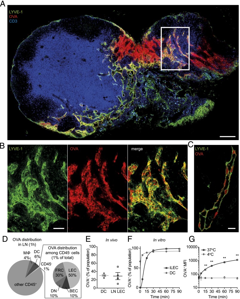
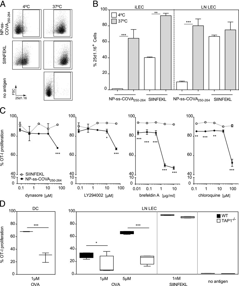
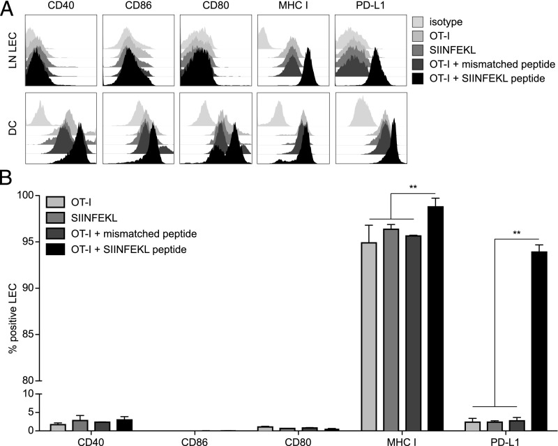
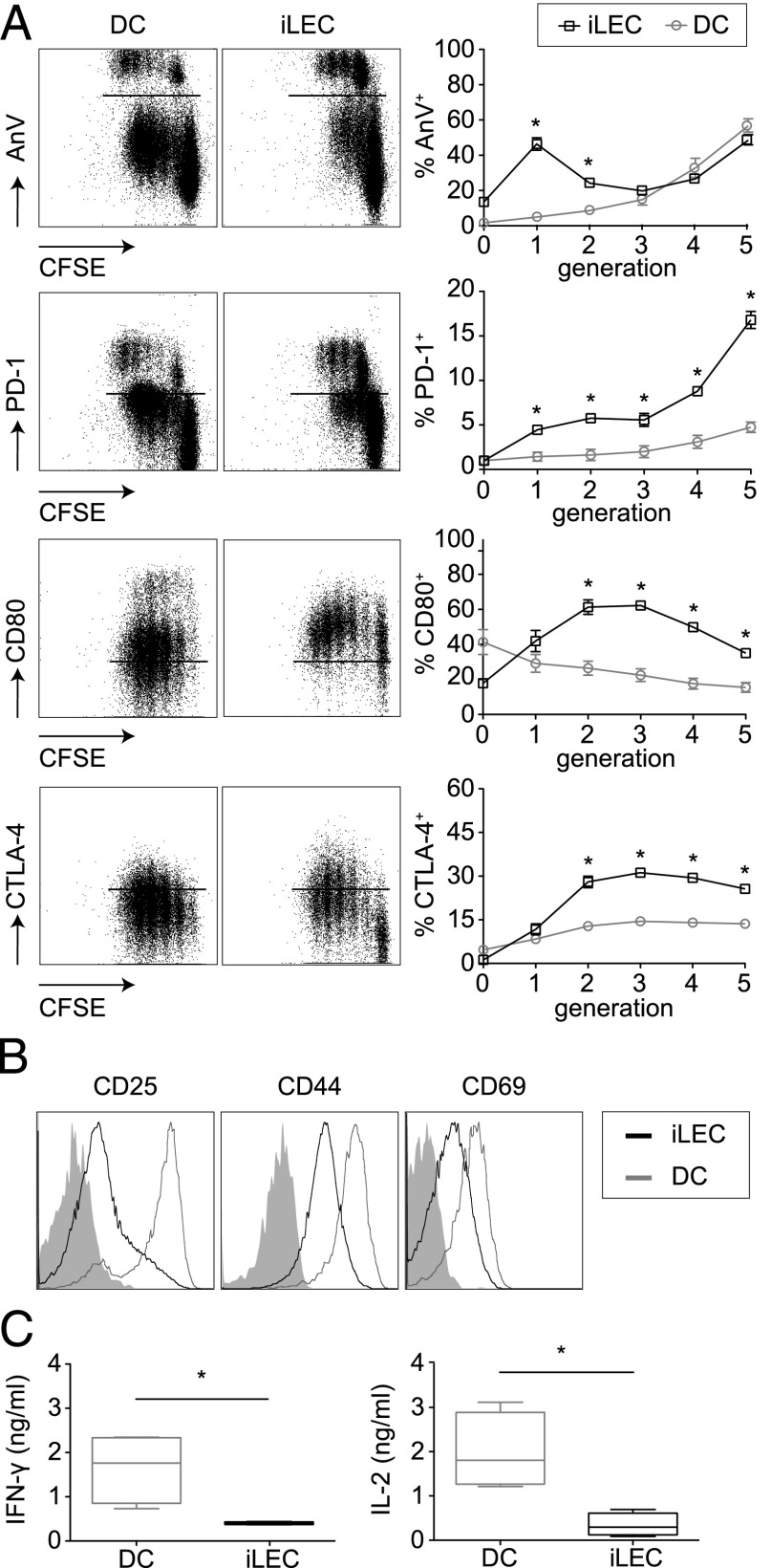
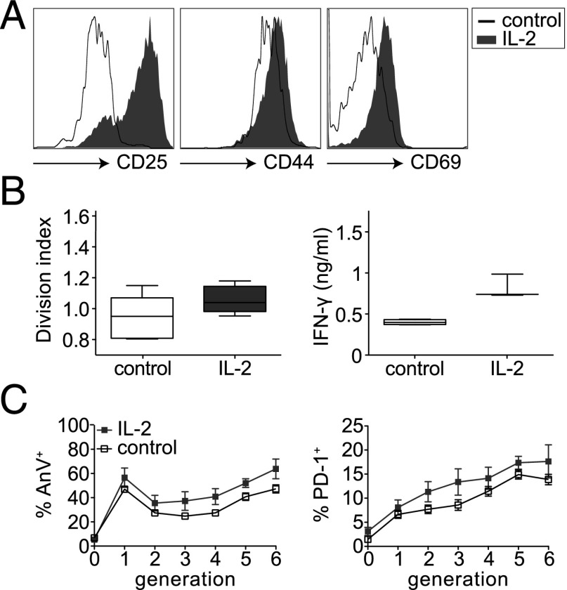
Until recently, the known roles of lymphatic endothelial cells (LECs) in immune modulation were limited to directing immune cell trafficking and passively transporting peripheral Ags to lymph nodes. Recent studies demonstrated that LECs can directly suppress dendritic cell maturation and present peripheral tissue and tumor Ags for autoreactive T cell deletion. We asked whether LECs play a constitutive role in T cell deletion under homeostatic conditions. In this study, we demonstrate that murine LECs under noninflamed conditions actively scavenge and cross-present foreign exogenous Ags to cognate CD8(+) T cells. This cross-presentation was sensitive to inhibitors of lysosomal acidification and endoplasmic reticulum-golgi transport and was TAP1 dependent. Furthermore, LECs upregulated MHC class I and the PD-1 ligand PD-L1, but not the costimulatory molecules CD40, CD80, or CD86, upon Ag-specific interactions with CD8(+) T cells. Finally, Ag-specific CD8(+) T cells that were activated by LECs underwent proliferation, with early-generation apoptosis and dysfunctionally activated phenotypes that could not be reversed by exogenous IL-2. These findings help to establish LECs as APCs that are capable of scavenging and cross-presenting exogenous Ags, in turn causing dysfunctional activation of CD8(+) T cells under homeostatic conditions. Thus, we suggest that steady-state lymphatic drainage may contribute to peripheral tolerance by delivering self-Ags to lymph node-resident leukocytes, as well as by providing constant exposure of draining peripheral Ags to LECs, which maintain tolerogenic cross-presentation of such Ags.


Figures

FIGURE 1.
FIGURE 1.
LECs scavenge exogenous protein, in vivo and in vitro. (AE) After i.d. injection into the footpads, LECs in the draining LN take up OVA rapidly. (A) Brachial LN section showing LEC (Lyve-1, green)–associated distribution of OVA-AF647 (OVA, red) after 90 min; T cells (CD3e, blue) are shown for orientation. Scale bar, 150 μm. (B) Close-up image of region indicated by white box in (A) shows colocalization of OVA with LYVE-1+ LECs. Scale bar, 75 μm. (C) LYVE-1+ lymphatic vessel shows OVA+ vesicles within the LECs. Scale bar, 10 μm. (D) Cellular distribution of OVA in the draining LN 90 min after i.d. injection, as analyzed by flow cytometry. Of all OVA+ cells, 6% were DCs (CD11b−/+CD11c+), and 4% were macrophages (MΦ; CD11b+CD11c), whereas 1% of OVA+ cells in the LN were stromal cells (CD45). Among these (inset), LECs (gp38+CD31+) scavenged the most compared with FRCs (gp38+CD31), BECs (gp38CD31+), and double-negative cells (DN; gp38CD31). (E) Shown as percentages of each LN cell population positive for OVA, LECs were similar to CD11bCD11c+MHCIImid+ DCs in their scavenging capabilities, and these two cell populations represented the highest percentage of OVA+ cells among all LN cell types. Data are from two independent experiments (n = 2 each). (F and G) To demonstrate in vitro OVA accumulation, iLECs and BMDCs were incubated over 90 min at 4 or 37°C with 1 μM OVA-AF647. Cells were washed and analyzed for OVA uptake by flow cytometric analysis. (F) Percentage of OVA-AF647+ cells plotted for gp38+CD31+-gated iLECs and CD11c+-gated DCs at 37°C over time. (G) Geometric mean of OVA fluorescence is plotted for iLECs at 4°C versus 37°C. The data are representative of two independent experiments (n = 3 each). **p < 0.01 using two-way ANOVA and Bonferroni posttest.
FIGURE 2.
FIGURE 2.
LECs process and cross-present exogenous Ag, resulting in priming naive CD8+ T cells. (A) Detection of the MHC class I–SIINFEKL complex using the Ab 25d1.16 on ex vivo–expanded LN LECs (CD45CD31+gp38+) after exposure to NP-ss-COVA250–264. Unlike the free peptide OVA257–264 (SIINFEKL), NP-ss-COVA250–264 cannot bind extracellularly to MHC class I; rather, the Ag must be processed intracellularly, as seen by the lack of presentation at 4°C. (B) Expression of OVA peptide (SIINFEKL)–MHC class I complex by LN LECs and cultured iLECs after 18 h of incubation with NP-ss-COVA250–264 or SIINFEKL at 2.5μ M for 18 h at 4 or 37°C. Data shown are from two independent experiments (n = 3 each). (C) Proliferation of CFSE-labeled OT-I CD8+ T cells after 3 d of coculture with iLECs is impaired in the presence of dynasore and LY294002, which block Ag uptake pathways, as well as with BFA and chloroquine, which block ER–golgi membrane trafficking and endosome acidification, respectively. A total of 1 nM SIINFEKL peptide or NP-ss-COVA250–264 was used as Ag; the data shown are representative of two experiments (n = 3 each). (D) The ability of LECs to cross-prime OT-I CD8+ T cells after OVA uptake depends on TAP1, which is required for intra-ER loading of peptides onto MHC class I molecules. Shown are percentages of proliferation of CFSE-labeled OT-I CD8+ T cells after 3 d of coculture with LN LECs or DCs derived from WT or TAP1-null mice in the presence of OVA or SIINFEKL. The data shown are representative of three independent experiments (n = 3 each). *p < 0.05, **p < 0.01, ***p < 0.001 using two-way ANOVA with a Bonferroni posttest.
FIGURE 3.
FIGURE 3.
Ag-specific interaction with naive CD8+ T cells results in upregulation of MHC class I and PD-L1 expression on LECs. (A and B) In the presence of Ag-specific CD8+ T cells in vitro, the LEC phenotype suggests coinhibitory signaling. Naive OVA-specific OT-I CD8+ T cells were cocultured with ex vivo–expanded LN LECs or BMDCs from C57BL/6 mice in the presence (OT-I + SIINFEKL peptide) or absence (OT-I) of 1 nM SIINFEKL, the immunodominant MHC class I peptide of OVA, or 1 nM AMQMLKETI peptide (OT-I + mismatched peptide). As an additional control, DCs or LN LECs also were incubated with 1 nM SIINFEKL in the absence of CD8+ T cells (SIINFEKL). After 24 h of T cell/LEC or T cell/DC coculture, the relative expression levels of costimulatory molecules CD40, CD86 (B7-2), CD80 (B7-1), MHC class I, and PD-L1 (or B7-H1) were determined by flow cytometric analysis. (A) Representative graphs for each marker are shown on gp38+CD31+-gated LN LECs or CD11c+-gated DCs incubated with OT-I + SIINFEKL, OT-I + mismatched peptide, SIINFEKL only, OT-I only, or isotype control. (B) Percentages of CD40+, CD86+, CD80+, MHC class I+, and PD-L1+ cells in gp38+CD31+-gated LN LECs in each case. Data are mean ± SD from one of two representative experiments (n = 3 each). **p < 0.01 using two-way ANOVA followed by Bonferroni posttest.
FIGURE 4.
FIGURE 4.
Cross-presentation by LECs induces impaired CD8+ T cell proliferation. Naive CFSE-labeled OVA-specific OT-I CD8+ T cells were cocultured with iLECs or BMDCs from C57BL/6 mice in the presence of NP-ss-COVA250–264 at 1 nM and analyzed after 3 d. (A) Phenotypes of OT-I CD8+ T cells after priming by cross-presenting DCs or iLECs, as analyzed by flow cytometric evaluation of annexin V, PD-1, CD80 (B7-1), and CTLA-4 surface marker expression. Representative dot plots from live-gated cells (left panels). Percentages of positive cells/generation from one representative of three to five independent experiments (n = 3–4 each) (right panels). (B) Representative flow cytometry graphs showing activation marker expression on OT-I CD8+ cells after 3 d of cross-priming by iLECs or DCs; shaded graphs show naive (noneducated) OT-I CD8+ T cells. (C) Cytokine secretion by iLEC- or DC-educated OT-I CD8+ T cells, as assessed by ELISA. Data are mean ± SD from one representative experiment of four (n = 4 each). *p < 0.05 using two-way ANOVA followed by Bonferroni posttest.
FIGURE 5.
FIGURE 5.
The LEC-educated T cell phenotype is only partially reversed by IL-2. Naive CFSE-labeled OVA-specific OT-I CD8+ T cells were cocultured with iLECs for 3 d in the presence of Ag (1 nM NP-ss-COVA250–264) and supplemented with 50 U/ml IL-2. (A) Representative flow cytometry graphs showing OT-I surface expression of activation markers after 3 d of priming by iLECs in the absence or presence of IL-2. Data are representative of three independent experiments (n = 4 each). (B) Division index of proliferating OT-I CD8+ T cells (left panel) and IFN-γ release (right panel) were affected only slightly by IL-2. Data are mean ± SD from one representative of four independent experiments (n = 4 each). (C) Percentages of annexin V+ and PD-1+ OT-I CD8+ T cells/generation after 3 d of coculture with iLECs are unaffected by IL-2. Data are mean ± SD from two independent experiments (n = 7 each).

References

    1. Oliver G., Alitalo K. 2005. The lymphatic vasculature: recent progress and paradigms. Annu. Rev. Cell Dev. Biol. 21: 457–483 - PubMed
    1. Tal O., Lim H. Y., Gurevich I., Milo I., Shipony Z., Ng L. G., Angeli V., Shakhar G. 2011. DC mobilization from the skin requires docking to immobilized CCL21 on lymphatic endothelium and intralymphatic crawling. J. Exp. Med. 208: 2141–2153 - PMC - PubMed
    1. Clement C. C., Cannizzo E. S., Nastke M.-D., Sahu R., Olszewski W., Miller N. E., Stern L. J., Santambrogio L. 2010. An expanded self-antigen peptidome is carried by the human lymph as compared to the plasma. PLoS ONE 5: e9863. - PMC - PubMed
    1. Fletcher A. L., Malhotra D., Turley S. J. 2011. Lymph node stroma broaden the peripheral tolerance paradigm. Trends Immunol. 32: 12–18 - PMC - PubMed
    1. Graham G. J. 2009. D6 and the atypical chemokine receptor family: novel regulators of immune and inflammatory processes. Eur. J. Immunol. 39: 342–351 - PubMed

Publication types

MeSH terms