Case Reports
. 2014 Nov 15;210(10):1595-9.
doi: 10.1093/infdis/jiu250. Epub 2014 May 1.

Identification of a novel polyomavirus in a pancreatic transplant recipient with retinal blindness and vasculitic myopathy

Affiliations
Case Reports

Identification of a novel polyomavirus in a pancreatic transplant recipient with retinal blindness and vasculitic myopathy

Nischay Mishra et al. J Infect Dis. .

Abstract

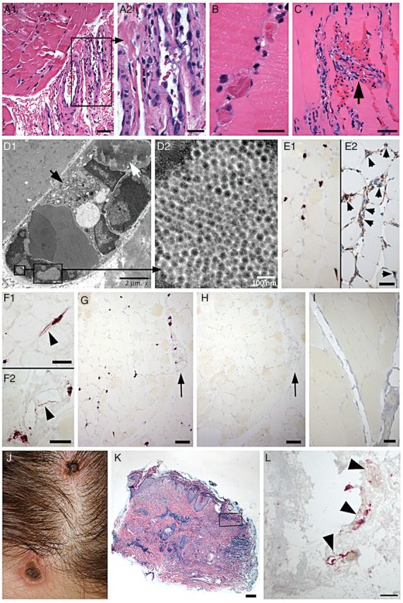
Background: A 33 year-old pancreatic transplant recipient developed weakness, retinal blindness, and necrotic plaques on her face, scalp, and hands.

Methods: A muscle biopsy was analyzed by light and electron microscopy and high-throughput nucleic acid sequencing.

Results: The biopsy revealed microthrombosis and viral particles in swollen endothelial cell nuclei. High-throughput sequencing of nucleic acid revealed a novel polyomavirus. In situ hybridization confirmed the presence of the polyomavirus in endothelial cells at sites of myositis and cutaneous necrosis.

Conclusions: New Jersey polyomavirus (NJPyV-2013) is a novel polyomavirus that may have tropism for vascular endothelial cells.

Keywords: immunosuppression; myositis; polyomavirus; transplantation; vasculitis; virus discovery.


Figures

Figure 1.
Figure 1.
Pathologic findings in deltoid muscle and skin lesions. AH, patient muscle; I, noninfected control muscle; JL, skin lesions. A, Prominent endothelial cell nuclear atypia in small perimysial venules (A1). Boxed area in A1 is enlarged in A2. There are only mild changes in the muscle in this focus, with mild fiber size variations and slightly increased cellularity in endomysium (A1), reflecting minimal inflammatory cell infiltration. B, Frequent intravascular microthrombi identified in endomysial capillaries. C, Endomysial capillary vascular destruction (arrow), with neutrophilic infiltration, nuclear fragmentation, and microhemorrhage. Marked endothelial cell atypia in adjacent capillaries and a prominent inflammatory cell infiltrate in endomysium. D, Electron microscopy demonstrates marked endomysial capillary damage, with atypical endothelial cell nuclei, swollen cytoplasm with numerous organelles (D1, black arrow), and an intravascular fibrin thrombus (D1, white arrow). Intranuclear viral inclusions were identified (boxed regions in D1), with a loose crystalloid arrangement (D2). Viral particles are spherical with a core of medium density, and measure 36–44 nm in diameter (D2). EI, In-situ hybridization with New Jersey polyomavirus (NJPyV-2013) probe (E, G, I) or enterovirus probe (H). NJPyV-2013 positive nuclei in endomysium (E1) are identified as endothelial cells by immunostain with CD34 in adjacent section (E2, small arrows). Labeled nuclei are often enlarged, and endothelial cells may have cytoplasmic labeling (arrowheads, F1, endomysial capillary; F2, perimysial venule). There is widespread vascular labeling with NJPyV-2013 probe (G) in endomysium and in perimysium (arrow, G). In situ hybridization with enterovirus probe (H) shows no labeling in an adjacent section (arrow, perimysial vessel, as in G). No labeling by NJPyV-2013 virus probe in muscle biopsy from noninfected patient (I). J, Blanching erythematous necrotic plaques on scalp and forehead. K, Scalp skin biopsy demonstrates epidermal erosion, multiple foci of dermal necrosis, and marked chronic perivascular inflammatory infiltrates. L, In situ hybridization with NJPyV-2013 probe (near boxed region in panel K) labels endothelial cell profiles (arrowheads) in the superficial dermis. Panels AC, K, Hematoxylin-eosin stain. Scale bars: A1, C, E1, E2, F1, F2, I, 50 µm; A2, B, L, 25 µm; G, H, 100 µm; K, 200 µm.
Figure 2.
Figure 2.
Molecular biology of New Jersey polyomavirus. A, Genome annotation. The double stranded DNA genome of NJPyV-2013 is 5108 base pairs in length. The coding sequences of the 3 capsid proteins VP1, VP2, and VP3 are represented by green hollow arrows. The reverse strand in the early region may have capacity for coding multiple transcripts by alternative pre-mRNA splicing. The coding sequences of several T antigens including large T (LT), small T (ST), and novel T (alternative T, AT) are indicated by blue hollow arrows. The expression of LT, ST, and AT transcripts were confirmed by PCR using RNA isolated from patient's muscle tissue. Positions of the primers TF1, TR1, and ELT/ST BF that were used for PCR detection were indicated with red bars inside the circular map. The overlapping palindrome repeats of “GAGGC” are indicated with an orange circle. B, Whole genome phylogenetic analysis. The phylogenetic tree was constructed using maximum likelihood method and bootstrap value 500 using the Tamura-Nei model. Results showed chimpanzee polyomavirus as the most closely related species. C, Early region transcription. Prediction of potential alternative transcripts of T antigens including large T (LT), small T (ST), extra-long T (ELT) and novel T (alternative T, AT). The predictions were made based on conserved splicing signals and amino acid sequences between chimpanzee polyomavirus and New Jersey polyomavirus. Red ovals indicate stop codons. D, Domain structure of NJPyV-2013T antigens. The potential functional domains and domain boundaries of LT, ST, and AT were predicted based on Clustal Omega alignments against the SV40 LT and ST sequences. E, Reverse transcription PCR amplification and DNA sequencing verification of LT, ST, and AT transcripts. Lane 1 and 6 DNA ladders. Lanes 3, 4, 5, and 8 (highlighted in red boxes) show PCR products corresponding to various T antigen transcripts. Lanes 2 and 7 are controls without DNase-treatment, which produced larger PCR products containing the unspliced intron. Abbreviations: mRNA, messenger RNA; PCR, polymerase chain reaction.

Similar articles

Cited by

References

    1. Abend JR, Joseph AE, Das D, Campbell-Cecen DB, Imperiale MJ. A truncated T antigen expressed from an alternatively spliced BK virus early mRNA. J Gen Virol. 2009;90:1238–45. - PMC - PubMed
    1. Trowbridge PW, Frisque RJ. Identification of three new JC virus proteins generated by alternative splicing of the early viral mRNA. J Neurovirol. 1995;1:195–206. - PubMed
    1. Pinto M, Dobson S. BK and JC virus: a review. J Infect. 2014;68 (suppl 1):S2–8. - PubMed
    1. Jiang M, Abend JR, Johnson SF, Imperiale MJ. The role of polyomaviruses in human disease. Virology. 2009;384:266–73. - PMC - PubMed
    1. Petrogiannis-Haliotis T, Sakoulas G, Kirby J, et al. BK-related polyomavirus vasculopathy in a renal-transplant recipient. N Engl J Med. 2001;345:1250–5. - PubMed

Publication types

MeSH terms

Associated data