

Regulating inflammation using acid-responsive electrospun fibrous scaffolds for skin scarless healing
- PMID: 24795507
- PMCID: PMC3984856
- DOI: 10.1155/2014/858045
Regulating inflammation using acid-responsive electrospun fibrous scaffolds for skin scarless healing
Abstract
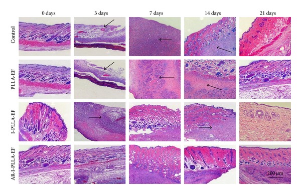
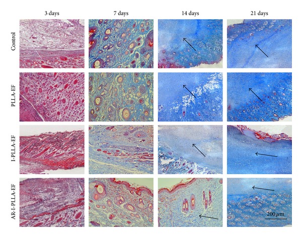
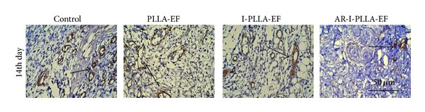
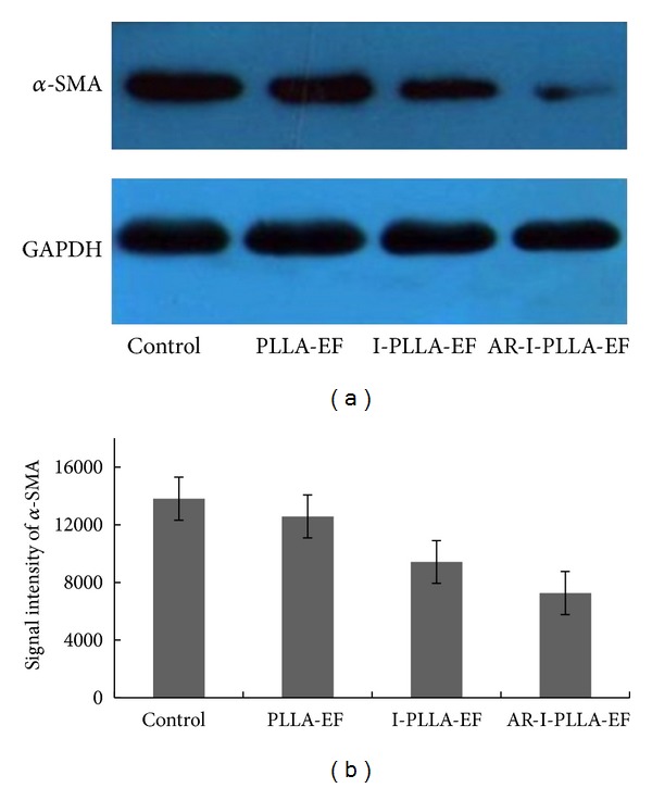
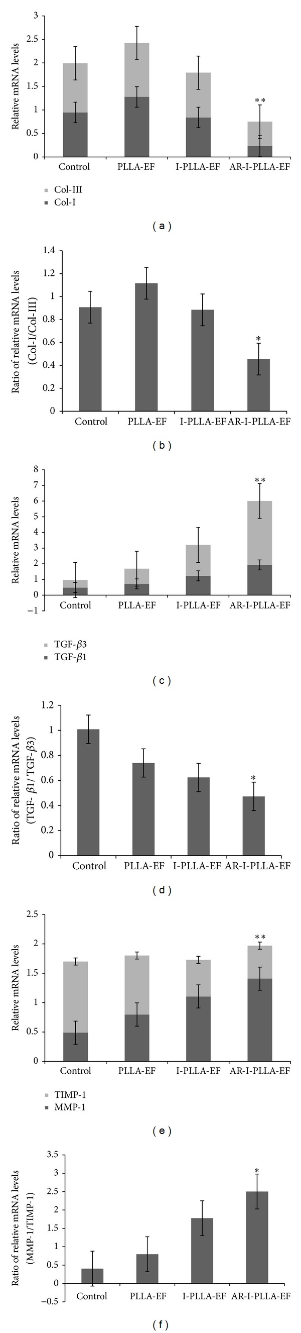
Skin injury in adult mammals brings about a series of events and inflammation in the wounded area is initiated first and provides lots of inflammatory factors, which is critical for the final scar formation. While the postinjured skin of fetus and nude mice heals scarlessly owing to the absence of inflammation or immunodeficient, we designed a feasible acid-responsive ibuprofen-loaded poly(L-lactide) (PLLA) fibrous scaffolds via doping sodium bicarbonate to prevent excessive inflammation and achieve scarless healing finally. The morphological results of in vivo experiments revealed that animals treated with acid-responsive ibuprofen-loaded PLLA fibrous scaffolds exhibited alleviative inflammation, accelerated healing process, and regulated collagen deposition via interference in the collagen distribution, the α-smooth muscle actin (α-SMA), and the basic fibroblast growth factor (bFGF) expression. The lower ratios of collagen I/collagen III and TGF-β1/TGF-β3 and higher ratio of matrix metalloproteinase-1 (MMP-1)/tissue inhibitor of metalloproteinase-1 (TIMP-1) in acid-responsive ibuprofen-loaded PLLA fibrous scaffolds group were confirmed by real-time qPCR as well. These results suggest that inhibiting the excessive inflammation will result in regular collagen distribution and appropriate ratio between the factors, which promote or suppress the scar formation, then decrease the scar area, and finally achieve the scarless healing.
Figures


Similar articles
-
Ibuprofen-loaded electrospun fibrous scaffold doped with sodium bicarbonate for responsively inhibiting inflammation and promoting muscle wound healing in vivo.Biomater Sci. 2014 Apr 4;2(4):502-511. doi: 10.1039/c3bm60198f. Epub 2013 Dec 23. Biomater Sci. 2014. PMID: 32481988
-
Tendon healing and anti-adhesion properties of electrospun fibrous membranes containing bFGF loaded nanoparticles.Biomaterials. 2013 Jun;34(19):4690-701. doi: 10.1016/j.biomaterials.2013.03.026. Epub 2013 Mar 26. Biomaterials. 2013. PMID: 23541108
-
Use of ginsenoside Rg3-loaded electrospun PLGA fibrous membranes as wound cover induces healing and inhibits hypertrophic scar formation of the skin.Colloids Surf B Biointerfaces. 2014 Mar 1;115:61-70. doi: 10.1016/j.colsurfb.2013.11.030. Epub 2013 Nov 24. Colloids Surf B Biointerfaces. 2014. PMID: 24333554
-
Functionalized gelatin/poly(l-lactide-co-ε-caprolactone) fibrous membrane promotes scarless wound healing by modulating inflammation and reducing fibrosis.Int J Biol Macromol. 2025 May;306(Pt 4):141785. doi: 10.1016/j.ijbiomac.2025.141785. Epub 2025 Mar 5. Int J Biol Macromol. 2025. PMID: 40054818
-
Advances in Skin Wound and Scar Repair by Polymer Scaffolds.Molecules. 2021 Oct 10;26(20):6110. doi: 10.3390/molecules26206110. Molecules. 2021. PMID: 34684690 Free PMC article. Review.
Cited by
-
Promotion of initial anti-tumor effect via polydopamine modified doxorubicin-loaded electrospun fibrous membranes.Int J Clin Exp Pathol. 2014 Aug 15;7(9):5436-49. eCollection 2014. Int J Clin Exp Pathol. 2014. PMID: 25337186 Free PMC article.
-
The capacity of exosomes derived from adipose-derived stem cells to enhance wound healing in diabetes.Front Pharmacol. 2023 Sep 21;14:1063458. doi: 10.3389/fphar.2023.1063458. eCollection 2023. Front Pharmacol. 2023. PMID: 37808198 Free PMC article. Review.
-
Tumor Responsive and Tunable Polymeric Platform for Optimized Delivery of Paclitaxel to Treat Glioblastoma.ACS Appl Mater Interfaces. 2020 Apr 29;12(17):19345-19356. doi: 10.1021/acsami.0c04102. Epub 2020 Apr 17. ACS Appl Mater Interfaces. 2020. PMID: 32252517 Free PMC article.
-
Optimizing type H vessels formation via short fibers 3D scaffolds with maintaining redox homeostasis for osteoporotic bone remodeling.Bioact Mater. 2025 Jul 23;53:417-432. doi: 10.1016/j.bioactmat.2025.07.030. eCollection 2025 Nov. Bioact Mater. 2025. PMID: 40735353 Free PMC article.
-
In vitro study of the mesenchymal stem cells-conditional media role in skin wound healing process: A systematic review.Int Wound J. 2022 Dec;19(8):2210-2223. doi: 10.1111/iwj.13796. Epub 2022 Apr 12. Int Wound J. 2022. PMID: 35412017 Free PMC article.
References
-
- Peng LH, Chen X, Chen L, Li N, Liang WQ, Gao JQ. Topical astragaloside IV-releasinghydrogel improves healing of skin wounds in vivo. Biological & Pharmaceutical Bulletin. 2012;35(6):881–888. - PubMed
-
- Lo DD, Zimmermann AS, Nauta A, Longaker MT, Lorenz HP. Scarless fetal skin wound healing update. Birth Defects Resarch C: Embryo Today. 2012;96(3):237–247. - PubMed
Publication types
MeSH terms
Substances
LinkOut - more resources
Full Text Sources
Other Literature Sources
Research Materials
Miscellaneous
