Review
. 2014 Apr 22:8:32.
doi: 10.3389/fnint.2014.00032. eCollection 2014.

The vestibular contribution to the head direction signal and navigation

Affiliations
Review

The vestibular contribution to the head direction signal and navigation

Ryan M Yoder et al. Front Integr Neurosci. .

Abstract

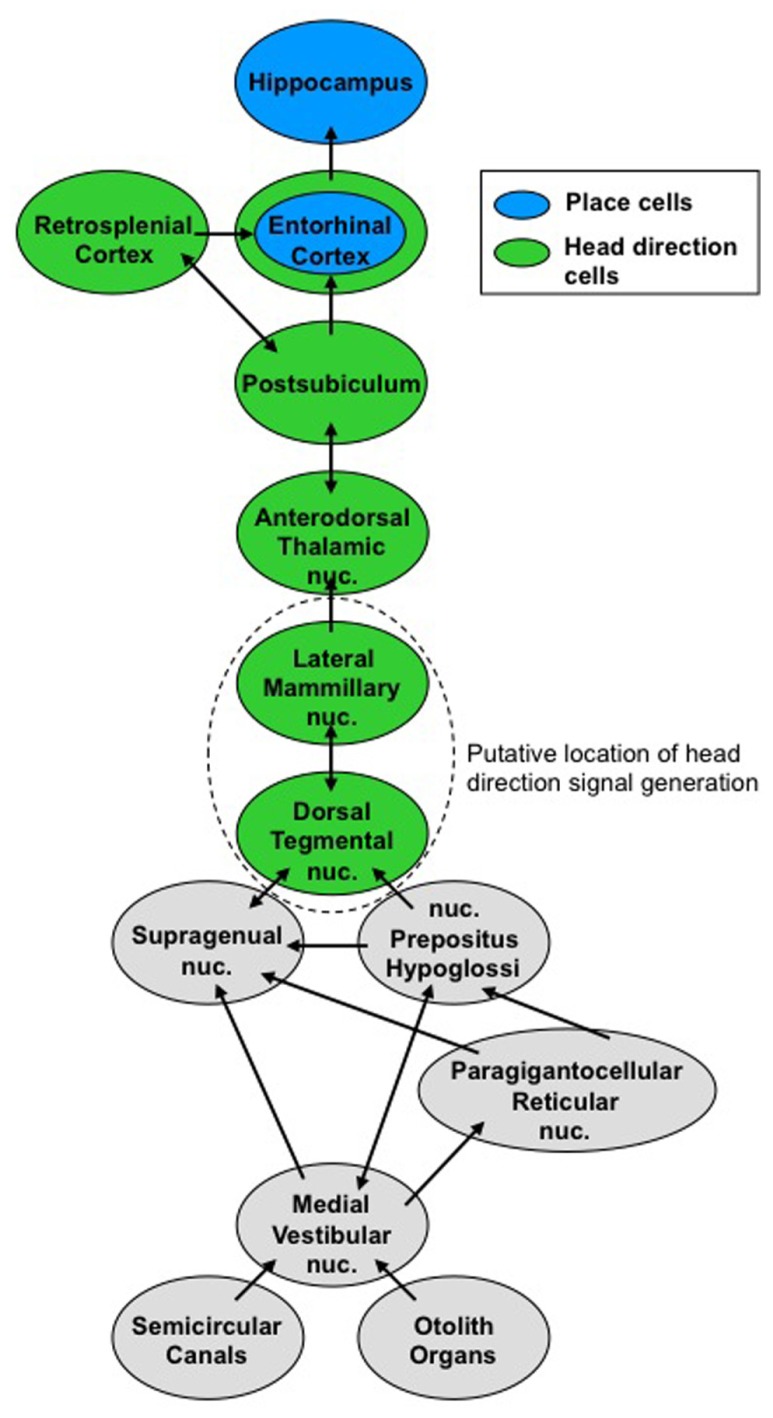
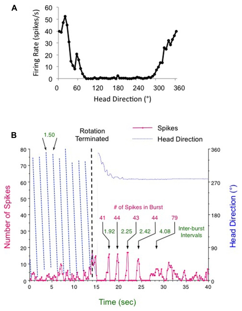
Spatial learning and navigation depend on neural representations of location and direction within the environment. These representations, encoded by place cells and head direction (HD) cells, respectively, are dominantly controlled by visual cues, but require input from the vestibular system. Vestibular signals play an important role in forming spatial representations in both visual and non-visual environments, but the details of this vestibular contribution are not fully understood. Here, we review the role of the vestibular system in generating various spatial signals in rodents, focusing primarily on HD cells. We also examine the vestibular system's role in navigation and the possible pathways by which vestibular information is conveyed to higher navigation centers.

Keywords: navigation; otolith organs; semicircular canals; spatial orientation; vestibular.


Figures

FIGURE 1
FIGURE 1
Vestibular pathways to the ascending head direction cell circuit. Signals from the semicircular canals and otolith organs converge on the medial vestibular nuclei. This vestibular information contributes to the head direction signal, which ascends to brain regions involved in navigation.
FIGURE 2
FIGURE 2
Head direction cell responses following (A) an active foraging session in the cylinder and (B) continuous clockwise rotation for 1 min (only the final 14 s are shown) followed by abrupt braking (vertical dashed line). After termination of rotation the HD cell fired in five periodic bursts over about 10 s. For this post-rotation period the amount of time in between each burst increased over each interval (times denoted in green for each inter-burst interval), although the number of spikes composing each burst (denoted in red) was about the same – particularly for the first four bursts. See text for further details.

References

    1. Aggleton J. P., Hunt P. R., Nagle S., Neave N. (1996). The effects of selective lesions within the anterior thalamic nuclei on spatial memory in the rat. Behav. Brain Res. 81 189–198 10.1016/S0166-4328(96)89080-2 - DOI - PubMed
    1. Aggleton J. P., Neave N., Nagle S., Hunt P. R. (1995). A comparison of the effects of anterior thalamic, mammillary body and fornix lesions on reinforced spatial alternation. Behav. Brain Res. 68 91–101 10.1016/0166-4328(94)00163-A - DOI - PubMed
    1. Allen K., Potvin O., Thibaudeau G., Dore F. Y., Goulet S. (2007). Processing idiothetic cues to remember visited locations: hippocampal and vestibular contributions to radial-arm maze performance. Hippocampus 17 642–653 10.1002/hipo.20304 - DOI - PubMed
    1. Avni R., Elkan T., Dror A. A., Shefer S., Eilam D., Avraham K. B., et al. (2009). Mice with vestibular deficiency display hyperactivity, disorientation, and signs of anxiety. Behav. Brain Res. 202 210–217 10.1016/j.bbr.2009.03.033 - DOI - PubMed
    1. Bassett J. P., Taube J. S. (2001). Neural correlates for angular head velocity in the rat dorsal tegmental nucleus. J. Neurosci. 21 5740–5751 - PMC - PubMed

LinkOut - more resources