

Human tumor xenograft models for preclinical assessment of anticancer drug development
- PMID: 24795792
- PMCID: PMC4007037
- DOI: 10.5487/TR.2014.30.1.001
Human tumor xenograft models for preclinical assessment of anticancer drug development
Abstract
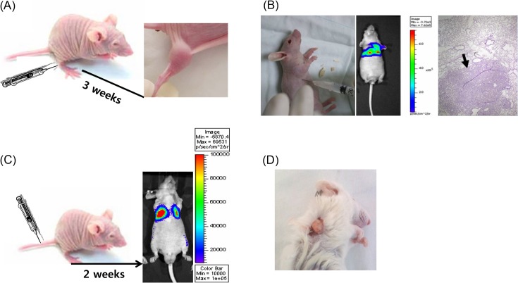
Xenograft models of human cancer play an important role in the screening and evaluation of candidates for new anticancer agents. The models, which are derived from human tumor cell lines and are classified according to the transplant site, such as ectopic xenograft and orthotopic xenograft, are still utilized to evaluate therapeutic efficacy and toxicity. The metastasis model is modified for the evaluation and prediction of cancer progression. Recently, animal models are made from patient-derived tumor tissue. The patient-derived tumor xenograft models with physiological characters similar to those of patients have been established for personalized medicine. In the discovery of anticancer drugs, standard animal models save time and money and provide evidence to support clinical trials. The current strategy for using xenograft models as an informative tool is introduced.
Keywords: Anticancer drug development; In vivo; Mouse; Xenograft model.
Figures
References
-
- Teicher B.A., Andrews P.A. Anticancer drug development guide; preclinical screening, clinical trials, and approval. 2nd edition. Humana Press; New Jersey: (2004). pp. 99–123.
-
- Suggitt M., Bibby M.C. 50 years of preclinical anticancer drug screening: empirical to target-driven approaches. Clin. Cancer Res. (2005);11:971–981. - PubMed
LinkOut - more resources
Full Text Sources
Other Literature Sources
