

Fracture healing and lipid mediators
- PMID: 24795811
- PMCID: PMC4007165
- DOI: 10.1038/bonekey.2014.12
Fracture healing and lipid mediators
Abstract
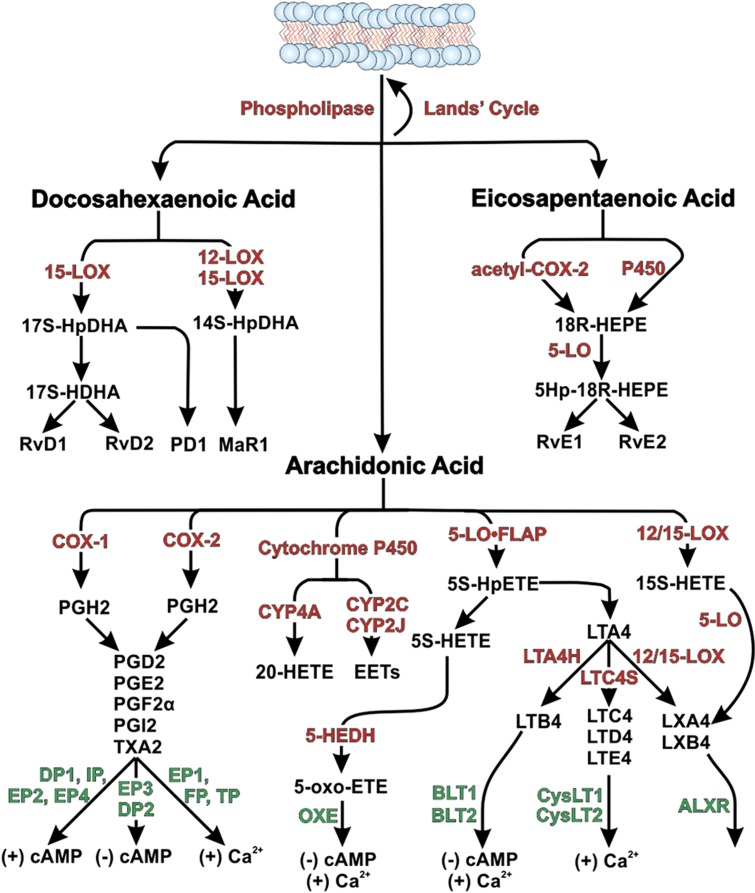
Lipid mediators regulate bone regeneration during fracture healing. Prostaglandins and leukotrienes are well-known lipid mediators that regulate inflammation and are synthesized from the Ω-6 fatty acid, arachidonic acid. Cyclooxygenase (COX-1 or COX-2) and 5-lipoxygenase (5-LO) catalyze the initial enzymatic steps in the synthesis of prostaglandins and leukotrienes, respectively. Inhibition or genetic ablation of COX-2 activity impairs fracture healing in animal models. Genetic ablation of COX-1 does not affect the fracture callus strength in mice, suggesting that COX-2 activity is primarily responsible for regulating fracture healing. Inhibition of cyclooxygenase activity with nonsteroidal anti-inflammatory drugs (NSAIDs) is performed clinically to reduce heterotopic ossification, although clinical evidence that NSAID treatment impairs fracture healing remains controversial. In contrast, inhibition or genetic ablation of 5-LO activity accelerates fracture healing in animal models. Even though prostaglandins and leukotrienes regulate inflammation, loss of COX-2 or 5-LO activity appears to primarily affect chondrogenesis during fracture healing. Prostaglandin or prostaglandin analog treatment, prostaglandin-specific synthase inhibition and prostaglandin or leukotriene receptor antagonism also affect callus chondrogenesis. Unlike the Ω-6-derived lipid mediators, lipid mediators derived from Ω-3 fatty acids, such as resolvin E1 (RvE1), have anti-inflammatory activity. In vivo, RvE1 can inhibit osteoclastogenesis and limit bone resorption. Although Ω-6 and Ω-3 lipid mediators have clear-cut effects on inflammation, the role of these lipid mediators in bone regeneration is more complex, with apparent effects on callus chondrogenesis and bone remodeling.
Conflict of interest statement
JPOC is an owner, board member and officer of Accelalox Inc., which is developing the use of 5-lipoxygenase inhibitors to accelerate fracture healing and promote bone formation. All other authors declare no conflict of interest.
Figures
References
-
- Schindeler A, McDonald MM, Bokko P, Little DG. Bone remodeling during fracture repair: The cellular picture. Semin Cell Dev Biol 2008;19:459–466. - PubMed
-
- Shimizu T. Lipid mediators in health and disease: enzymes and receptors as therapeutic targets for the regulation of immunity and inflammation. Annu Rev Pharmacol Toxicol 2009;49:123–150. - PubMed
-
- Stables MJ, Gilroy DW. Old and new generation lipid mediators in acute inflammation and resolution. Prog Lipid Res 2011;50:35–51. - PubMed
Publication types
Grants and funding
LinkOut - more resources
Full Text Sources
Other Literature Sources
Research Materials