. 2014 May 5;9(5):e92960.
doi: 10.1371/journal.pone.0092960. eCollection 2014.

Involvement of interleukin-17A-induced hypercontractility of intestinal smooth muscle cells in persistent gut motor dysfunction

Affiliations

Involvement of interleukin-17A-induced hypercontractility of intestinal smooth muscle cells in persistent gut motor dysfunction

Hirotada Akiho et al. PLoS One. .

Abstract

Background and aim: The etiology of post-inflammatory gastrointestinal (GI) motility dysfunction, after resolution of acute symptoms of inflammatory bowel diseases (IBD) and intestinal infection, is largely unknown, however, a possible involvement of T cells is suggested.

Methods: Using the mouse model of T cell activation-induced enteritis, we investigated whether enhancement of smooth muscle cell (SMC) contraction by interleukin (IL)-17A is involved in postinflammatory GI hypermotility.

Results: Activation of CD3 induces temporal enteritis with GI hypomotility in the midst of, and hypermotility after resolution of, intestinal inflammation. Prolonged upregulation of IL-17A was prominent and IL-17A injection directly enhanced GI transit and contractility of intestinal strips. Postinflammatory hypermotility was not observed in IL-17A-deficient mice. Incubation of a muscle strip and SMCs with IL-17A in vitro resulted in enhanced contractility with increased phosphorylation of Ser19 in myosin light chain 2 (p-MLC), a surrogate marker as well as a critical mechanistic factor of SMC contractility. Using primary cultured murine and human intestinal SMCs, IκBζ- and p38 mitogen-activated protein kinase (p38MAPK)-mediated downregulation of the regulator of G protein signaling 4 (RGS4), which suppresses muscarinic signaling of contraction by promoting inactivation/desensitization of Gαq/11 protein, has been suggested to be involved in IL-17A-induced hypercontractility. The opposite effect of L-1β was mediated by IκBζ and c-jun N-terminal kinase (JNK) activation.

Conclusions: We propose and discuss the possible involvement of IL-17A and its downstream signaling cascade in SMCs in diarrheal hypermotility in various GI disorders.


Conflict of interest statement

Competing Interests: YT, KS, MN, SN, KT, KO and MY are the employees of Tsumura & Co. whose company partly funded this study. There are no patents, products in development or marketed products to declare. This does not alter the authors' adherence to all the PLOS ONE policies on sharing data and materials.

Figures

Figure 1
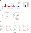
Figure 1. T cell activation induces GI hypermotility after resolution of inflammation, with sustained upregulation of IL-17A protein.
(A) GI transit distribution histograms and calculated geometric center (GC) values from the histograms. GC has been frequently and reliably used to estimate GI transit. Stom, stomach; S1–S10, small intestine segments 1–10; C, caecum; L1–2, Large intestine segments 1–2. Control (PBS-treated), day 1, 3 and 7 after αCD3 injection (n = 3 for each group of mice). (B) Carbachol (CCh)-stimulated dose-response curves of contractile response of SI strips prepared from day 7 with or without αCD3 treatment (n = 7). CCh was added cumulatively. (C) CCh-stimulated dose-response curves of contractile response of SI longitudinal SMCs prepared from day 7 with or without αCD3 treatment. The respective concentration of CCh was added separately (n = 4). (D) Time-dependent changes of mRNA and protein levels of various Th-cytokines in the intestinal tissue measured by quantitative RT-PCR and Bio-plex assay, respectively (n = 4∼8). Numerical data represent means ± s.e.m. *P<0.05, **P<0.01 versus control/PBS-treated, repeated measure ANOVA (B), Student's t-test (A, C, D) with Bonferroni correction for multiple comparison (A).
Figure 2
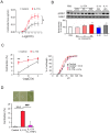
Figure 2. A possible involvement of IL-17A in αCD3-induced hypermotility.
(A) Calculated GCs (left panel) and contractile response induced by 10−5 M CCh (right panel) of IL-17A-injected mice (n = 11∼15). Saline or IL-17A (10 µg/mouse/day) was intraperitoneally injected three times. GC was measured on day 5. SI strips was isolated on day 5 and the contractility was immediately measured. (B) Calculated GCs of αCD3-injected mice in the recovery phase. The effect of IL-17R-FcChimera (twice i.p. at 1.5 and 3 hrs after αCD3-injection; n = 13–15) was demonstrated. (C) Calculated GCs of control and αCD3-injected IL-17 KO mice in the day 1 inflammatory phase (n = 3) and in the day 7 recovery phase (n = 11∼13). Numerical data represent means ± s.e.m. *P<0.05 versus control/PBS-treated, Student's t-test (A-C) with Bonferroni correction for multiple comparison (B).
Figure 3
Figure 3. IL-17A induces hypercontractility of SI longitudinal MS and SMCs.
(A) CCh-stimulated dose-response curves of MS contractility induced by IL-17A treatment for 3 days. CCh was added cumulatively (n = 9). (B) Immunoblot analysis of phosphorylated Ser19 of myosin light chain 2 (p-MLC) in samples of MS treated with or without IL-17A, IL-1β or IL-4 and quantitation of band intensity (n = 3∼4). The ratio of p-MLC to total MLC was calculated as described in Materials and Methods and normalized to untreated samples. N means the number of mice. Upper panels are representative images. (C) The Left panel shows CCh-stimulated dose-dependent changes of SMC contractility induced by IL-17A treatment for 24 hr. The respective concentration of CCh was added separately (n = 5). The right panel shows the distribution of cell length of 10−11 M CCh-stimulated SMCs with or without IL-17A treatment for 24 hr (n = 4). Y axis represents accumulating percentage of the number of the cells whose length are equal to, or smaller than, the length indicated in the x axis. The graph suggests that almost all of the analyzed SMCs responded to CCh stimulation. (D) Contractile response to CCh of SMCs treated with or without L-17A and/or IL-17R-Fc-Chimera (n = 6∼7). Upper panels are representative phase-contrast images. Scale bar, 50 µm. Numerical data represent means ± s.e.m. *P<0.05, **P<0.01 ***P<0.001 versus control/PBS-treated, ###P<0.001 versus IL-17A-treated. Repeated measure ANOVA (A), Student's t-test (B-D) with the closed testing procedure (B) and Bonferroni correction (D) for multiple comparisons.
Figure 4
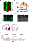
Figure 4. A possible involvement of the IκBζ pathway in IL-17A-induced hypercontractility.
(A) Heatmap of genes expressed in MS after 24 h IL-17A treatment, assessed by microarray (n = 6). (B) Summary of changes of mRNA levels of the genes of IL-17A signalling cascade assessed by microarray (n = 6). Genes with significant changes in expression are coloured in red (increased) or blue (decreased). (C) Immunohistochemical staining for NFκB p65 protein in MS treated with or without IL-17A and IL-1β for 1 hr. Scale bars, 50 µm. (D) Immunofluorescence staining of NFκB p65 protein in primary cultured murine SMCs after 30 min treatment with IL-17A or IL-1β. Lower panels represent p65 immunosignal in the nuclei picked up automatically by the cell imaging system Celaview. Scale bar, 25 µm. (E) Relative intensity of NFκB p65 immunosignal accumulated in the nucleus treated with IL-17R-Fc-Chimera or IL17RC siRNA (n = 3∼6). (F) Time-dependent changes in mRNAs of NFκB proteins evaluated by real-time RT-PCR in mouse cultured SMCs (n = 6). Numerical data represent means ± s.e.m. *P<0.05, **P<0.01, ***p<0.001, Student's t-test (E, F) under the closed testing procedure for multiple comparison (E).
Figure 5
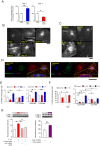
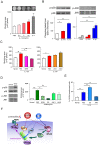
Figure 5. The effect of IL-17A on IκBζ - RGS4 signaling.
(A) RGS4 activity in samples of MS isolated from the mice treated with PBS or αCD3. RGS4 activity was estimated as the ratio of membrane RGS4 to cytosolic RGS4 as described in Materials and Methods. In the inflammatory phase (Day 1) RGS4 activity was enhanced while in the recovery phase (Day 7) the activity was decreased (n = 8). (B) Immunofluorescence staining of RGS4 protein. The days from the beginning of the culture are indicated. Scale bar, 50 µm. (C) Immunofluorescence staining of RGS4 protein after treatment with IL-17A, IL-1β or IL-4 for 24 hrs. Scale bar, 50 µm. (D) Three dimensional reconstruction of laser confocal microscopic images of cultured SMCs treated with IL-1β (right panel) and control (left panel). Cross-sectional images are included. Scale bar, 50 µm. (E) RGS4 activity was assessed by calculating the ratio of the cells with punctate RGS4 immunosignals. The effect of IL-17A or IL-1β with or without L-17RFcChimera or IL-17RC siRNA was examined (n = 4∼5). (F) The effect of IL-17A, IL-1β, or IL-4 on RGS 4 activity was assessed with or without SN50, a p65 translocation inhibitor, or IκBζ siRNA (n = 4∼5). (G) Quantitation of p-MLC by immunoblot analysis. The ratio of p-MLC to total MLC was calculated as described in Materials and Methods. N means the number of wells prepared from a mixed single lot of SMCs derived from 24 mice. The experiment was repeated and essentially the same results were obtained. The left panel shows the effect of SN50 or IκBζ siRNAs on the IL-17A-induced increase in p-MLC (n = 3∼4). The right panel shows the effect of RGS4 siRNAs on the p-MLC (n = 3). Numerical data represent means ± s.e.m. *P<0.05, **P<0.01, Student's t-test under the closed testing procedure for multiple comparison (A, E–F).
Figure 6
Figure 6. Involvement of p38MAPK activation in IL-17A-mediated hypercontractility in primary cultured murine SI SMCs.
(A) Contractility assay of IL-17A-treated SMCs. SMCs were cultured with IL-17A or IL-4 for 4 days using the temperature-responsive cell culture surface, UpCell plates. After addition of 10−11 M CCh, the cell layers became detached from the well during incubation at room temperature. Cell shrinkage was calculated by measuring the area of cell layers (n = 3∼6). Upper panels are representative images. (B) Quantitation of activation of p38MAPK (left panel) and JNK (right panel) by immunoblot analysis. The amount of phosphorylated form of p38MAPK (p-p38) and JNK (p-JNK) was measured (n = 4). (C) Contractility assay of IL-17A-treated SMCs. The effects of p38MAPK inhibitors (left panel) and JNK inhibitors (right panel) are shown. SB203580 (SB, 1 µM), BIRB796 (BIRB, 10 nM), SP600125 (JNK II, 100 nM) and JNK inhibitor III (JNK III, 100 nM) were added 15 min before addition of IL-17A (50 ng/ml). Contractility was measured as described in Fig. 6A (n = 8). (D) Effect of MAPK inhibitors on on anisomycin-induced contractility. The activation of p38MAPK by anisomycin was demonstrated by the representative images of the immunoblot in the left panel. The right panel shows the effects of p38MAPK and JNK inhibitors on anisomycin-induced contractility. Contractility was measured the day after addition of anisomycin (n = 4). (E) The effects of p38MAPK and JNK inhibitors on IL-1β-induced RGS4 translocation. RGS4 activity was evaluated by Celaview analysis as described in Fig. 5D (n = 4). Numerical data represent means ± s.e.m. *P<0.05, **P<0.01, Student's t-test under the closed testing procedure for multiple comparison (A–E). (F) Scheme of IL-17A and IL-1β signalling leading to hyper- and hypo-contractility induced by IL-17A and IL-1β, respectively. IL-17A induces p38 MAPK phosphorylation and NFκB activation (i.e., translocation of p65 RelA protein into the nucleus) independently within 15 min. Inhibition of p38 MAPK activation had no effect on RelA translocation and conversely, inhibition of NFκB activation had no effect on MAPK phosphorylation (data not shown). The expression of IκBζ, which is virtually absent without NFκB activation was massively induced after 30 min. The decrease in translocation of RGS4 protein to the cell membrane, where RGS4 suppresses muscarinic receptor-mediated signalling via interaction with G proteins coupled to muscarinic receptor, was observed between day 1 and day 4. A significant hypercontractility of SMCs began to be detected after day 2 and continued through to day 4. Inhibition of p38MAPK and NFκB/IκBζ activation both suppressed IL-17-induced hypercontractility and the changes of RGS4 translocation. By contrast, IL-1β activates JNK as well as NFκB/IκBζ and p38MAPK. Inhibition of NFκB/IκBζ and JNK abrogated the effect of IL-1β while p38MAPK inhibition enhanced it. Thus the balance in the relative activity levels of JNK and p38MAPK is critical for determining the direction of contractility. NFκB-IκBζ signalling regulates the movement of MAPK-triggered molecular events toward/against hypercontractility of SI SMC.
Figure 7
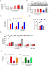
Figure 7. IL-17A-induced hypercontractility and its signal transduction in cultured human colonic SMCs.
(A) Contractility assay of IL-17A-treated human SMCs. SMCs were cultured with IL-17A and IL-4 for 5 days and contractility was evaluated as described in Figure? 6A. (B) Immunoblot analysis of p-MLC and quantitation of band intensity in samples of colonic SMCs treated with or without IL-17A, IL-1β or IL-4 (n = 6). The cells were treated with 10−11 M CCh for 3 min. The ratio of p-MLC to total MLC was calculated as described in Materials and Methods and normalized to untreated samples. N refers to the number of experiments. Upper panels are representative images. (C) Relative intensity of NFκB p65 immunosignal accumulated in the nucleus was measured in cultured colonic SMCs after 30 min treatment with PBS, IL-17A or IL-1β in the absence or presence of anti-IL-17RC antibody or siRNAs to control and IL-17RC. (D) The effect of IL-17A and IL-1β on RGS 4 activity in human SMCs on day 4 was assessed (n = 4). RGS4 activity was evaluated as described in Materials and Methods. (E) Effect of anti-IL-17RC antibody, SN50 and IκBζ siRNA on IL-17A-induced contractility in human SMCs. Contractility was measured on day 4 (n = 3–6) as described in Fig.6A. (F) The effects of MAPK inhibitors on IL-17A- and anisomycin-induced hypercontractility on day 4 (n = 3–6). The left and right panels show the effects MAPK inhibitors on IL-17A-, and anisomycin-induced contractility, respectively. Contractility was measured as described in Fig. 6A (n = 3). Numerical data represent means ± s.e.m. *P<0.05, **P<0.01, Student's t-test under the closed testing procedure for multiple comparison (A–F).

Similar articles

Cited by

References

    1. Brand S (2009) Crohn's disease: Th1, Th17 or both? The change of a paradigm: new immunological and genetic insights implicate Th17 cells in the pathogenesis of Crohn's disease. Gut 58: 1152–1167. - PubMed
    1. Anderson CA, Massey DC, Barrett JC, Prescott NJ, Tremelling M, et al. (2009) Investigation of Crohn's disease risk loci in ulcerative colitis further defines their molecular relationship. Gastroenterology 136: 523–529 e523. - PMC - PubMed
    1. Khan WI, Collins SM (2006) Gut motor function: immunological control in enteric infection and inflammation. Clin Exp Immunol 143: 389–397. - PMC - PubMed
    1. Facco M, Brun P, Baesso I, Costantini M, Rizzetto C, et al. (2008) T cells in the myenteric plexus of achalasia patients show a skewed TCR repertoire and react to HSV-1 antigens. Am J Gastroenterol 103: 1598–1609. - PubMed
    1. Frasko R, Maruna P, Gurlich R, Trca S (2008) Transcutaneous electrogastrography in patients with ileus. Relations to interleukin-1beta, interleukin-6, procalcitonin and C-reactive protein. Eur Surg Res 41: 197–202. - PubMed

Publication types

MeSH terms