

Molecular and cellular mechanisms of heterotopic ossification
- PMID: 24796520
- PMCID: PMC4372050
- DOI: 10.14670/HH-29.1281
Molecular and cellular mechanisms of heterotopic ossification
Abstract
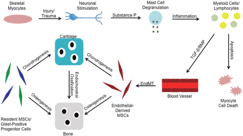
Heterotopic ossification (HO) is a debilitating condition in which cartilage and bone forms in soft tissues such as muscle, tendon, and ligament causing immobility. This process is induced by inflammation associated with traumatic injury. In an extremely rare genetic disorder called fibrodysplasia ossificans progessiva (FOP), a combination of inflammation associated with minor soft tissue injuries and a hereditary genetic mutation causes massive HO that progressively worsens throughout the patients' lifetime leading to the formation of an ectopic skeleton. An activating mutation in the BMP type I receptor ALK2 has been shown to contribute to the heterotopic lesions in FOP patients, yet recent studies have shown that other events are required to stimulate HO including activation of sensory neurons, mast cell degranulation, lymphocyte infiltration, skeletal myocyte cell death, and endothelial-mesenchymal transition (EndMT). In this review, we discuss the recent evidence and mechanistic data that describe the cellular and molecular mechanisms that give rise to heterotopic bone.
Figures
References
-
- Connor JM, Evans DA. Fibrodysplasia ossificans progressiva. The clinical features and natural history of 34 patients. J Bone Joint Surg Br. 1982;64:76–83. - PubMed
-
- Furuya H, Ikezoe K, Wang L, Ohyagi Y, Motomura K, Fujii N, Kira J, Fukumaki Y. A unique case of fibrodysplasia ossificans progressiva with an ACVR1 mutation, G356D, other than the common mutation (R206H) Am J Med Genet A. 2008;146A:459–463. - PubMed
Publication types
MeSH terms
Grants and funding
LinkOut - more resources
Full Text Sources
Other Literature Sources
