

Alterations in regulatory T cell subpopulations seen in preterm infants
- PMID: 24796788
- PMCID: PMC4010410
- DOI: 10.1371/journal.pone.0095867
Alterations in regulatory T cell subpopulations seen in preterm infants
Abstract
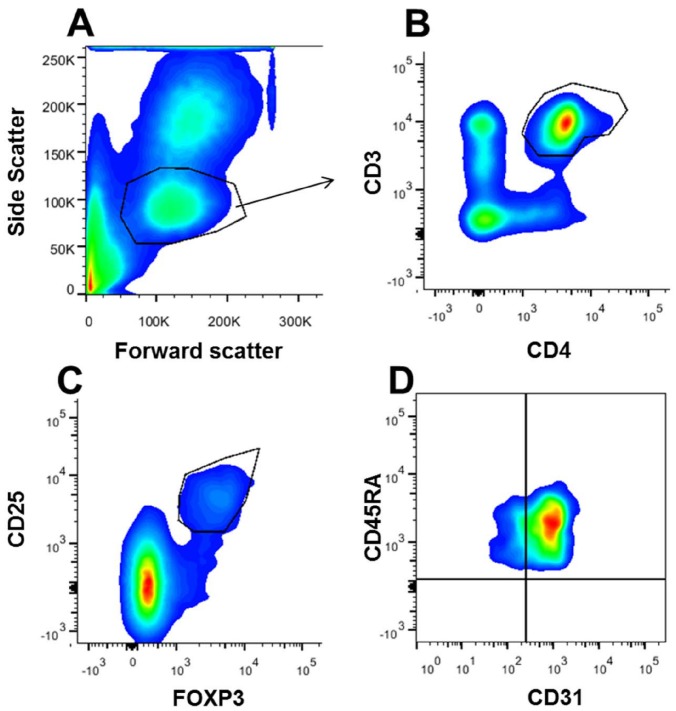
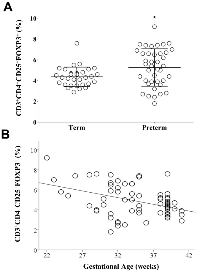
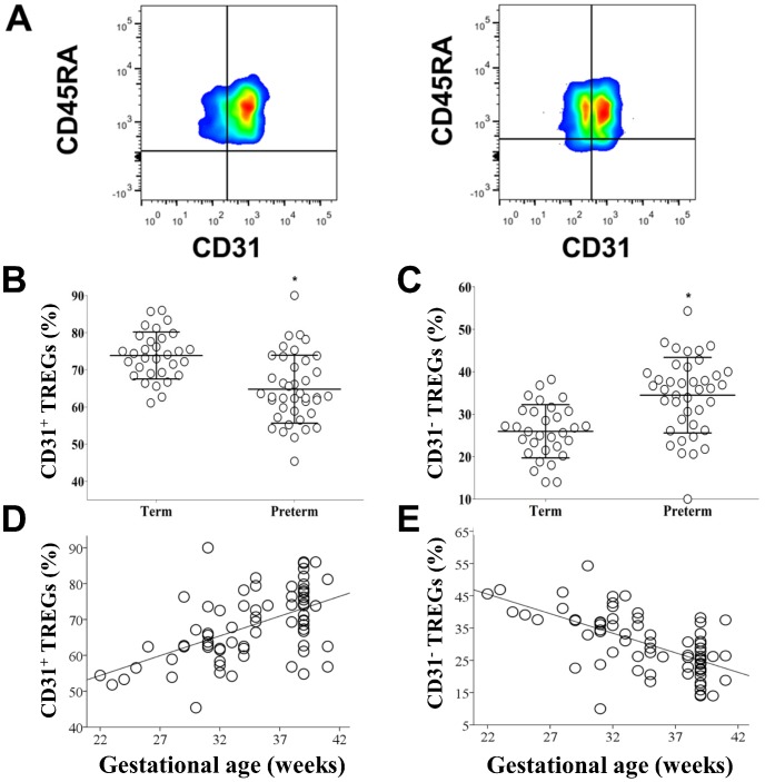
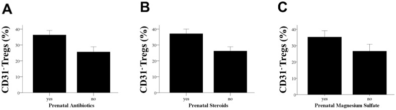
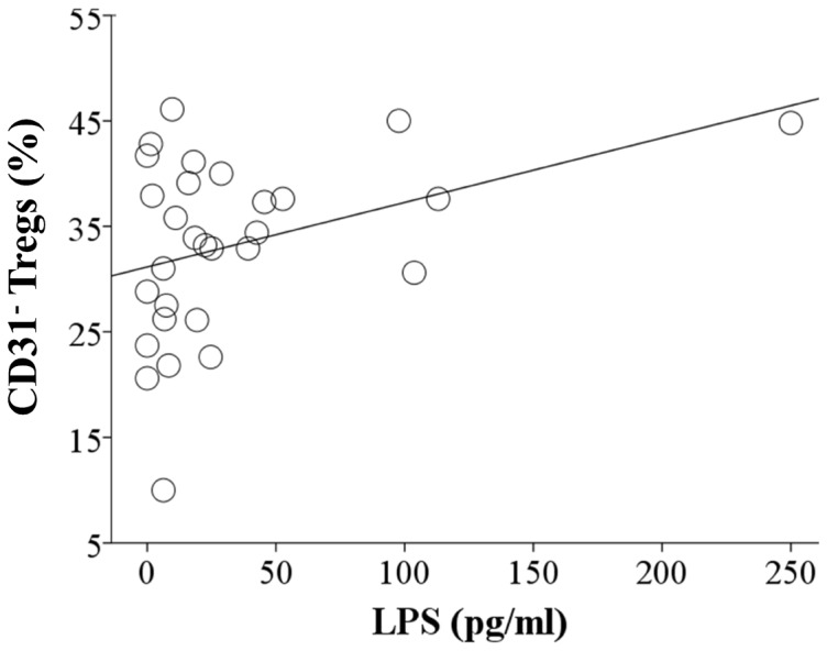
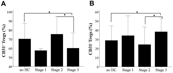
Regulatory T cells are a population of CD4+ T cells that play a critical role in peripheral tolerance and control of immune responses to pathogens. The purpose of this study was to measure the percentages of two different regulatory T cells subpopulations, identified by the presence or absence of CD31 (Recent thymic emigrants and peripherally induced naïve regulatory T cells), in term and preterm infant cord blood. We report the association of prenatal factors, intrauterine exposure to lipopolysaccharide and inflammation and the percentages of these regulatory T cell subpopulations in term and preterm infants. Cord blood samples were collected from both term and preterm infants and mononuclear cells isolated over a Ficoll-Hypaque cushion. Cells were then stained with fluorochrome-labeled antibodies to characterize regulatory T cell populations and analyzed with multi-color flow cytometry. Cord blood plasma C-reactive protein, and lipopolysaccharide were also measured. Placental pathology was also examined. We report a gestational age-dependent difference in the percentage of total regulatory T cells, in which preterm infants of lower gestational ages have an increased percentage of regulatory T cells. We report the presence of two populations of regulatory T cells (CD31+ and CD31-) in cord blood of term and preterm infants and their association with different maternal and fetal characteristics. Factors associated with differences in the percentage of CD31- Tregs included the use of prenatal antibiotics, steroids and magnesium sulfate. In addition, the percentage of CD31- Tregs was significantly higher in cord blood of preterm pregnancies associated with inflammation and prenatal lipopolysaccharide exposure. The peripheral Treg pool of preterm infants could be altered by prenatal exposure to inflammation and chorioamnionitis; however, the clinical implications of this finding are not yet understood.
Conflict of interest statement
Figures
Similar articles
-
Characterization of Regulatory T Cells in Preterm and Term Infants.Arch Immunol Ther Exp (Warsz). 2019 Feb;67(1):49-54. doi: 10.1007/s00005-018-0530-x. Epub 2018 Oct 29. Arch Immunol Ther Exp (Warsz). 2019. PMID: 30374518
-
Preterm cord blood CD4⁺ T cells exhibit increased IL-6 production in chorioamnionitis and decreased CD4⁺ T cells in bronchopulmonary dysplasia.Hum Immunol. 2015 May;76(5):329-338. doi: 10.1016/j.humimm.2015.03.007. Epub 2015 Mar 20. Hum Immunol. 2015. PMID: 25797206 Free PMC article.
-
Maturation of CD4+ regulatory T lymphocytes and of cytokine secretions in infants born prematurely.J Clin Immunol. 2013 Aug;33(6):1126-33. doi: 10.1007/s10875-013-9911-4. Epub 2013 Jun 21. J Clin Immunol. 2013. PMID: 23793781
-
Expression of S100A Alarmins in Cord Blood Monocytes Is Highly Associated With Chorioamnionitis and Fetal Inflammation in Preterm Infants.Front Immunol. 2020 Jun 16;11:1194. doi: 10.3389/fimmu.2020.01194. eCollection 2020. Front Immunol. 2020. PMID: 32612607 Free PMC article.
-
The Immunomodulatory Role of Regulatory T Cells in Preterm Birth and Associated Pregnancy Outcomes.Int J Mol Sci. 2024 Nov 5;25(22):11878. doi: 10.3390/ijms252211878. Int J Mol Sci. 2024. PMID: 39595948 Free PMC article. Review.
Cited by
-
Regulatory T cell frequencies are increased in preterm infants with clinical early-onset sepsis.Clin Exp Immunol. 2016 Aug;185(2):219-27. doi: 10.1111/cei.12810. Epub 2016 Jun 12. Clin Exp Immunol. 2016. PMID: 27163159 Free PMC article.
-
Myeloid-Derived Suppressor Cells in Pregnancy and the Neonatal Period.Front Immunol. 2020 Oct 9;11:584712. doi: 10.3389/fimmu.2020.584712. eCollection 2020. Front Immunol. 2020. PMID: 33162999 Free PMC article. Review.
-
Extremely Preterm Infants Have Significant Alterations in Their Conventional T Cell Compartment during the First Weeks of Life.J Immunol. 2020 Jan 1;204(1):68-77. doi: 10.4049/jimmunol.1900941. Epub 2019 Dec 4. J Immunol. 2020. PMID: 31801814 Free PMC article. Clinical Trial.
-
Granulocytic myeloid-derived suppressor cells (GR-MDSC) accumulate in cord blood of preterm infants and remain elevated during the neonatal period.Clin Exp Immunol. 2018 Mar;191(3):328-337. doi: 10.1111/cei.13059. Epub 2017 Oct 26. Clin Exp Immunol. 2018. PMID: 28963753 Free PMC article.
-
Developmental origins of inflammatory and immune diseases.Mol Hum Reprod. 2016 Aug;22(8):858-65. doi: 10.1093/molehr/gaw036. Epub 2016 May 25. Mol Hum Reprod. 2016. PMID: 27226490 Free PMC article. Review.
References
-
- Feuerer M, Hill JA, Mathis D, Benoist C (2009) Foxp3+ regulatory T cells: differentiation, specification, subphenotypes. Nat Immunol 10: 689–695. - PubMed
-
- Apostolou I, Sarukhan A, Klein L, von Boehmer H (2002) Origin of regulatory T cells with known specificity for antigen. Nat Immunol 3: 756–763. - PubMed
-
- Levings MK, Sangregorio R, Galbiati F, Squadrone S, de Waal Malefyt R, et al. (2001) IFN-alpha and IL-10 induce the differentiation of human type 1 T regulatory cells. J Immunol 166: 5530–5539. - PubMed
-
- Haas J, Fritzsching B, Trubswetter P, Korporal M, Milkova L, et al. (2007) Prevalence of newly generated naive regulatory T cells (Treg) is critical for Treg suppressive function and determines Treg dysfunction in multiple sclerosis. J Immunol 179: 1322–1330. - PubMed
Publication types
MeSH terms
Substances
Grants and funding
LinkOut - more resources
Full Text Sources
Other Literature Sources
Medical
Research Materials
