Comparative Study
. 2015 Mar;7(2):250-259.
doi: 10.1111/1753-0407.12164. Epub 2014 Jul 15.

Alterations in connexin 43 during diabetic cardiomyopathy: competition of tyrosine nitration versus phosphorylation

Affiliations
Comparative Study

Alterations in connexin 43 during diabetic cardiomyopathy: competition of tyrosine nitration versus phosphorylation

Mandar S Joshi et al. J Diabetes. 2015 Mar.

Abstract

Background: Cardiac conduction abnormalities are observed early in the progression of type 1 diabetes (T1D), but the mechanism(s) involved are undefined. Connexin 43, a critical component of ventricular gap junctions, depends on tyrosine phosphorylation status to modulate channel conductance; changes in connexin 43 content, distribution, and/or phosphorylation status may be involved in cardiac rhythm disturbances. We tested the hypothesis that cardiac content and/or distribution of connexin 43 is altered in a rat model of T1D cardiomyopathy, investigating a mechanistic role for tyrosine.

Methods: Electrocardiographic analyses were conducted during the progression of diabetic cardiomyopathy in rats dosed with streptozotocin (STZ; 65 mg/kg) 3, 7, and 35 days after the induction of diabetes. Following functional analyses, we conducted immunohistochemical and immunoprecipitation studies to assess alterations in connexin 43.

Results: There was significant evidence of ventricular conduction abnormalities (QRS complex, Q-T interval) as early as 7 days after STZ, persisting throughout the study. Connexin 43 levels were increased 7 days after STZ and remained elevated throughout the study. Connexin 40 content was unchanged relative to controls throughout the study. Changes in connexin 43 distribution were also observed: connexin 43 staining was dispersed from myocyte short axis junctions. Connexin 43 tyrosine phosphorylation declined during the progression of diabetes, with concurrent increases in tyrosine nitration.

Conclusions: The data suggest that changes in connexin 43 content and distribution occur during experimental diabetes and likely contribute to alterations in cardiac function, and that oxidative modification of tyrosine-mediated signaling may play a mechanistic role.

Keywords: cardiomyopathy; connexins; diabetes; oxidative stress; signal transduction; 关键词:心肌病,间隙连接蛋白,糖尿病,氧化应激,信号转导.


Figures

Figure 1
Figure 1. Representative signal-averaged ECGs from control and diabetic rats
Three lead ECGs were collected in anesthetized rats at 3, 7, and 35 days following treatment with STZ or vehicle control. Waveforms were signal averaged over 150-200 beats. Panel A Representative signal averaged waveform, illustrating the start and endpoints for each ECG parameter collected. Panel B Representative signal-averaged waveforms from control rats and diabetic rats at 7 and 35 days post-STZ showing altered P-wave morphology, abnormal S-T segment shape and QRS broadening at advanced stages of diabetic cardiomyopathy.
Figure 2
Figure 2. Cardiac electrophysiological parameters
Average data from control and diabetic animals at 0, 3, 7, and 35 days post-STZ. At 35 days of diabetes, P-waves were undetectable in many of the waveforms studied, and these intervals were not calculated. QTc represents Q-T interval corrected for heart rate by Fridericia's method [Q-Tc = Q-T/(R-R interval)1/3]. In control animals, no age-dependent effects (0 vs. 35 days) were observed in any parameter studied. ○, control ; ●, diabetic. *, p<0.05 vs. pooled control (0 and 35 days).
Figure 3
Figure 3. Selective alterations in cardiac connexin isoform content during experimental diabetes
Connexin isoform content for Cx40 and Cx43 was determined by immunohistochemistry, in left ventricular cross-sections from control and diabetic animals. Panel A Representative photomicrographs of connexin immunohistochemistry for Cx40 (800x magnification, left panels) and Cx43 (400x magnification, right panels), brown staining indicates connexin immunoprevalence. Panel B Average integrated optical densities for Cx40 (left panel) and Cx43 (right panel) by digital image analysis. In control animals, no age-dependent effects (0 vs. 35 days) were observed in any parameter studied, these values were pooled and represented as Time 0. Cx43 content was significantly increased by 7 days of diabetes, and remained elevated throughout the study, while no changes in Cx40 were observed at any time point studied. *, p<0.05 vs. pooled control (plotted as time 0).
Figure 4
Figure 4. Cardiac connexin 43 distributional changes during experimental diabetes
Cx43 distribution was studied from histological cross-sections in myocytes that demonstrated longitudinal alignment. Control tissues were used to define normal ranges for short axis Cx43 staining; mid-myocyte regions describe staining that fell out of the 95% confidence interval for short axis regions in control hearts (see Methods). Panel A Representative photomicrographs of single cardiac myocytes obtained from histological cross-sections. Control myocytes exhibit Cx43 staining that is strictly confined to myocyte short axes; diabetic hearts show significant increases in mid-myocyte staining, away from short axis regions. Panel B Digital image analysis for Cx43 staining in short axis versus midmyocyte regions. Short axis staining was increased 4-5 fold by 35 days diabetes (consistent with whole heart histology, Figure 4). Increases in mid-myocyte staining was much more dramatic, with 40-50 fold increases in Cx43 integrated optical densities observed at 7 and 35 days post-STZ. The average percentage of total Cx43 staining that exhibited mid-myocyte localization (mid-myocyte IOD/total myocyte IOD) for each time point is shown in the lower panel. *, p<0.05 vs. pooled control (plotted as time 0).
Figure 5
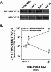
Figure 5. Cardiac connexin 43 tyrosine status was significantly altered in diabetic hearts
Cx43 was immunoprecipitated from cardiac left ventricular homogenates, then probed for tyrosine phosphorylation and nitration. Panel A Representative western blots from control and diabetic hearts at 3, 7, and 35 days post-STZ. Panel B Digital image analysis for band intensities, expressed as a percent of control staining. IOD, integrated optical density (band intensity x band area). *, p<0.05 vs. control (plotted as time 0).
Figure 6
Figure 6. Connexins are gap junction proteins that facilitate impulse conduction in the heart
Connexins are critical components of cardiac gap junctions. These channels consist of multiple isoforms; Cx43 and Cx40 predominate in cardiac tissue. Cardiac conduction is modulated by channel composition & distribution; connexin size, number, and spatial distribution determine ion flow and thus the conduction in the heart.

Similar articles

Cited by

References

    1. Garcia MJ, McNamara PM, Gordon T, Kannel WB. Morbidity and mortality in diabetics in the Framingham population. Sixteen year follow-up study. Diabetes. 1974;23:105–11. - PubMed
    1. Gu K, Cowie CC, Harris MI. Diabetes and decline in heart disease mortality in US adults. Jama. 1999;281:1291–7. - PubMed
    1. Kannel WB, Hjortland M, Castelli WP. Role of diabetes in congestive heart failure: the Framingham study. Am J Cardiol. 1974;34:29–34. - PubMed
    1. Pieske B, Wachter R. Impact of diabetes and hypertension on the heart. Current opinion in cardiology. 2008;23:340–9. - PubMed
    1. Hayat SA, Patel B, Khattar RS, Malik RA. Diabetic cardiomyopathy: mechanisms, diagnosis and treatment. Clin Sci (Lond) 2004;107:539–57. - PubMed

Publication types

MeSH terms