. 2014 Sep;99(9):3450-9.
doi: 10.1210/jc.2014-1026. Epub 2014 May 5.

Transforming growth factor-β induced Warburg-like metabolic reprogramming may underpin the development of peritoneal endometriosis

Affiliations

Transforming growth factor-β induced Warburg-like metabolic reprogramming may underpin the development of peritoneal endometriosis

Vicky J Young et al. J Clin Endocrinol Metab. 2014 Sep.

Abstract

Context: TGF-β is believed to play a major role in the etiology of peritoneal endometriosis. In tumors, TGF-β induces the metabolic conversion of glucose to lactate via glycolysis, a process referred to as the "Warburg effect." Lactate increases cell invasion, angiogenesis, and immune suppression, all crucial steps in the development of endometriosis.

Objective: The aim of this study was to determine whether TGF-β induces a "Warburg-like" effect in peritoneal endometriosis.

Design: The study was informed by human tissue analysis and cel culture.

Setting: The study was conducted at the university research institute.

Patients or other participants: We studied women undergoing surgical investigation for endometriosis.

Interventions: Concentrations of lactate and TGF-β1 in peritoneal fluid (n = 16) were measured by commercial assay. Expression of genes implicated in glycolysis was measured in endometrial and peritoneal biopsies (n = 31) by quantitative RT-PCR and immunohistochemistry. The effect of TGF-β1 on primary human peritoneal mesothelial cells (n = 6) and immortalized mesothelial (MeT-5A) cells (n = 3) was assessed by quantitative RT-PCR, Western blot, and commercial assays.

Main outcome measures: Lactate, TGF-β1, and markers of glycolysis were measured.

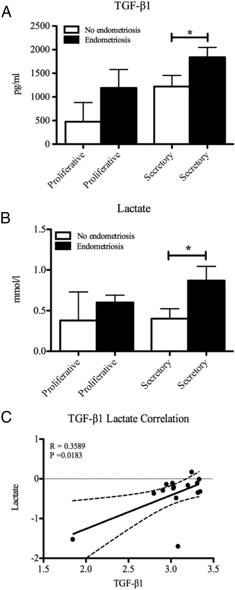
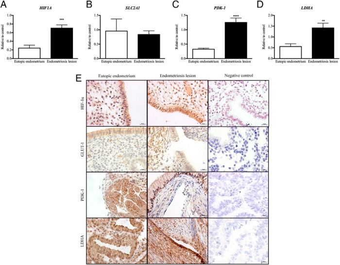
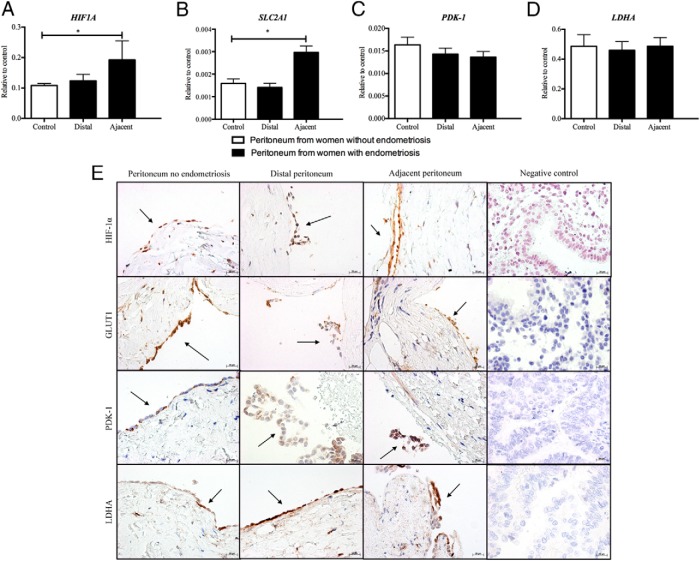
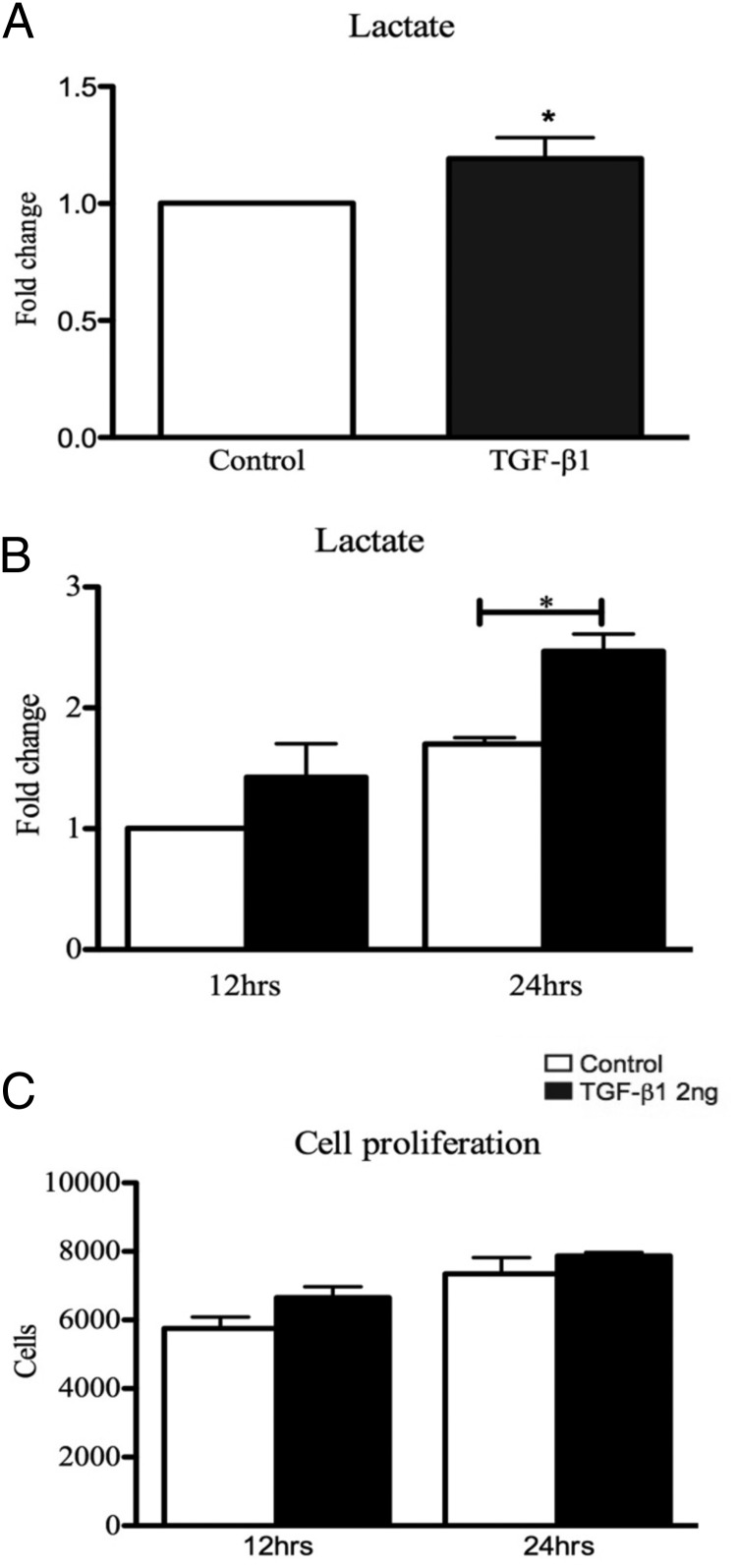
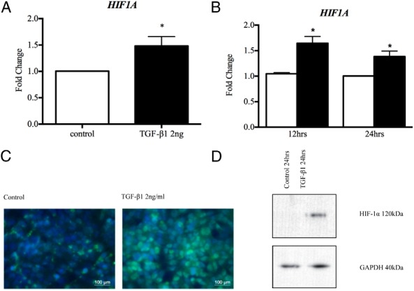
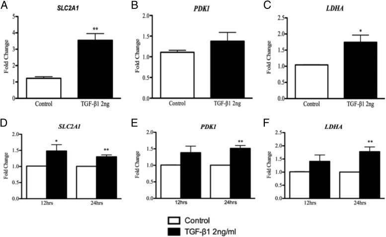
Results: Concentrations of lactate in peritoneal fluid paralleled those of TGF-β1, being significantly higher in women with endometriosis compared to women without (P < .05). Endometriosis lesions expressed higher levels of glycolysis-associated genes HIF1A, PDK1, and LDHA than eutopic endometrium, and adjacent peritoneum had higher levels of HIF1A and SLC2A1 than peritoneum from women without disease (P < .05 to P < .001). Exposure of mesothelial cells to TGF-β1 increased production of lactate (P < .05), increased HIF1A mRNA (P < .05), and protein, and increased concentrations of mRNAs encoded by glycolysis-associated genes (LDHA, PDK1, SLC2A1; P < .05).

Conclusions: A change in the metabolic phenotype of endometriosis lesions and peritoneal mesothelium in women with endometriosis may favor development of endometriosis.


Figures

Figure 1.
Figure 1.
A and B, TGF-β1 (A) and lactate (B) levels were increased in peritoneal fluid from women with endometriosis compared to women without endometriosis during the secretory phase of the menstrual cycle. C, There was a significant positive correlation across the menstrual cycle between levels of TGF-β1 and lactate in women with and without endometriosis. *, P < .05; R2 = 0.3589 (n = 2 each proliferative group, n = 6 each secretory group).
Figure 2.
Figure 2.
HIF1A (A), PDK1 (C), and LDHA (D) mRNA concentrations were significantly increased in endometriosis lesions when compared to endometrium from women with endometriosis. B, There was no significant change in SLC2A1 mRNA expression between endometriosis lesions and endometrium from women with disease. E, HIF-1α, GLUT-1, PDK-1, and LDHA were expressed in glandular epithelial cells and surrounding stromal cells of endometriosis lesions and endometrium. There was no staining in sections probed with control antibodies. **, P < .01; ***, P < .001; ****, P < .0001 (n = 7 each group mRNA analysis, n = 2 each group immunohistochemical analysis). Original magnification, ×400.
Figure 3.
Figure 3.
A and B, HIF1A (A) and SLC2A1 (B) mRNA concentrations were significantly increased in peritoneum adjacent to endometriosis lesions when compared to peritoneum of women without disease. There was no significant change in HIF1A and SLC2A1 expression from sites of peritoneum distal to endometriosis lesions and peritoneum from women without endometriosis. C and D, PDK1 (C) and LDHA (D) were expressed at all sites of peritoneum biopsied; however, there was no change in expression between women with and without endometriosis. E, HIF-1α, GLUT-1, PDK-1, and LDHA were expressed in PMCs of peritoneum from women with and without endometriosis. There was no staining in sections probed with control antibodies. *, P < .05 (n = 13 peritoneal control, n = 11 peritoneum distal, and n = 4 peritoneal adjacent, for mRNA analysis; n = 2 in each group for immunohistochemical analysis). Original magnification, ×400.
Figure 4.
Figure 4.
PMCs and MeT-5A cells were exposed to 2 ng/mL TGF-β1 for 12 hours or for 12 and 24 hours, respectively. A and B, TGF-β1 treatment resulted in a significant increase in lactate expression at 12 hours in PMCs (A) and 24 hours in MeT-5A cells (B). Lactate levels significantly increased over time in both the TGF-β1 treatment and the control group. C, Exposure of MeT-5A cells to TGF-β1 (2 ng/mL) showed no change in cell number compared with control. *, P < .05 (n = 5 PMCs, n = 3 MeT-5a).
Figure 5.
Figure 5.
A and B, TGF-β1 treatment (2 ng/mL) significantly up-regulated HIF-1α gene expression in PMCs (A) and MeT-5A cells (B) after 12 hours. C, MeT-5A cells treated with TGF-β1 for 24 hours showed increased HIF-1α levels and localization within the nucleus by green immunofluorescence staining. D, MeT-5A cells treated with TGF-β1 in normoxic conditions for 24 and 48 hours showed stable expression of HIF-1a protein compared to controls. *, P < .05 (n = 6 PMCs, n = 3 MeT-5A).
Figure 6.
Figure 6.
TGF-β1 treatment (2 ng/mL) up-regulated LDHA and SLC2A1 mRNA expression at 12 hours in PMCs (A and C) and at 24 hours in MeT-5A cells (D and F). PDK-1 was up-regulated by TGF-β1 in PMCs at 12 hours (B), but this was not significant; however, PDK-1 was significantly up-regulated by TGF-β1 in MeT-5A cells at 24 hours (E). *, P < .05; **, P < .001 (n = 6 PMCs, n = 3 MeT-5A cells).

References

    1. Giudice LC. Clinical practice. Endometriosis. N Engl J Med. 2010;362:2389–2398. - PMC - PubMed
    1. Simoens S, Dunselman G, Dirksen C, et al. The burden of endometriosis: costs and quality of life of women with endometriosis and treated in referral centres. Hum Reprod. 2012;27:1292–1299. - PubMed
    1. Jacobson TZ, Duffy JM, Barlow D, Koninckx PR, Garry R. Laparoscopic surgery for pelvic pain associated with endometriosis. Cochrane Database Syst Rev. 2009;4:CD001300. - PubMed
    1. Sampson J. Peritoneal endometriosis due to the menstrual dissemination of endometrial tissue into the peritoneal cavity. Am J Obstet Gynecol. 1927;14:422–469.
    1. Young VJ, Brown JK, Saunders PT, Horne AW. The role of the peritoneum in the pathogenesis of endometriosis. Hum Reprod Update. 2013;19:558–569. - PubMed

Publication types

MeSH terms