. 2014 Jul;34(14):2624-34.
doi: 10.1128/MCB.00073-14.

Functional amyloids in the mouse sperm acrosome

Functional amyloids in the mouse sperm acrosome

Benoit Guyonnet et al. Mol Cell Biol. 2014 Jul.

Abstract

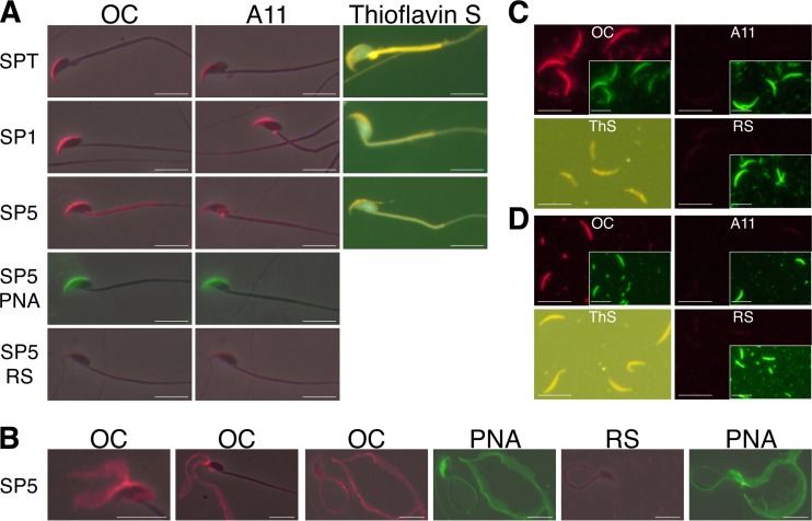
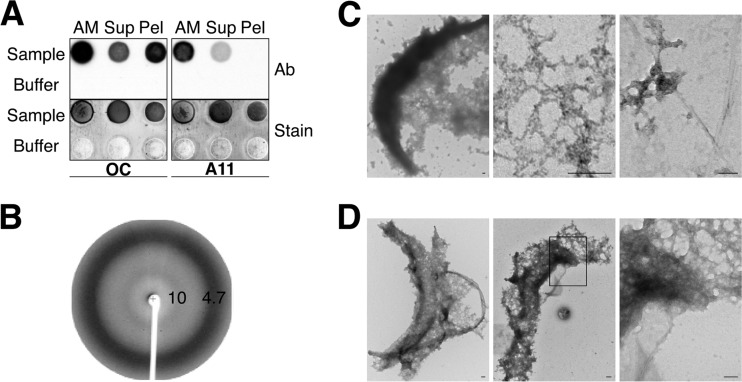
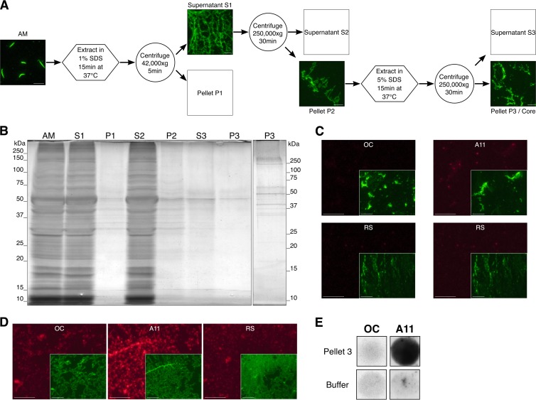
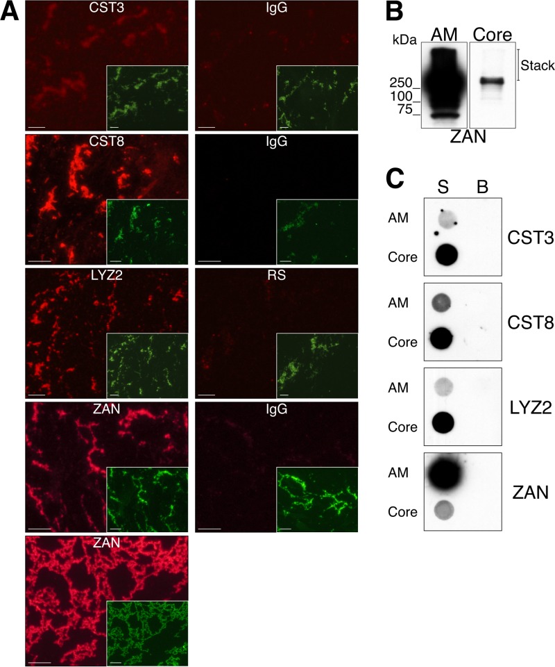
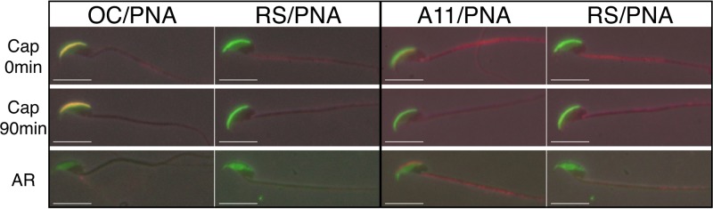
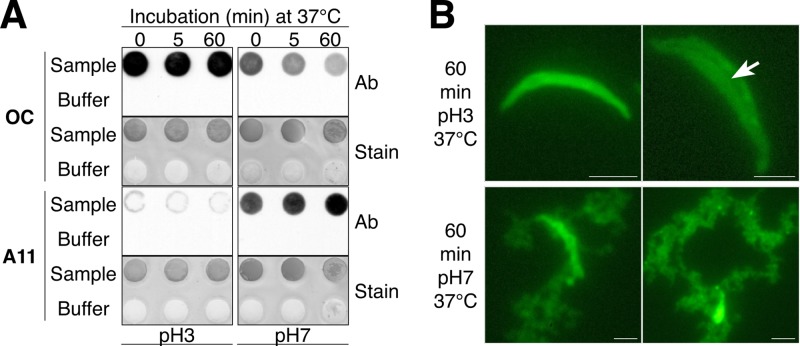
The acrosomal matrix (AM) is an insoluble structure within the sperm acrosome that serves as a scaffold controlling the release of AM-associated proteins during the sperm acrosome reaction. The AM also interacts with the zona pellucida (ZP) that surrounds the oocyte, suggesting a remarkable stability that allows its survival despite being surrounded by proteolytic and hydrolytic enzymes released during the acrosome reaction. To date, the mechanism responsible for the stability of the AM is not known. Our studies demonstrate that amyloids are present within the sperm AM and contribute to the formation of an SDS- and formic-acid-resistant core. The AM core contained several known amyloidogenic proteins, as well as many proteins predicted to form amyloid, including several ZP binding proteins, suggesting a functional role for the amyloid core in sperm-ZP interactions. While stable at pH 3, at pH 7, the sperm AM rapidly destabilized. The pH-dependent dispersion of the AM correlated with a change in amyloid structure leading to a loss of mature forms and a gain of immature forms, suggesting that the reversal of amyloid is integral to AM dispersion.


Figures

FIG 1
FIG 1
Amyloids are present in mouse sperm acrosomes. Amyloids were detected by IIF analysis with OC and A11 antiserum (red fluorescence) and by ThS staining. Normal RS was used as a control. All slides were costained with FITC-PNA (green fluorescence) as a marker for acrosomal material. Phase-contrast and epifluorescence images were merged informatically. Scale bars, 10 μm. (A) Intact spermatozoa from the testis (SPT), caput (SP1), and cauda (SP5) epididymis. (B) Cauda epididymal spermatozoa (SP5) with mechanically disrupted acrosomal shrouds in various states of detachment and dispersion. (C and D) Isolated AM (total) from caput epididymal (C) and cauda epididymal (D) spermatozoa. Insets show FITC-PNA staining shown at a 40% reduction.
FIG 2
FIG 2
Purified AM are composed of amyloids. (A) Dot blot analysis with OC and A11 antibodies (Ab) of total AM, soluble AM (Sup), and insoluble AM (Pel) fractions isolated from cauda epididymal spermatozoa. Buffer only served as a control. Colloidal gold staining (Stain) was performed after dot blot analysis to confirm the presence of protein in each spot. (B) X-ray fiber diffraction analysis of AM isolated from cauda epididymal spermatozoa. (C and D) Negative-staining electron microscopy of AM isolated from caput (C) and cauda (D) epididymal spermatozoa. The boxed area in the middle section of panel D is magnified in the right panel. Scale bars, 10 μm.
FIG 3
FIG 3
The AM contains an amyloid-rich core structure. Purified AM were exposed to a two-step extraction to sequentially strip off soluble proteins (A and B). The presence of amyloid in the remaining insoluble material (core) was determined by IIF analysis (C and D) and dot blot analysis (E) with OC and A11 antibodies. (A) Isolated AM were incubated in 1% SDS in 20 mM SA (pH 3) for 15 min at 37°C and then centrifuged at 42,000 × g for 5 min to pellet nonextracted AM (P1). The supernatant (S1) containing the extracted AM and solubilized proteins was centrifuged at 250,000 × g for 30 min. The pellet (P2) was then extracted in either 5% SDS or 70% formic acid for 15 min at 37°C, and samples were centrifuged at 250,000 × g. The pellet (P3) represented the AM core. AM, S1, P2, and P3 were stained with FITC-PNA. Scale bars, 10 μm. (B) Silver-stained SDS-PAGE of proteins sequentially extracted from the AM during purification of the core. Lanes were equally loaded with proteins from 9 × 10⁶ AM equivalents. Proteins were solubilized in 8 M urea–100 mM DTT before the addition of reducing Laemmli buffer and electrophoresis. The second P3 lane contains proteins from 4 × 10⁷ AM equivalents. (C and D) P3 obtained by extraction in 5% SDS (C) or 70% formic acid (D) was examined by IIF analysis with OC and A11 antibodies (red fluorescence). Normal RS served as a control antibody. Insets, P3 costained with FITC-PNA (green fluorescence) and shown at a 40% reduction. Scale bars, 10 μm. (E) P3 obtained by extraction in 5% SDS was dotted onto nitrocellulose membrane and incubated with OC and A11 antibodies in a dot blot analysis.
FIG 4
FIG 4
Immunodetection of proteins in the AM core. (A) The AM core obtained by extraction with 5% SDS was spread on slides and immunostained with CST3, CST8, LYZ2, and ZAN antibodies (red fluorescence). Last panel, AM core obtained by extraction with 70% formic acid and immunostained with ZAN antibody. Control staining was carried out with normal rabbit IgG or serum (RS). Insets, costaining with FITC-PNA shown at a 50% reduction. Scale bars, 10 μm. (B) Western blot analysis of ZAN in total AM and AM core fractions. Proteins from 5 × 10⁶ and 6 × 10⁷ AM equivalents were loaded into the total AM and AM core lanes, respectively. (C) Dot blot analysis of CST3, CST8, LYZ2, and ZAN in total AM and AM core fractions. The AM and AM core proteins were dotted onto nitrocellulose membrane and incubated with the relevant antibodies. Proteins from 1 × 10⁶ and 3 × 10⁷ AM equivalents were dotted for AM and AM core, respectively. S, sample, B, buffer.
FIG 5
FIG 5
Examination of sperm acrosomal amyloid during capacitation and AR. IIF analysis was carried out with OC and A11 antibodies (red fluorescence) to examine acrosomal amyloid after incubation of cauda epididymal spermatozoa under capacitating conditions at 0 and 90 min and following induction of the AR by the addition of progesterone. Normal RS served as a control antiserum. Acrosomal integrity was determined by costaining with FITC-PNA (green fluorescence). Phase-contrast and epifluorescence images were merged informatically. Scale bars, 10 μm.
FIG 6
FIG 6
A pH-dependent dispersion of the AM is associated with amyloid reversal. (A) Total AM were incubated for 0, 5, or 60 min at 37°C in 20 mM SA at pH 3 or 7. At each time point, a sample was removed for FITC-PNA staining while the remaining material (5 × 10⁶ AM) was spotted onto nitrocellulose membrane for dot blot analysis with OC and A11 antibodies (Ab). Buffer only served as a negative control. Colloidal gold staining of the dot blots was performed to confirm the presence of protein in each spot (Stain). (B) AM integrity after incubation at pH 3 or 7 was determined by staining with FITC-PNA. The arrow shows a longitudinal fissure that was observed in some AM that were beginning to disperse. Scale bars, 2.5 μm.

Similar articles

Cited by

References

    1. Buffone MG, Foster JA, Gerton GL. 2008. The role of the acrosomal matrix in fertilization. Int. J. Dev. Biol. 52:511–522. 10.1387/ijdb.072532mb - DOI - PubMed
    1. Hardy DM, Oda MN, Friend DS, Huang TT. 1991. A mechanism for differential release of acrosomal enzymes during the acrosome reaction. Biochem. J. 275(Pt 3):759–766 - PMC - PubMed
    1. Kim K-S, Gerton GL. 2003. Differential release of soluble and matrix components: evidence for intermediate states of secretion during spontaneous acrosomal exocytosis in mouse sperm. Dev. Biol. 264:141–152. 10.1016/j.ydbio.2003.08.006 - DOI - PubMed
    1. Florman HM, Storey BT. 1982. Mouse gamete interactions: the zona pellucida is the site of the acrosome reaction leading to fertilization in vitro. Dev. Biol. 91:121–130. 10.1016/0012-1606(82)90015-X - DOI - PubMed
    1. Storey BT, Lee MA, Muller C, Ward CR, Wirtshafter DG. 1984. Binding of mouse spermatozoa to the zonae pellucidae of mouse eggs in cumulus: evidence that the acrosomes remain substantially intact. Biol. Reprod. 31:1119–1128. 10.1095/biolreprod31.5.1119 - DOI - PubMed

Publication types