

Digital subtraction MR angiography roadmapping for magnetic steerable catheter tracking
- PMID: 24797218
- PMCID: PMC4223000
- DOI: 10.1002/jmri.24651
Digital subtraction MR angiography roadmapping for magnetic steerable catheter tracking
Abstract
Purpose: To develop a high temporal resolution MR imaging technique that could be used with magnetically assisted remote control (MARC) endovascular catheters.
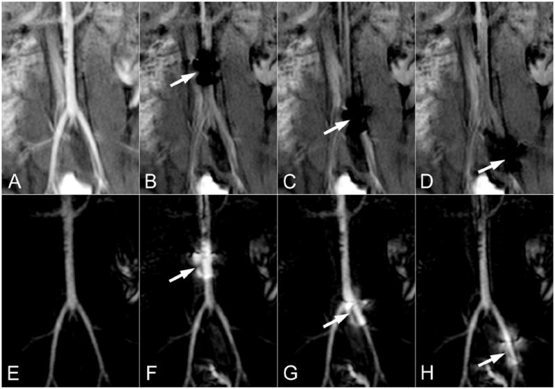
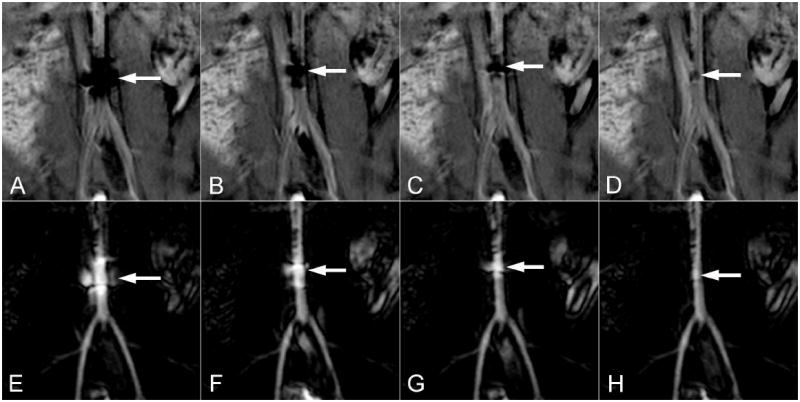
Materials and methods: A technique is proposed based on selective intra-arterial injections of dilute MR contrast at the beginning of a fluoroscopic MR angiography acquisition. The initial bolus of contrast is used to establish a vascular roadmap upon which MARC catheters can be tracked. The contrast to noise ratio (CNR) of the achieved roadmap was assessed in phantoms and in a swine animal model. The ability of the technique to permit navigation of activated MARC catheters through arterial branch points was evaluated.
Results: The roadmapping mode proved effective in phantoms for tracking objects and achieved a CNR of 35.7 between the intra- and extra-vascular space. In vivo, the intra-arterial enhancement strategy produced roadmaps with a CNR of 42.0. The artifact produced by MARC catheter activation provided signal enhancement patterns on the roadmap that experienced interventionalists could track through vascular structures.
Conclusion: A roadmapping approach with intra-arterial contrast-enhanced MR angiography is introduced for navigating the MARC catheter. The technique mitigates the artifact produced by the MARC catheter, greatly limits the required specific absorption rate, permits regular roadmap updates due to the low contrast agent requirements, and proved effective in the in vivo setting. Inc.
Keywords: digital subtraction angiography; endovascular procedure; interventional MR; magnetic resonance angiography.
© 2014 Wiley Periodicals, Inc.
Figures
Similar articles
-
New-Generation Laser-lithographed Dual-Axis Magnetically Assisted Remote-controlled Endovascular Catheter for Interventional MR Imaging: In Vitro Multiplanar Navigation at 1.5 T and 3 T versus X-ray Fluoroscopy.Radiology. 2015 Dec;277(3):842-52. doi: 10.1148/radiol.2015142648. Epub 2015 Jun 1. Radiology. 2015. PMID: 26030659 Free PMC article.
-
Endovascular MR-guided Renal Embolization by Using a Magnetically Assisted Remote-controlled Catheter System.Radiology. 2016 Oct;281(1):219-28. doi: 10.1148/radiol.2016152036. Epub 2016 Mar 28. Radiology. 2016. PMID: 27019290 Free PMC article.
-
[MR angiography of peripheral vessels with automatic tracking table technique at 1.0 in comparison with intra-arterial digital subtraction angiography].Rofo. 1999 Sep;171(3):240-3. doi: 10.1055/s-1999-245. Rofo. 1999. PMID: 10520335 German.
-
MR angiography with three-dimensional MR digital subtraction angiography.Top Magn Reson Imaging. 1996 Dec;8(6):366-88. Top Magn Reson Imaging. 1996. PMID: 9402678 Review.
-
Selection of patients for carotid thromboendarterectomy: the role of magnetic resonance angiography.J Comput Assist Tomogr. 1999 Nov;23 Suppl 1:S91-4. doi: 10.1097/00004728-199911001-00012. J Comput Assist Tomogr. 1999. PMID: 10608403 Review.
Cited by
-
Magnetic Resonance-Guided Passive Catheter Tracking for Endovascular Therapy.Magn Reson Imaging Clin N Am. 2015 Nov;23(4):591-605. doi: 10.1016/j.mric.2015.05.003. Epub 2015 Aug 12. Magn Reson Imaging Clin N Am. 2015. PMID: 26499277 Free PMC article. Review.
-
MRI-guided endovascular intervention: current methods and future potential.Expert Rev Med Devices. 2022 Oct;19(10):763-778. doi: 10.1080/17434440.2022.2141110. Expert Rev Med Devices. 2022. PMID: 36373162 Free PMC article. Review.
References
-
- Bock M, Wacker FK. MR-guided intravascular interventions: techniques and applications. J Magn Reson Imaging. 2008 Feb;27(2):326–338. - PubMed
-
- Roberts TP, Hassenzahl WV, Hetts SW, Arenson RL. Remote control of catheter tip deflection: an opportunity for interventional MRI. Magn Reson Med. 2002 Dec;48(6):1091–1095. - PubMed
Publication types
MeSH terms
Grants and funding
LinkOut - more resources
Full Text Sources
Other Literature Sources
