Review
. 2015 Jun 18:296:80-91.
doi: 10.1016/j.neuroscience.2014.04.048. Epub 2014 May 2.

A tale of two species: Neural integration in zebrafish and monkeys

Affiliations
Review

A tale of two species: Neural integration in zebrafish and monkeys

M Joshua et al. Neuroscience. .

Abstract

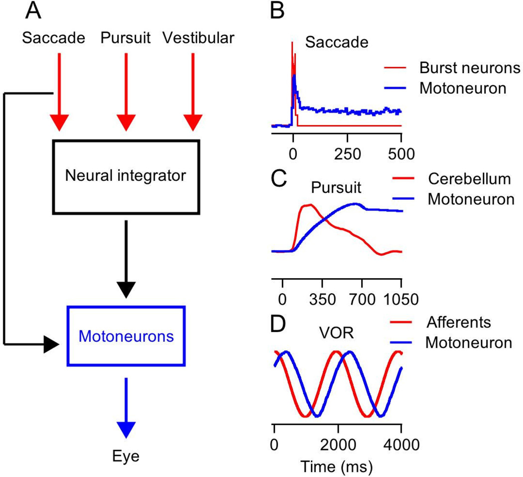
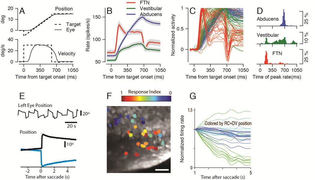
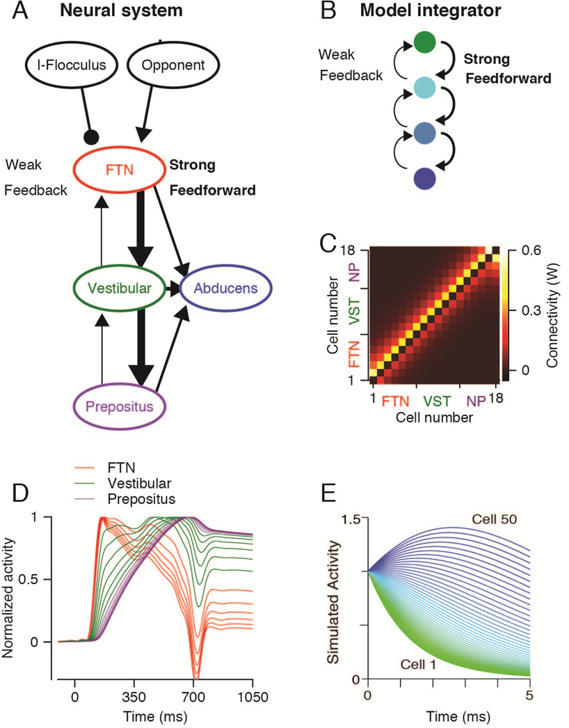
Selection of a model organism creates tension between competing constraints. The recent explosion of modern molecular techniques has revolutionized the analysis of neural systems in organisms that are amenable to genetic techniques. Yet, the non-human primate remains the gold-standard for the analysis of the neural basis of behavior, and as a bridge to the operation of the human brain. The challenge is to generalize across species in a way that exposes the operation of circuits as well as the relationship of circuits to behavior. Eye movements provide an opportunity to cross the bridge from mechanism to behavior through research on diverse species. Here, we review experiments and computational studies on a circuit function called "neural integration" that occurs in the brainstems of larval zebrafish, primates, and species "in between". We show that analysis of circuit structure using modern molecular and imaging approaches in zebrafish has remarkable explanatory power for details of the responses of integrator neurons in the monkey. The combination of research from the two species has led to a much stronger hypothesis for the implementation of the neural integrator than could have been achieved using either species alone.

Keywords: animal models; brainstem; eye movements; monkey; neural integrator; zebrafish.


Figures

Figure 1
Figure 1. Rationale for an oculomotor neural integrator
A: Schematic representation of the neuron integrator hypothesis. Commands for saccades, smooth pursuit, and the vestibulo-ocular reflex all signal the desired eye velocity, and therefore require neural integration to create the eye position signal that dominates the activity of extraocular motoneurons. The “Neural integrator” converts its input signals into commands for maintaining the eye in an eccentric position. The discharge of “Motoneurons” is assembled as a combination of the inputs and outputs of the integrator, the inputs to move the eye to a new position and the outputs to hold the eye stable in the final position. B–D: Red and blue traces represent the inputs to the integrator and motoneuron activity during saccades (B), smooth pursuit eye movement (C) and the vestibulo-ocular reflex (D). The traces were scaled arbitrarily to allow easier comparison of their temporal dynamics.
Figure 2
Figure 2. Eye movements and time-varying waveforms of neural integrator activity in zebrafish and monkeys
A: Top and bottom superimposed traces show horizontal eye position (top) and velocity (bottom) during smooth pursuit eye movements. Solid and dashed lines show eye and target motion parameters. B–C: Each trace shows the time-varying firing rate in the monkey brainstem during the pursuit behavior illustrated in A. Panel B shows averages across functionally identified neuron types and panel C shows firing rate averages from individual neurons. Red, green, and blue traces denote the activity of FTNs, other, non-FTN neurons in the vestibular nucleus, and Abducens neurons. D: Histograms plot the time of peak firing rate using the same color code as in B and C. E: Eye movements of a larva zebrafish. Top, an eye position trace from 100 seconds of spontaneous movements. Bottom, representative eye position trajectories before and after saccades that take eye position toward (black) or away from (green) the side of the integrator under study. F: Image of 29 identified integrator neurons in the brainstem of the larval zebrafish. Neurons are color coded according to how strongly the calcium responses of pairs of neurons co-varied with eye position versus eye velocity. Red versus blue coloring indicates neurons that co-varied strongly with eye position versus eye velocity. G. Each trace shows the estimate of the time varying firing rate of an individual neuron in the zebrafish neural integrator from 1 to 5 seconds after a saccade. Traces are colored according to the location of the cell, which was defined as the sum of the rostrocaudal (RC) and dorsoventral (DV) coordinates. Panels B–D are adapted with permission from Joshua et al. (2013). Panels E–G are adapted with permission from Miri et al. (2011).
Figure 3
Figure 3. A network model that integrates and reproduces the time-varying firing rates of neurons in the oculomotor neural integrator of monkeys and zebrafish
A: Schematic representation of the brainstem pursuit network as defined by recordings from functionally identified neurons in monkeys. Each ellipse represents a population of neurons, arrows are excitatory connections, and lines ending with a circle represent inhibitory connections. Thick and thin arrows indicate strong versus weak connections. B. The architecture of a neural integrator model. Each circle represents a single neuron or group of neurons, and the arrows represent connections. Each neuron is connected strongly the next neuron and weakly to the previous neuron. C: The connectivity matrix used for simulation of monkey brainstem. The colors indicate the connection weights in the connection matrix (W) between neurons for a network with stronger feed-forward versus feedback connections. D–E. Each trace shows the time-varying firing rate of an individual model neuron in the neural networks used by the two laboratories to simulate neural integration in monkey (D) and zebrafish (E). In panel D, the colors of the traces indicate the functional group assigned to the three groups of 6 neurons in the model integrator. Panels A–D are adapted with permission from Joshua et al. (2013). Panel E is adapted with permission from Miri et al. (2011).

References

    1. Adelsberger H, Zainos A, Alvarez M, Romo R, Konnerth A. Local domains of motor cortical activity revealed by fiber-optic calcium recordings in behaving nonhuman primates. Proc Natl Acad Sci U S A. 2014;111:463–468. - PMC - PubMed
    1. Aksay E, Baker R, Seung HS, Tank DW. Anatomy and discharge properties of pre-motor neurons in the goldfish medulla that have eye-position signals during fixations. J Neurophysiol. 2000;84:1035–1049. - PubMed
    1. Aksay E, Baker R, Seung HS, Tank DW. Correlated discharge among cell pairs within the oculomotor horizontal velocity-to-position integrator. J Neurosci. 2003;23:10852–10858. - PMC - PubMed
    1. Aksay E, Gamkrelidze G, Seung HS, Baker R, Tank DW. In vivo intracellular recording and perturbation of persistent activity in a neural integrator. Nat Neurosci. 2001;4:184–193. - PubMed
    1. Aksay E, Olasagasti I, Mensh BD, Baker R, Goldman MS, Tank DW. Functional dissection of circuitry in a neural integrator. Nat Neurosci. 2007;10:494–504. - PMC - PubMed

Publication types

LinkOut - more resources