. 2014 Oct 1;31(19):1647-63.
doi: 10.1089/neu.2013.3320. Epub 2014 Jul 16.

Acute reduction of microglia does not alter axonal injury in a mouse model of repetitive concussive traumatic brain injury

Affiliations

Acute reduction of microglia does not alter axonal injury in a mouse model of repetitive concussive traumatic brain injury

Rachel E Bennett et al. J Neurotrauma. .

Abstract

The pathological processes that lead to long-term consequences of multiple concussions are unclear. Primary mechanical damage to axons during concussion is likely to contribute to dysfunction. Secondary damage has been hypothesized to be induced or exacerbated by inflammation. The main inflammatory cells in the brain are microglia, a type of macrophage. This research sought to determine the contribution of microglia to axon degeneration after repetitive closed-skull traumatic brain injury (rcTBI) using CD11b-TK (thymidine kinase) mice, a valganciclovir-inducible model of macrophage depletion. Low-dose (1 mg/mL) valganciclovir was found to reduce the microglial population in the corpus callosum and external capsule by 35% after rcTBI in CD11b-TK mice. At both acute (7 days) and subacute (21 days) time points after rcTBI, reduction of the microglial population did not alter the extent of axon injury as visualized by silver staining. Further reduction of the microglial population by 56%, using an intermediate dose (10 mg/mL), also did not alter the extent of silver staining, amyloid precursor protein accumulation, neurofilament labeling, or axon injury evident by electron microscopy at 7 days postinjury. Longer treatment of CD11b-TK mice with intermediate dose and treatment for 14 days with high-dose (50 mg/mL) valganciclovir were both found to be toxic in this injury model. Altogether, these data are most consistent with the idea that microglia do not contribute to acute axon degeneration after multiple concussive injuries. The possibility of longer-term effects on axon structure or function cannot be ruled out. Nonetheless, alternative strategies directly targeting injury to axons may be a more beneficial approach to concussion treatment than targeting secondary processes of microglial-driven inflammation.

Keywords: axon injury; concussion; microglia.


Figures

<b>FIG. 1.</b>
FIG. 1.
Experimental design. CD11b-TK+/− or CD11b-TK−/− controls were treated with either valganciclovir or saline by intracerebroventricular osmotic pump for 7 days preceding injury. Pumps were then removed, and a closed-skull or sham injury was performed (day 0). Twenty-four hours later, a second injury was delivered. Pumps were immediately reimplanted, and drug or saline treatment was resumed for another 7 (acute time point) or 21 days (subacute time point). TK, thymidine kinase.
<b>FIG. 2.</b>
FIG. 2.
Treatment of CD11b-TK+/− mice with intracerebroventricular low-dose valganciclovir (1 mg/mL) reduces microglia, but does not affect silver staining 7 days after repetitive closed-skull traumatic brain injury. Iba-1 staining in treated CD11b-TK−/− (A and C) and CD11b-TK+/− (B and D) mice. (C and D) Higher magnification views of Iba-1 staining in corpus callosum ipsilateral to injury (region boxed in A, outlined with red dashed line). Silver staining in valganciclovir-treated CD11b-TK−/− (E) and CD11b-TK+/− (F) mice. (G) Stereological quantification of Iba-1-positive microglia in CD11b-TK+/− mice shows a ∼35% reduction, compared to controls (one-tailed Student's t-test, **p<0.01; dashed line indicates sham levels). (H) Silver staining in white matter was unchanged. A.U., arbitrary units; n.s., not significant. Error bars represent standard error of the mean. TK, thymidine kinase. Color image is available online at www.liebertpub.com/neu
<b>FIG. 3.</b>
FIG. 3.
Low-dose valganciclovir (1 mg/mL), but not saline, reduces microglia and has no effect on silver staining 7 days after injury in an independent cohort of mice. (A) Iba-1 staining and (B) silver staining in corpus callosum and external capsule of CD11b+/− and CD11b−/− mice receiving either drug or vehicle treatment. A.U., arbitrary units; n.s., not significant. *p<0.05; **p<0.01. Dashed line indicates sham levels. Error bars represent standard error of the mean. TK, thymidine kinase. Color image is available online at www.liebertpub.com/neu
<b>FIG. 4.</b>
FIG. 4.
High-dose (50 mg/mL) valganciclovir treatment was toxic in CD11b-TK+/− mice 7 days postinjury. (A) An Iba-1-stained section near the cannula site (dashed line) shows depletion of microglia, but also numerous microhemorrhages (arrows). (B) Cresyl violet staining in a section posterior to valganciclovir injection shows extensive tissue loss in the hippocampus and thalamus (dashed oval). TK, thymidine kinase. Color image is available online at www.liebertpub.com/neu
<b>FIG. 5.</b>
FIG. 5.
Valganciclovir dose response in CD11b-TK mice 7 days postinjury. Iba-1-stained whole sections (A, C, and E) and higher-magnification images (box A) corresponding to the region of interest assessed for iba-1 depletion by stereology (outlined with red dashed line; B, D, and F). CD11b-TK+/− mice treated with 2 (A and B), 5 (C and D), or 10 mg/mL (E and F) of valganciclovir. Microglial depletion is most prominent ipsilateral to cannulation and drug delivery (right). Arrowheads indicate region of microglial reduction in cortex of hemisphere opposite drug infusion. TK, thymidine kinase. Color image is available online at www.liebertpub.com/neu
<b>FIG. 6.</b>
FIG. 6.
Acute intermediate-dose valganciclovir (10 mg/mL) reduces Iba-1, but not silver, staining in CD11b-TK mice 7 days after repetitive closed-skull traumatic brain injury. Iba-1 staining in corpus callosum (outlined with red dashed line) in treated CD11b-TK−/− (A) and CD11b-TK+/− (B) mice. Silver staining in adjacent sections from CD11b-TK−/− (C) and CD11b-TK+/− (D) mice. (E) Iba-1 was reduced by 56% in CD11b-TK+/− mice, compared to controls (one-tailed Student's t-test, ***p<0.001; dashed line indicates sham levels). (F) No change in silver staining was observed. A.U., arbitrary units; n.s., not significant. Error bars represent standard error of the mean. TK, thymidine kinase. Color image is available online at www.liebertpub.com/neu
<b>FIG. 7.</b>
FIG. 7.
Amyloid precursor protein (APP) and neurofilament (RM014) labeling in an injured CD11b-TK−/− mouse treated with 10 mg/mL of valganciclovir and sacrificed 7 days postinjury. APP and labeling was prominent at the site of cannulation (dashed line) −0.5 mm relative to bregma (A and B). Only sparse axonal swellings were detectable in the contralateral hemisphere (injured side) in the corpus callosum and external capsule corresponding to the region of interest assessed for axonal injury in these experiments (C and D). Insets show higher magnification of labeling in boxed regions in (A) and (C). TK, thymidine kinase. Color image is available online at www.liebertpub.com/neu
<b>FIG. 8.</b>
FIG. 8.
Normal axonal ultrastructure in a cannulated sham CD11b-TK−/− mouse treated with 10 mg/mL of valganciclovir and sacrificed 7 days post–sham injury. (A and B) Axons in the corpus callosum near the cingulum display organized cytoskeletal elements and intact myelin sheaths. (C) Region containing axons cut parallel to the predominant direction of the corpus callosum. Asterisk (*) indicates a swollen unmyelinated axon with abnormal organelle accumulation. Note nearby axons appear relatively intact, however. TK, thymidine kinase.
<b>FIG. 9.</b>
FIG. 9.
Ultrastructural abnormalities in an injured CD11b-TK−/− littermate control mouse treated with 10 mg/mL of valganciclovir and sacrificed 7 days postinjury. (A) Arrowheads indicate regions of end-stage axonal degeneration in corpus callosum near the cingulum where the axolemma has completely collapsed. Asterisks (*) denote examples of myelinated and unmyelinated axons with organelle accumulation. (B) Myelinated axon undergoing Wallerian degeneration (asterisk). (C) Region of relatively healthy appearing axons in corpus callosum. rcTBI, repetitive closed-skull traumatic brain injury; TK, thymidine kinase.
<b>FIG. 10.</b>
FIG. 10.
Ultrastructural abnormalities in an injured CD11b-TK+/− mouse treated with 10 mg/mL of valganciclovir and sacrificed 7 days postinjury. (A) Arrowhead indicates an example of end-stage axon degeneration in corpus callosum near the cingulum where the axolemma has completely collapsed. Asterisk (*) denotes a swollen axon undergoing late-stage degeneration, and arrows indicate examples of axons containing densely packed intracellular aggregates. (B) Arrow indicates an axon cut parallel to the predominant direction of the corpus callosum with abnormal accumulation of electron-dense intra-axoplasmic material in a region of relatively normal axons (asterisk). (C) Asterisk indicates an axon undergoing axolemma collapse. rcTBI, repetitive closed-skull traumatic brain injury; TK, thymidine kinase.
<b>FIG. 11.</b>
FIG. 11.
Treatment with low-dose valganciclovir (1 mg/mL), but not saline, reduces Iba-1, but not silver staining in corpus callosum 21 days after repetitive closed-skull traumatic brain injury. Iba-1 staining in valganciclovir-treated CD11b-TK−/− (A) and CD11b-TK+/− (B) mice. Silver staining in valganciclovir-treated CD11b-TK−/− (C) and CD11b-TK+/− (D) mice. (E) Iba-1 was reduced by 35% in CD11b-TK+/− mice, compared to CD11b-TK–/– controls (one-tailed Student's t-test; **p<0.01). (F) No change in silver staining was observed. A.U., arbitrary units; n.s., not significant. Error bars represent standard error of the mean. TK, thymidine kinase. Color image is available online at www.liebertpub.com/neu
<b>FIG. 12.</b>
FIG. 12.
Subacute treatment with intermediate-dose valganciclovir is toxic in injured CD11b-TK mice. Twenty-eight days total treatment resulted in tissue loss, as visualized by cresyl violet, in hippocampus and thalamus of injured CD11b-TK+/− (A), but not CD11b-TK−/− (B), mice. Iba-1 staining shows extent of microglial depletion in CD11b-TK+/− (C) and not in a CD11b-TK−/− (D) littermate control mouse subjected to repetitive closed-skull traumatic brain injury. TK, thymidine kinase. Color image is available online at www.liebertpub.com/neu
<b>FIG. 13.</b>
FIG. 13.
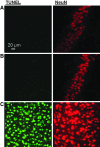
Double immunofluorescence for TUNEL (green) and NeuN (red) in the hippocampal CA3 does not indicate neuronal apoptosis in injured mice treated with 10 mg/mL of valganciclovir and sacrificed 7 days postinjury. TUNEL labeling was not observed in NeuN+ cells in the hippocampal CA3 of CD11b−/− (A) or CD11b+/− (B) mice. A positive control for TUNEL labeling was generated by incubating a more anterior section (cortical region) in DNAse I to induce double-stranded DNA breaks (C). All images were acquired using the same settings on a laser scanning confocal microscope. TUNEL, terminal deoxynucleotidyl transferase dUTP nick end labeling; NeuN, neuronal nuclear antigen. Color image is available online at www.liebertpub.com/neu
<b>FIG. 14.</b>
FIG. 14.
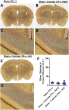
Right-sided cerebroventricular cannulation does not contribute to silver staining in the left corpus callosum region of interest assessed for axonal injury. Silver staining was performed in sections from CD11b-TK−/− mice that underwent the sham procedure alone (A and C) or the sham procedure plus cannulation and treatment with saline (NaCl; B and D). Silver staining was also performed in sections from CD11b-TK+/− mice that underwent the sham procedure plus cannulation and treatment with 10 mg/mL of valganciclovir (E and G). All mice were sacrificed 7 days post–sham injury. (F) No difference in silver staining was observed between groups. A.U., arbitrary units; TK, thymidine kinase. Color image is available online at www.liebertpub.com/neu
<b>FIG. 15.</b>
FIG. 15.
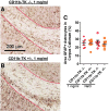
Astrocytes 7 days after repetitive closed-skull traumatic brain injury in mice treated with 1 mg/mL of valganciclovir or NaCl. GFAP staining in treated CD11b-TK−/− (A) and CD11b-TK+/− (B) mice. (C) No differences in GFAP optical density was observed between groups. GFAP, glial fibrillary acidic protein; TK, thymidine kinase. Color image is available online at www.liebertpub.com/neu
<b>FIG. 16.</b>
FIG. 16.
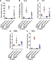
qPCR measurement of inflammatory gene expression. Relative expression of (A) Ccl2, (B) Il1β, (C) Il6, (D) iNOS, and (E) Tnfα in each mouse was normalized first to the geometric mean of 3 reference genes (Hprt, Pgk1, Gapdh) and then to arithmetic mean of the uninjured, naïve mice (n=4; dashed line). CCL2, chemokine (C-C motif ) ligand 2; IL, interleukin; iNOS, inducible nitric oxide synthetase; TNF-α, tumor necrosis factor alpha; rcTBI, repetitive closed-skull traumatic brain injury; TK, thymidine kinase. Color image is available online at www.liebertpub.com/neu

Similar articles

Cited by

References

    1. Corsellis J.A., Bruton C.J., and Freeman-Browne D. (1973). The aftermath of boxing. Psychol. Med. 3, 270–303 - PubMed
    1. Mortimer J.A. (1985). Epidemiology of post-traumatic encephalopathy in boxers. Minn. Med. 68, 299–300 - PubMed
    1. Roberts G.W., Allsop D., and Bruton C. (1990). The occult aftermath of boxing. J. Neurol. Neurosurg. Psychiatry 53, 373–378 - PMC - PubMed
    1. Geddes J.F., Vowles G.H., Nicoll J.A., and Revesz T. (1999). Neuronal cytoskeletal changes are an early consequence of repetitive head injury. Acta Neuropathol. 98, 171–178 - PubMed
    1. Guo Z, Cupples LA, Kurz A, Auerbach SH, Volicer L, Chui H, Green RC, Sadovnick AD, Duara R, DeCarli C, Johnson K, Go RC, Growdon JH, Haines JL, Kukull WA, and Farrer L.A. (2000). Head injury and the risk of AD in the MIRAGE study. Neurology 54, 1316–1323 - PubMed

Publication types

LinkOut - more resources