

Restoring systemic GDF11 levels reverses age-related dysfunction in mouse skeletal muscle
- PMID: 24797481
- PMCID: PMC4104429
- DOI: 10.1126/science.1251152
Restoring systemic GDF11 levels reverses age-related dysfunction in mouse skeletal muscle
Abstract
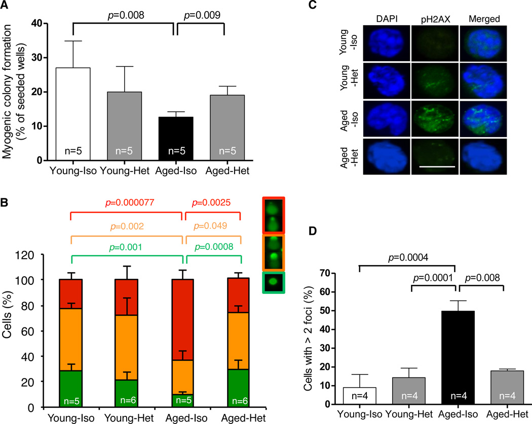
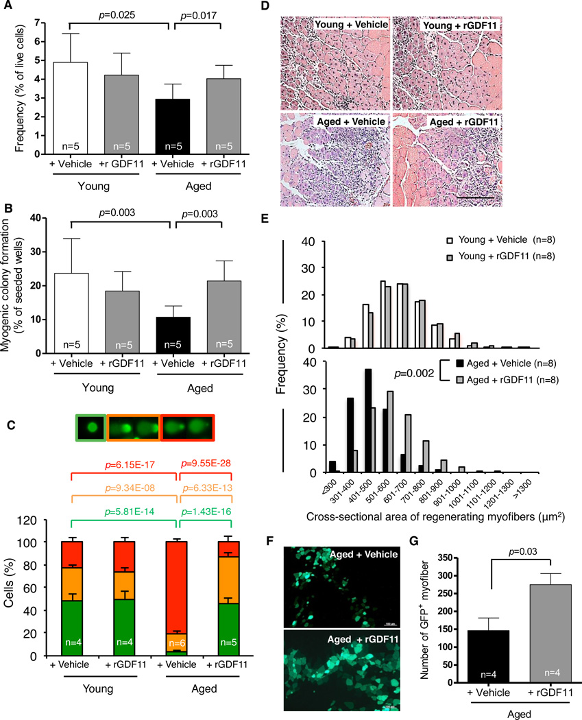
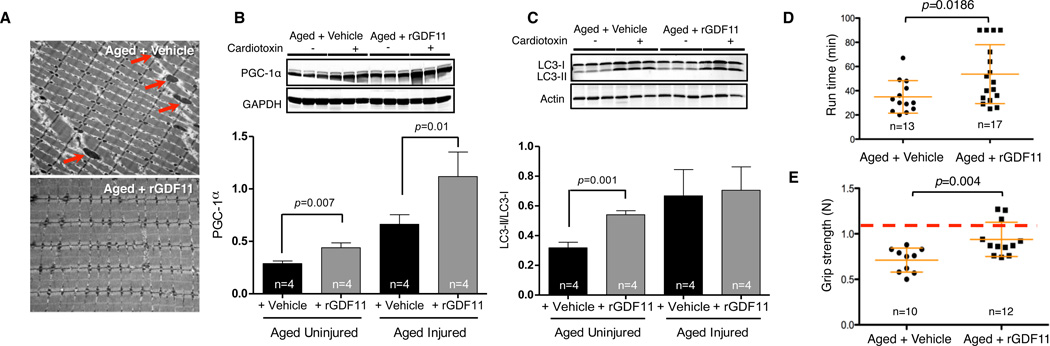
Parabiosis experiments indicate that impaired regeneration in aged mice is reversible by exposure to a young circulation, suggesting that young blood contains humoral "rejuvenating" factors that can restore regenerative function. Here, we demonstrate that the circulating protein growth differentiation factor 11 (GDF11) is a rejuvenating factor for skeletal muscle. Supplementation of systemic GDF11 levels, which normally decline with age, by heterochronic parabiosis or systemic delivery of recombinant protein, reversed functional impairments and restored genomic integrity in aged muscle stem cells (satellite cells). Increased GDF11 levels in aged mice also improved muscle structural and functional features and increased strength and endurance exercise capacity. These data indicate that GDF11 systemically regulates muscle aging and may be therapeutically useful for reversing age-related skeletal muscle and stem cell dysfunction.
Figures
Comment in
-
Brain ageing: Blood-derived rejuvenation.Nat Rev Neurosci. 2014 Jun;15(6):352-3. doi: 10.1038/nrn3763. Nat Rev Neurosci. 2014. PMID: 24840795 No abstract available.
References
-
- Jang YC, Sinha M, Cerletti M, Dall'osso C, Wagers AJ. Skeletal Muscle Stem Cells: Effects of Aging and Metabolism on Muscle Regenerative Function. Cold Spring Harb Symp Quant Biol. 2011 Sep 29; - PubMed
-
- Sherwood RI, et al. Isolation of adult mouse myogenic progenitors: functional heterogeneity of cells within and engrafting skeletal muscle. Cell. 2004 Nov 12;119:543. - PubMed
Publication types
MeSH terms
Substances
Grants and funding
- R01 AR042238/AR/NIAMS NIH HHS/United States
- 5U01 HL100402/HL/NHLBI NIH HHS/United States
- 1R01 AG033053/AG/NIA NIH HHS/United States
- 1R01 AG040019/AG/NIA NIH HHS/United States
- DP2 OD004345/OD/NIH HHS/United States
- 1DP2 OD004345/OD/NIH HHS/United States
- R01 AR42238/AR/NIAMS NIH HHS/United States
- T32 DE007057/DE/NIDCR NIH HHS/United States
- R01 AG033053/AG/NIA NIH HHS/United States
- R01 AG032977/AG/NIA NIH HHS/United States
- HHMI/Howard Hughes Medical Institute/United States
- R01 AG040019/AG/NIA NIH HHS/United States
- U01 HL100402/HL/NHLBI NIH HHS/United States
- P30 DK036836/DK/NIDDK NIH HHS/United States
LinkOut - more resources
Full Text Sources
Other Literature Sources
Medical
Molecular Biology Databases
