. 2014 May 5;9(5):e96657.
doi: 10.1371/journal.pone.0096657. eCollection 2014.

Poldip2 knockout results in perinatal lethality, reduced cellular growth and increased autophagy of mouse embryonic fibroblasts

Affiliations

Poldip2 knockout results in perinatal lethality, reduced cellular growth and increased autophagy of mouse embryonic fibroblasts

David I Brown et al. PLoS One. .

Abstract

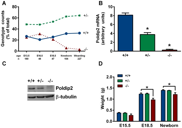
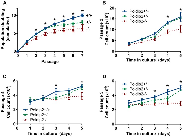
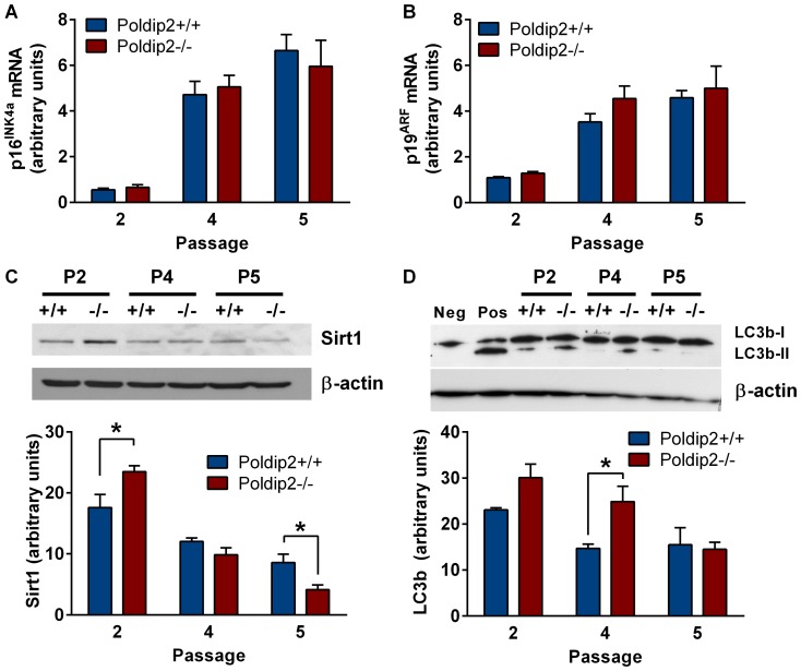
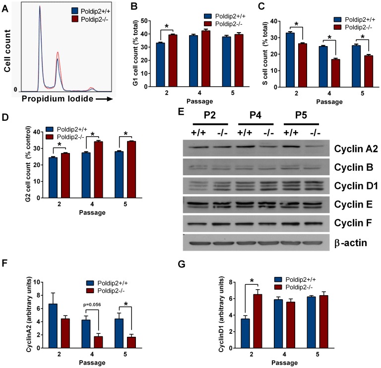
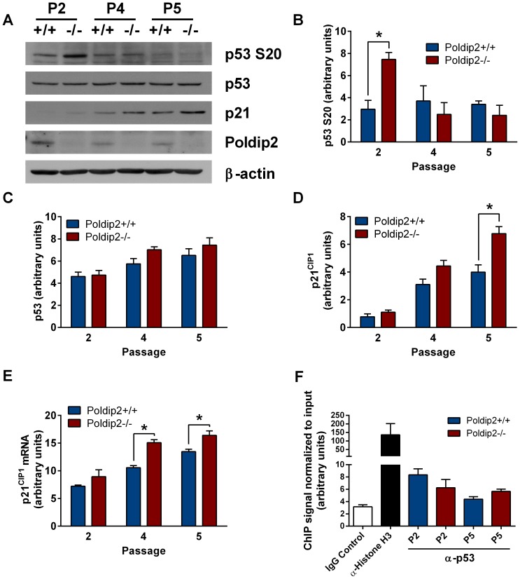
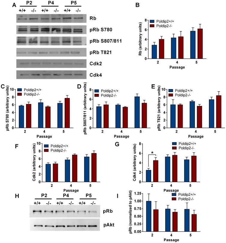
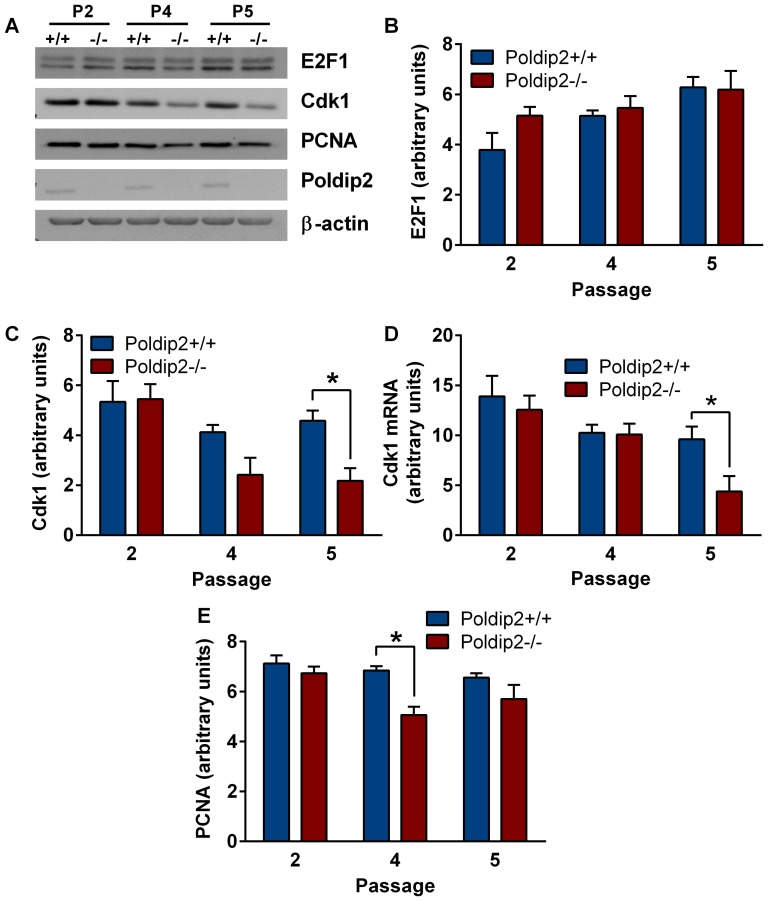
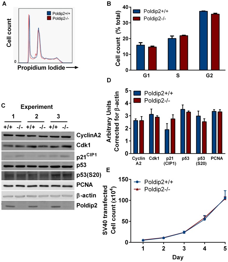
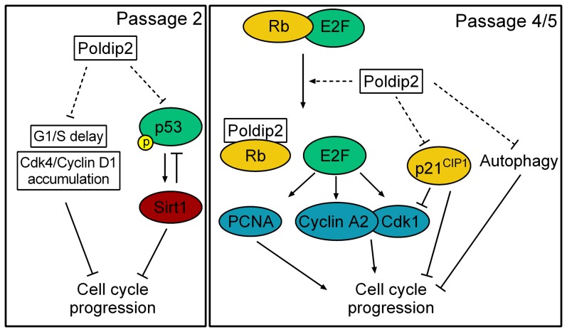
Polymerase-δ interacting protein 2 (Poldip2) is an understudied protein, originally described as a binding partner of polymerase delta and proliferating cell nuclear antigen (PCNA). Numerous roles for Poldip2 have been proposed, including mitochondrial elongation, DNA replication/repair and ROS production via Nox4. In this study, we have identified a novel role for Poldip2 in regulating the cell cycle. We used a Poldip2 gene-trap mouse and found that homozygous animals die around the time of birth. Poldip2-/- embryos are significantly smaller than wild type or heterozygous embryos. We found that Poldip2-/- mouse embryonic fibroblasts (MEFs) exhibit reduced growth as measured by population doubling and growth curves. This effect is not due to apoptosis or senescence; however, Poldip2-/- MEFs have higher levels of the autophagy marker LC3b. Measurement of DNA content by flow cytometry revealed an increase in the percentage of Poldip2-/- cells in the G1 and G2/M phases of the cell cycle, accompanied by a decrease in the percentage of S-phase cells. Increases in p53 S20 and Sirt1 were observed in passage 2 Poldip2-/- MEFs. In passage 4/5 MEFs, Cdk1 and CyclinA2 are downregulated in Poldip2-/- cells, and these changes are reversed by transfection with SV40 large T-antigen, suggesting that Poldip2 may target the E2F pathway. In contrast, p21CIP1 is increased in passage 4/5 Poldip2-/- MEFs and its expression is unaffected by SV40 transfection. Overall, these results reveal that Poldip2 is an essential protein in development, and underline its importance in cell viability and proliferation. Because it affects the cell cycle, Poldip2 is a potential novel target for treating proliferative conditions such as cancer, atherosclerosis and restenosis.


Conflict of interest statement

Competing Interests: The authors have declared that no competing interests exist.

Figures

Figure 1
Figure 1. Poldip2−/− embryos exhibit perinatal lethality and reduced weight.
(A) Progeny from heterozygote x heterozygote crosses were genotyped at different days post-conception and after birth. Mouse embryonic fibroblast Poldip2 (B) mRNA and (C) protein were measured to verify successful knockout. (D) Progeny at various stages of development were weighed and genotyped. Bars represent mean ± SEM of 3–4 independent mRNA experiments or 6–62 embryos or pups. * P<0.05 comparing Poldip2+/+ with Poldip2−/−.
Figure 2
Figure 2. Reduced growth in Poldip2 null cells.
Mouse embryonic fibroblasts were derived from Poldip2+/+, +/− and −/− E13.5 embryos. (A) Growth was assessed by counting cells at each passage and recorded as a cumulative population doubling. Additionally, growth was assessed by performing a growth curve at (B) passage 2, (C) passage 4, and (D) passage 5. Error bars represent mean ± SEM of 3–4 independent experiments. * P<0.05 comparing Poldip2+/+ with Poldip2−/−.
Figure 3
Figure 3. Poldip2 deletion increases Sirt1 expression and LC3b conversion but does not affect expression of senescence markers.
(A) p16INK4a and (B) p19ARF mRNA expression was compared between Poldip2+/+ (blue) and Poldip2−/− (red) cells by qRT-PCR and corrected for PPIA. (C) Sirt1 and (D) LC3b-1 and -II protein levels were assessed by western blot in Poldip2+/+ and Poldip2−/− MEFs at passages 2, 4 and 5 and normalized to β-actin. Error bars represent mean ± SEM of 3 independent experiments. * P<0.05 comparing Poldip2+/+ with Poldip2−/−.
Figure 4
Figure 4. Poldip2−/− MEFs exhibit altered cell cycle characteristics.
Asynchronous MEFs were collected at passage 2, 4 and 5 and stained with propidium iodide for FACS analysis of the cell cycle. (A) Passage 4 data is shown as an example histogram of DNA content as measured by flow cytometry and fit to the Dean-Jett-Fox model to calculate the percentage of cells in (B) G1, (C) S and (D) G2/M. (E) Key cell cycle protein expression was measured by immunoblotting. Protein levels were quantified by densitometry and corrected to β-actin expression for (F) CyclinA2 and (G) CyclinD1. All three bands were used in the quantification of CyclinD1. Error bars represent mean ± SEM of 3–4 independent experiments. * P<0.05 comparing Poldip2+/+ with Poldip2−/−.
Figure 5
Figure 5. Poldip2 inhibits the p53 pathway.
(A) Immunoblotting was performed using lysates from Poldip2+/+ and Poldip2−/− MEFs in passages 2, 4 and 5. The blots were probed with antibodies against β-actin, Poldip2, p53, phospho-p53(S20), and p21CIP1. Densitometry was performed and corrected to β-actin (B, C, E). (D) p21CIP1 mRNA levels were assessed by RT-qPCR and corrected for the housekeeping gene PPIA. (F) ChIP was performed on Poldip2+/+ (blue) and Poldip2−/− (red) MEFs using p53 antibody and p21CIP1 promoter primers. Poldip2+/+ cells were used for the IgG negative and Histone H3 antibody positive controls. All samples were normalized to input DNA. Error bars represent mean ± SEM of 3–4 independent experiments. * P<0.05 comparing Poldip2+/+ with Poldip2−/−.
Figure 6
Figure 6. Poldip2 deletion does not affect Rb expression/phosphorylation.
(A) Immunoblotting was performed using lysates from Poldip2+/+ and Poldip2−/− MEFs in passages 2, 4 and 5. The blots were probed with antibodies against Rb, pRb S780, pRb S807/811, pRb T821, Cdk2, and Cdk4. Densitometry was performed and corrected to β-actin run in parallel (shown in Figure 7A) (B–G). (H) Phosphorylated proteins from Poldip2+/+ and Poldip2−/− cells in passages 2, 4 and 5 were purified with a phosphoprotein binding column. Phosphorylated Rb was measured by immunoblot and normalized to pAkt levels (I). Error bars represent mean ± SEM of 3–4 independent experiments. * P<0.05 comparing Poldip2+/+ with Poldip2−/−.
Figure 7
Figure 7. Poldip2 activates the E2F1 pathway.
(A) Protein levels of E2F1 target genes were measured by immunoblot. Lysates from Poldip2+/+ and Poldip2−/− cells in passages 2, 4 and 5 were probed with antibodies against β-actin, Poldip2, E2F1, Cdk1 and PCNA. Densitometry was performed and corrected to β-actin (B–D). (E) Cdk1 mRNA levels were measured by RT-qPCR and corrected with PPIA. Error bars represent mean ± SEM of 3–4 independent experiments. * P<0.05 comparing Poldip2+/+ with Poldip2−/−.
Figure 8
Figure 8. SV40 immortalization of Poldip2−/− MEFs restores growth and cell cycle distribution.
Poldip2 +/+ and −/− MEFs were immortalized with SV40 large T-antigen. (A, B) Cell cycle analysis was performed as in Figure 4 using flow cytometry. (C) Expression of the indicated proteins was assessed by western blot in 3 independent batches of immortalized cells. (D) Densitometry was performed and corrected to β-actin. (E) A growth curve was performed to compare Poldip2+/+ (blue line) to Poldip2−/− (red line) MEFs. Error bars represent mean ± SEM of 3 experiments.
Figure 9
Figure 9. Proposed mechanism by which Poldip2 promotes cell cycle progression.
Left: Passage 2. Poldip2 promotes progression though G1/S by preventing the accumulation of Cdk4/Cyclin D1 that occurs in Poldip2−/− cells. Poldip2 also limits the activation of p53 by phosphorylation at S20, which results in reduced Sirt1 expression. Reduced Sirt1 negatively regulates p53 by de-acetylation and reduces cell cycle progression. Right: Passage 4/5. Poldip2 activates the transcription of E2F target genes such as Cyclin A, Cdk1 and PCNA. These act to promote cell cycle progression. Poldip2 also reduces expression of the cell cycle inhibitor p21CIP1. Reduced p21CIP1 promotes cell cycle progression by relieving inhibition of the activity of Cyclin/Cdk complexes. We propose that Poldip2, like SV40 immortalization, inhibits p53 activity and sequesters Rb away from E2F, promoting cell cycle progression. Finally, Poldip2 inhibits autophagy, which results in increased growth.

References

    1. Liu L, Rodriguez-Belmonte EM, Mazloum N, Xie B, Lee MY (2003) Identification of a novel protein, PDIP38, that interacts with the p50 subunit of DNA polymerase delta and proliferating cell nuclear antigen. J Biol Chem 278: 10041–10047. - PubMed
    1. Klaile E, Kukalev A, Obrink B, Muller MM (2008) PDIP38 is a novel mitotic spindle-associated protein that affects spindle organization and chromosome segregation. Cell Cycle 7: 3180–3186. - PubMed
    1. Tissier A, Janel-Bintz R, Coulon S, Klaile E, Kannouche P, et al. (2010) Crosstalk between replicative and translesional DNA polymerases: PDIP38 interacts directly with Poleta. DNA Repair (Amst) 9: 922–928. - PubMed
    1. Cheng X, Kanki T, Fukuoh A, Ohgaki K, Takeya R, et al. (2005) PDIP38 associates with proteins constituting the mitochondrial DNA nucleoid. J Biochem 138: 673–678. - PubMed
    1. Arakaki N, Nishihama T, Kohda A, Owaki H, Kuramoto Y, et al. (2006) Regulation of mitochondrial morphology and cell survival by Mitogenin I and mitochondrial single-stranded DNA binding protein. Biochim Biophys Acta 1760: 1364–1372. - PubMed

Publication types

MeSH terms

Substances