

Gene expression profiles reveal that chondrogenic progenitor cells and synovial cells are closely related
- PMID: 24797716
- PMCID: PMC6415308
- DOI: 10.1002/jor.22641
Gene expression profiles reveal that chondrogenic progenitor cells and synovial cells are closely related
Abstract
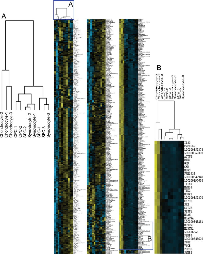
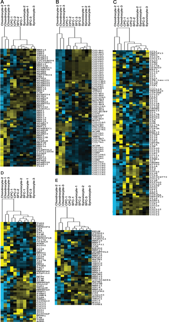
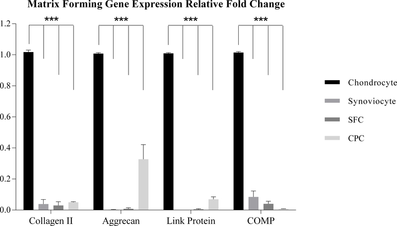
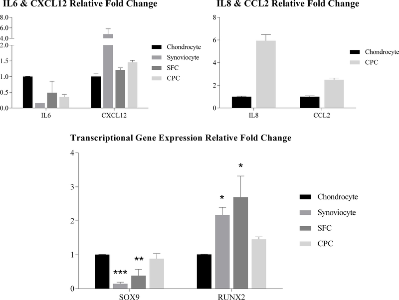
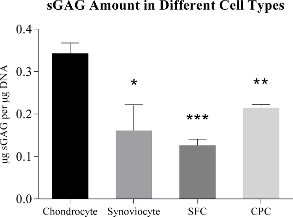
We showed previously that chondrogenic progenitor cells (CPCs) from the superficial zone of articular cartilage respond vigorously to cartilage wounding by responding chemotactically to cell debris, but the physiologic functions of CPCs remain unclear. To help bridge this knowledge gap we undertook a comparative analysis of gene expression in bovine CPCs, chondrocytes, synovial fibroblasts (synoviocytes), and cells isolated from synovial fluid (SFCs). Analysis of microarrays parsed the four cell types into two distinct groups, one composed only of chondrocytes and the other of CPCs, synoviocytes, and SFCs. The groups differed with respect to metalloendopeptidase, collagen, and cytokine gene expression. Quantitative PCR showed that, relative to chondrocytes, all other cells under-expressed cartilage matrix genes. CPCs significantly over-expressed genes encoding the chemokines interleukin 8 (IL8), and C-C motif ligand 2, while synoviocytes over-expressed the chemokine C-X-C motif Ligand 12. Sulfated glycosaminoglycan deposition in pellet cultures by CPCs was intermediate between chondrocytes and synoviocytes/SFCs. These results indicate that the CPC phenotype more closely resembles synoviocytes and SFCs than chondrocytes. CPCs show a tendency to over-express chemokines that promote immune cell chemotaxis, suggesting they mediate inflammation in response to cartilage wounding.
Keywords: chondrocytes; chondrogenic progenitor cells (CPCs); gene expression; microarray.
© 2014 Orthopaedic Research Society. Published by Wiley Periodicals, Inc.
Figures
References
-
- Brooks PM. 2002. Impact of osteoarthritis on individuals and society: how much disability? Social consequences and health economic implications. Current opinion in rheumatology 14:573–577. - PubMed
-
- Martin JA, Brown TD, Heiner AD, et al. 2004. Chondrocyte senescence, joint loading and osteoarthritis. Clinical orthopaedics and related research:S 96–103. - PubMed
-
- Wolf J 1975. Blood supply and nutrition of articular cartilage. Folia morphologica 23:197–209. - PubMed
-
- Villiger PM, Terkeltaub R, Lotz M. 1992. Production of monocyte chemoattractant protein-1 by inflamed synovial tissue and cultured synoviocytes. Journal of immunology 149:722–727. - PubMed
-
- Winchester R, Su F, Ritchlin C. 1993. Alteration of synoviocytes by inflammation--the source of a persistent non-immunologic drive in synovitis: analysis of levels of mRNA expression by a simple multi-gene assay. Clinical and experimental rheumatology 11 Suppl 8:S87–90. - PubMed
Publication types
MeSH terms
Substances
Grants and funding
LinkOut - more resources
Full Text Sources
Other Literature Sources
Medical
Miscellaneous
