Review
. 2014 Oct;1839(10):939-950.
doi: 10.1016/j.bbagrm.2014.04.019. Epub 2014 May 4.

Structure and function of preQ1 riboswitches

Affiliations
Review

Structure and function of preQ1 riboswitches

Catherine D Eichhorn et al. Biochim Biophys Acta. 2014 Oct.

Abstract

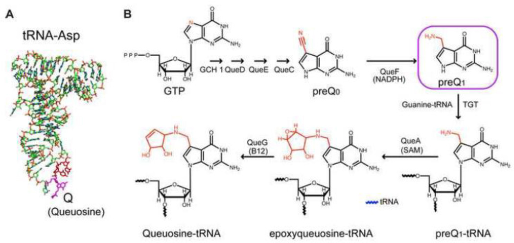
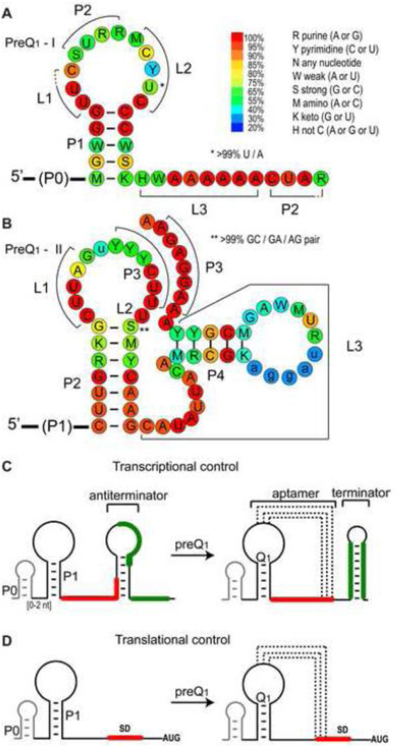
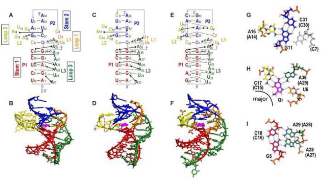
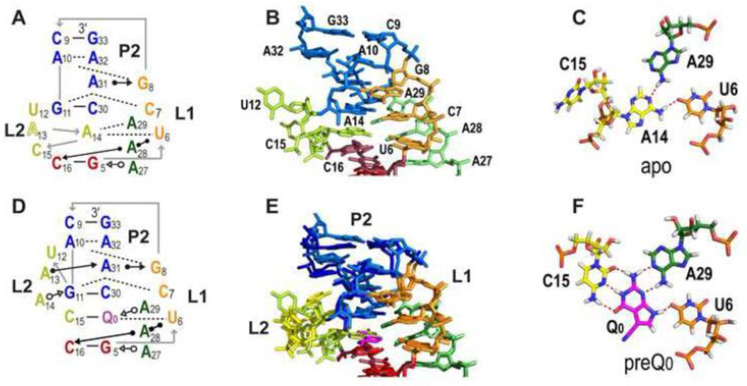
PreQ1 riboswitches help regulate the biosynthesis and transport of preQ1 (7-aminomethyl-7-deazaguanine), a precursor of the hypermodified guanine nucleotide queuosine (Q), in a number of Firmicutes, Proteobacteria, and Fusobacteria. Queuosine is almost universally found at the wobble position of the anticodon in asparaginyl, tyrosyl, histidyl and aspartyl tRNAs, where it contributes to translational fidelity. Two classes of preQ1 riboswitches have been identified (preQ1-I and preQ1-II), and structures of examples from both classes have been determined. Both classes form H-type pseudoknots upon preQ1 binding, each of which has distinct unusual features and modes of preQ1 recognition. These features include an unusually long loop 2 in preQ1-I pseudoknots and an embedded hairpin in loop 3 in preQ1-II pseudoknots. PreQ1-I riboswitches are also notable for their unusually small aptamer domain, which has been extensively investigated by NMR, X-ray crystallography, FRET, and other biophysical methods. Here we review the discovery, structural biology, ligand specificity, cation interactions, folding, dynamics, and applications to biotechnology of preQ1 riboswitches. This article is part of a Special Issue entitled: Riboswitches.

Keywords: NMR; PreQ(0); Queuine; Queuosine; X-ray crystallography; tRNA modification.


Figures

Figure 1
Figure 1
Structure and synthesis of queuosine in bacteria. (A) Structure of aspartyl tRNA with queuosine at the wobble position of the anticodon highlighted in magenta. (B) Pathway for synthesis of queuosine, highlighting the structures of preQ0 and PreQ1.
Figure 2
Figure 2
Predicted secondary structure, sequence conservation, and gene regulatory mechanism of PreQ1 riboswitches. (A,B) Sequence conservation of riboswitch aptamer domains of PreQ1-I (A) and PreQ1-II (B). Sequence conservation is from 894 sequences from 647 species for PreQ1-I and 429 sequences from 423 species for PreQ1-II from the Rfam database (1/31/2014) http://rfam.sanger.ac.uk/. (C,D) Schematic of gene regulation by PreQ1-I riboswitches for (C) transcription or (D) translation. The antiterminator in (C) and mechanism for regulation were revealed from structural studies [45, 46].
Figure 3
Figure 3
Structures of PreQ1-I riboswitch aptamers. (A-B) Solution NMR structure of Bsu (C12,15U) [PDB ID 2L1V), (C-D) X-ray crystal structure of Bsu WT [PDB ID 3FU2] (E-F) X-ray crystal structure of Tte WT [PDB ID 3Q50]. (A,C,E) Schematics of secondary structures with base interactions illustrated using the symbols from Leontis and Westhof notation [87]. (B, D, F) Three-dimensional structures. Nucleotides C12 and U13 (shown as a dotted line) and the base of C15 (marked with an asterisk) are missing from the electron density in the Bsu WT crystal structure (D). Mg2+ and SO42− are depicted as green, and yellow and red spheres, respectively. (G, H, I) Stick representation of the three layers of the PreQ1-I binding pocket (from Bsu WT) showing (G) ‘ceiling’: loop 1-stem 2 A-G-C triple (A-G-C-C quartet for Tte, the loop 1 C shown in gray). (H) ‘binding core’: PreQ1-loop 2-loop 3-loop 1 PreQ1-C-A-U quartet, and (I) ‘floor’: loop 3-stem 1-loop 3 A-C-G-A quartet. The corresponding residue numbers for Tte WT are given in parentheses. For all structures, residues are colored as follows: P1 (red), P2 (dark blue), L1 (orange), L2 (gold), L3 (green), PreQ1 (magenta). For the PreQ1 binding core, hydrogen is white, nitrogen is blue, oxygen is red, phosphorus is orange, and carbon follows the main color scheme. For ease of comparison, structures are numbered starting with the first nucleotide in P1; note that this differs by two from the numbering used for the NMR structure of Bsu aptamer in references [45, 50].
Figure 4
Figure 4
X-ray crystal structures of the apo and preQ0 bound Tte PreQ1-I riboswitch. (A-C) apo Tte WT: (A) Schematic of secondary structures and base interactions at and above PreQ1 binding pocket. (B) Stick representation of structure of the regions shown in (A). (C) The loop 2-loop 3-loop 1 A14-A29-U6 base triple formed in the apo structure, where A14 replaces preQ0 or PreQ1 in the binding core. C15 is flipped out of the binding pocket. (D-F) Tte WT with preQ0: (D) Schematic of secondary structure at and above PreQ1 binding pocket. (E) Superposition of the apo and preQ0 bound structures, to highlight the similarities and differences (F) The preQ0-loop 2-loop 3-loop 1 preQ0-C15-A29-U6 quartet.
Figure 5
Figure 5
Schematic of PreQ1-I riboswitch folding pathway (A) (i) The Bsu riboswitch initially folds into a hairpin followed by a flexible ssRNA tail. (ii) The ssRNA tail, in the presence of divalent cations, dynamically samples a variety of conformations (illustrated in gray), some of which bring L3 close to P1. (iii) On addition of PreQ1, the P2 stem forms and L3 docks into the P1 minor groove. L1 and L2 show dynamics on the µs-ms and ps-ns timescales, respectively (open arrows). (iv) Divalent cations bind to L1, quenching local flexibility while retaining L2 motions. (B) Alternate P1A hairpin observed in Bsu NMR construct as a result of additional 5’ GG residues. (C) A palindromic sequence commonly observed overlapping with the P2 site in the P1 loop gives rise to dimers. (D) Effect of Bsu loop mutagenesis on apparent PreQ1 binding. +++ better than WT, ++ equivalent to WT, + and +/− worse than WT, − no detectable binding. Multiple mutations or mutations to L3 are inset. Mutations in Bsu L2var are shown in gray.
Figure 6
Figure 6
Structures of PreQ1-II riboswitches (A-B) Solution NMR structure of Spn WT [PDB ID 2MIY), (C-D) X-ray crystal structure of Lra WT' [PDB ID 4JF2]. (A, C) Schematics of secondary structures and base interactions. (B, D) Stick representation of three-dimensional structures. Mg2+, Cs+ are depicted as green and purple spheres, respectively. (E) Schematic illustrating the secondary structure of the Spn riboswitch in the absence of PreQ1 and the divalent cation and PreQ1 induced folding. The curved arrow above P4 indicates the rotation of P4 required to position the two As (dark and light green bars) in the binding pocket. The gray P4 hairpins illustrate that the position of P4 is dynamic. (F) Stick representations of interactions of PreQ1 in the binding core showing the PreQ1-loop 1-loop 2-loop 3 PreQ1-C-U-A quartet from Lra WT' For all structures, residues are colored as follows: P2 (red), P3 (dark blue), L1 (orange), L2 (gold), L3 (green, with P4 hairpin insert in purple), PreQ1 (magenta). Note that P2 is stem 1 and P3 is stem 2 in standard H-type pseudoknot nomenclature.
Figure 7
Figure 7
The PreQ1 binding pocket for PreQ1-I and PreQ1-II riboswitch aptamers. (A-C) PreQ1-I structures of Bsu WT X-ray (A) and Bsu (C12,15U) NMR (B-C) and (D-F) PreQ1-II structures of Lra WT' crystal (D) and Spn WT NMR (E-F), comparing the PreQ1-1 and PreQ1-II binding cores (A,D), ceiling of the binding pockets (B,E), and binding pockets (C,F). (A,D) Sphere representation of Bsu WT PreQ1-loop 2-loop 3-loop 1 PreQ1-C-A-U quartet (A) and Lra WT' PreQ1-loop 1-loop 2-loop 3 PreQ1-C-U-A quartet (D), illustrating hydrogen bond and van der Waals interactions, especially between A50-PreQ1 and A50-U19. The corresponding residue numbers for Spn WT are given in parentheses. The exocyclic methylamine projects from the major groove in PreQ1-I and from the minor groove in PreQ1-II riboswitches. (B,E) Overlay of Bsu loop 2-stem 2 A-G-C triple on PreQ1-C-A-U quartet (B) and Spn loop 1-stem 2-loop 3 U-A-U-A quartet on PreQ1-C-U-A quartet (E). PreQ1 quartets are gray. (C,F) Stick representations of the three layers of binding pocket from Bsu (C) and Spn (F) NMR structures, with G-C bp below, PreQ1 binding core, and base triple or quartet above. The exocyclic amine protons point down toward the G-C pair below and have van der Waals contact with O6 and N7 of G5 (C), and point up toward the base quartet above, with possible interactions with O2 of U9 or phosphate backbone of A51 (F).
Figure 8
Figure 8
Comparison of PreQ1-II vs PreQ1-I riboswitch structures. Sphere representation of (A,B) PreQ1-I Bsu (C12,15U) and (C,D) PreQ1-II Spn WT. (B, D) are the 180 degree rotation of (A, C), respectively. Loop 3 (green) lies along the minor groove in PreQ1-I aptamers (A) while shows loop 3 does not insert in a groove but spans the major groove in PreQ1-II aptamers (B). The exocyclic amine group of PreQ1 projects out from the major groove of PreQ1-I aptamers (A) and from the minor groove of PreQ1-II aptamers.

References

    1. Kim JN, Breaker RR. Purine sensing by riboswitches. Biology of the cell / under the auspices of the European Cell Biology Organization. 2008;100:1–11. - PubMed
    1. Batey RT. Structure and mechanism of purine-binding riboswitches. Q Rev Biophys. 2012;45:345–381. - PMC - PubMed
    1. Edmonds CG, Crain PF, Gupta R, Hashizume T, Hocart CH, Kowalak JA, Pomerantz SC, Stetter KO, McCloskey JA. Posttranscriptional modification of tRNA in thermophilic archaea (Archaebacteria) Journal of bacteriology. 1991;173:3138–3148. - PMC - PubMed
    1. Harada F, Nishimura S. Possible anticodon sequences of tRNA His, tRNA Asm, and tRNA Asp from Escherichia coli B. Universal presence of nucleoside Q in the first postion of the anticondons of these transfer ribonucleic acids. Biochemistry. 1972;11:301–308. - PubMed
    1. Morris RC, Elliott MS. Queuosine modification of tRNA: a case for convergent evolution. Mol Genet Metab. 2001;74:147–159. - PubMed