. 2015 Mar;89(3):459-83.
doi: 10.1007/s00204-014-1264-0. Epub 2014 May 6.

Mechanisms of acute kidney injury induced by experimental Lonomia obliqua envenomation

Affiliations

Mechanisms of acute kidney injury induced by experimental Lonomia obliqua envenomation

Markus Berger et al. Arch Toxicol. 2015 Mar.

Abstract

Lonomia obliqua caterpillar envenomation causes acute kidney injury (AKI), which can be responsible for its deadly actions. This study evaluates the possible mechanisms involved in the pathogenesis of renal dysfunction. To characterize L. obliqua venom effects, we subcutaneously injected rats and examined renal functional, morphological and biochemical parameters at several time points. We also performed discovery-based proteomic analysis to measure protein expression to identify molecular pathways of renal disease. L. obliqua envenomation causes acute tubular necrosis, which is associated with renal inflammation; formation of hematic casts, resulting from intravascular hemolysis; increase in vascular permeability and fibrosis. The dilation of Bowman's space and glomerular tuft is related to fluid leakage and intra-glomerular fibrin deposition, respectively, since tissue factor procoagulant activity increases in the kidney. Systemic hypotension also contributes to these alterations and to the sudden loss of basic renal functions, including filtration and excretion capacities, urinary concentration and maintenance of fluid homeostasis. In addition, envenomed kidneys increase the expression of proteins involved in cell stress, inflammation, tissue injury, heme-induced oxidative stress, coagulation and complement system activation. Finally, the localization of the venom in renal tissue agrees with morphological and functional alterations, suggesting also a direct nephrotoxic activity. In conclusion, the mechanisms of L. obliqua-induced AKI are complex involving mainly glomerular and tubular functional impairment and vascular alterations. These results are important to understand the mechanisms of renal injury and may suggest more efficient ways to prevent or attenuate the pathology of Lonomia's envenomation.


Figures

Figure 1
Figure 1. Functional parameters during L. obliqua-induced AKI
Rats were injected subcutaneously with PBS (controls - CTRL) or LOBE (1.5 or 1.0 mg/kg). After different times post administration the following parameters were determined: (A) Body weight, (B) water intake, (C) urine outoput, (D) glomerular filtration rate (GFR), (E) urinary density, (F) urine osmolality (urine osm), (G) plasma osmolality (plasma osm), (H) osmolar clearance (Cosm), (I) water free clearance (CH2O), (J) fractional water excretion (FEH2O), (K) fractional sodium excretion (FENa+) and (L) fractional potassium excretion (FEK+). Data are presented as means ± SE (n=6/group). Significant differences: *p < 0.05 vs. CTRL and §p < 0.05 vs. LOBE (1.0 mg/kg, s.c).
Figure 2
Figure 2. Proteinuria
(A) Rats were injected subcutaneously with PBS (controls - CTRL) or LOBE (1.5 or 1.0 mg/kg). After different times post administration the protein levels in urine were measured. Data are presented as means ± SE (n=6/group). Statistical differences of *p < 0.05 vs. CTRL and §p < 0.05 vs. LOBE (1.0 mg/kg, s.c) were considered significant. (B) Representative urine samples from CTRL and rats treated with LOBE (1.5 mg/kg, s.c.) were analyzed by SDS-PAGE (8-20 %) under reducing conditions. (C) Toxins excreted in urine were detected by western-blot. Samples of urine from CTRL and rats treated with LOBE (1.5 mg/kg, s.c.) at 6 and 12 h post-venom injection were separated by SDS-PAGE and different toxins were detected by immunoreaction with polyclonal antibodies against LOBE. Toxins present in crude bristle extract are also showed (LOBE). The arrows indicate bands detected in urine samples at 6 and 12 h of envenomation. Molecular weight (MW) standards were shown on the left of figure B and C.
Figure 3
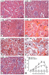
Figure 3. L. obliqua envenomation induces acute tubular necrosis
Representative kidney sections from control (CTRL) or envenomed animals (injected with 1.5 mg/kg, s.c.) are presented (A-G). Note the normal morphology of kidney from CTRL animal (A) in comparison to the progressive degenerative lesions of venom-treated rats (B-G). Increased acidophilia, dilation of renal tubules, loss of proximal brush border, cytoplasm vacuolation, nuclear pyknosis and desquamation of necrotic cells can be observed (D-F). Hyaline (arrowheads in C, D and G) and hematic (arrows in D) casts are also present inside renal tubules. Arrowheads in E indicate necrotic cells. Asterisks in D and F indicate the presence of a hyaline material within the Bowman’s space and an inflammatory cell infiltrate and edema, respectively. All sections were stained with H&E. Magnification: 10 ×. (H) Levels of urinary γ-glutamyl transferase (γ-GT) activity were measured in rats injected with PBS or LOBE (1.5 or 1.0 mg/kg) at different times post-administration. Data are presented as means ± SE (n=6/group). Statistical differences of *p < 0.05 vs. CTRL and §p < 0.05 vs. LOBE (1.0 mg/kg, s.c) were considered significant.
Figure 4
Figure 4. L. obliqua envenomation induces renal tubular obstruction
Representative kidney sections from animals injected with LOBE (1.5 mg/kg, s.c.) showing details of tubular obstruction by hyaline and hematic casts and cellular debris at 12 h (A) and 24 h (B) post-venom injection. Hyaline casts (black arrowheads) are formed by a protein-rich material (predominantly serum albumin and hemoglobin), while hematic casts (black arrows) are formed by fragmented or intact erythrocytes. Due to tubular necrosis, the basement membrane in some tubules is disrupted, resulting in detachment of necrotic cells into the lumen (white arrowheads). All sections were stained with H&E. Magnification: 4 × (panel A) and 20 × (panel B).
Figure 5
Figure 5. L. obliqua envenomation induces renal inflammation and fibrosis
Light micrographs showing a marked inflammatory cell infiltrate (arrowheads) in the tubulo-interstitial region at 96 h (B) and glomerulus at 48 h (C) after LOBE injection (1.5 mg/kg, s.c.). There were no signs of inflammation in control (CTRL) animals (A). It was also observed an extensive peritubular (E) and interglomerular (F) collagen deposition at 96 h (regions stained in red), indicating fibrosis. Arrowheads in these panels indicate inflammatory infiltrate. There were no signs of fibrosis in CTRL rats (D). Stain: H&E (panels A-C) and picrosirius (panels D-F). Magnification: 10 × (panel A, B, D and E), 20 × (panel F) and 40 × (panel C).
Figure 6
Figure 6. Glomerular alterations
A. Light micrographs showing a time dependent increase in the deposition of a PAS positive material in glomerular capillaries of LOBE-injected animals (1.5 mg/kg, s.c.) in comparison to controls. Also note the increase in glomerular size. All sections were stained with PAS. Magnification: 40 ×. B. Thirty glomeruli from each animal injected with the dose of 1.5 mg/kg were used to quantify the mean area of glomerulus, glomerular tuft and Bowman’s space of animals treated with the dose of 1.5 mg/kg. Data are presented as means ± SE (n=6/group). Statistical differences of *p < 0.05 in comparison to the respective control (C) were considered significant. C. Renal tissue factor activity was measured in control (C) and envenomed (1.5 mg/kg, s.c.) animals by generation of factor Xa (FXa). Data are presented as means ± SE (n=6/group). Statistical differences of *p < 0.05 were considered significant in comparison to the respective control.
Figure 7
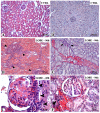
Figure 7. Renal vascular permeability
Representative micrographs of a kidney blood vessel (A) and glomerulus (B) from animals injected with LOBE (1.5 mg/kg, s.c.) after 12 h of envenomation. Note the vascular leakage and edema (asterisks) and migration of inflammatory cells to damaged tissue (arrowheads). Also, the presence of a hyaline material inside the Bowman’s space (asterisk in B) was associated with the increase in glomerular area observed at this time. All sections were stained with H&E. Magnification: 10 × (panel A) and 40 × (panel B). C. Evaluation of changes in renal vascular permeability were assessed by Evans blue dye extravasation. Results are expressed as μg Evans blue dye per 100 mg of renal tissue from control (CTRL) and LOBE-treated (1.5 mg/kg, s.c.) rats at 12 and 24 h post-venom administration. Data are presented as means ± SE (n=6/group). Statistical comparisons are indicated.
Figure 8
Figure 8. Immunohistochemical detection of L. obliqua venom in renal tissue
Positive immunohistochemical reaction was found in cortical and medullar regions of kidneys from rats injected with 1.5 mg/kg of LOBE (B-E). Venom was detected in glomerular capillaries (arrows in B), Bowman’s capsule (arrow in the inset B), tubular brush border (arrows in the inset C), in intra-tubular casts (arrowheads in C) and also was present in cells of tubules in degeneration (arrowheads in the insets C and D). After 96 h the immunoreactivity for venom was weak and mainly localized in tubules (arrows in E). There was no immunoreactivity in the renal structures of control (CTRL) rats (A). Magnification: 10 × (panels A-E) and 40 × (insets in B-D) F. The amounts of venom detected in renal tissue was estimated by the area of positive immunohistochemical reaction. Thirty sections per rat were analyzed as described in material and methods. Data are presented as means ± SE (n=6/group).
Figure 9
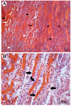
Figure 9. Immunohistochemical detection of L. obliqua venom in renal vascular tissue
Positive immunohistochemical reaction was detected in renal arteries (A) and veins (B) of rats injected with LOBE (1.5 mg/kg, s.c.) at 6 h of envenomation. Note the presence of venom in perivascular connective tissue (arrows) and endothelium and smooth muscle cells (arrowheads). Magnification: 10 × (panel B) and 40 × (panel A).
Figure 10
Figure 10. Kidney proteins differentially expressed during L. obliqua envenomation
Unique or differentially expressed kidney proteins from control and envenomed (1.5 mg/kg, s.c.) animals were identified at 24 h post-venom injection by proteomic analysis. Exclusive and common proteins in each condition, as well as the total number of proteins identified are showed via a Venn diagram (A). Top canonical pathways of differentially expressed and unique proteins identified in control (CTRL) (B) and LOBE-treated (C) kidneys are shown. Those proteins functionally related to renal disease were also categorized accordingly to their roles in different types of renal pathologies (D). Each functional annotation is assigned to a significance score represented as P value (Fisher exact test) determining the probability that the association between the proteins in the data set and the canonical pathway or function in disease is explained by chance alone. The number of identified proteins (NIP) that belong to a particular canonical pathway or play a role in renal pathology is shown.

Similar articles

Cited by

References

    1. Abbate M, Zoja C, Remuzzi G. How does proteinuria cause progressive renal damage? J Am Soc Nephrol. 2006;17:2974–2984. - PubMed
    1. Alvarez-Flores MP, Fritzen M, Reis CV, Chudzinski-Tavassi AM. Losac, a factor X activator from Lonomia obliqua bristle extract: its role in the pathophysiological mechanisms and cell survival. Biochem Biophys Res Commun. 2006;343:1216–1223. - PubMed
    1. Ambatipudi KS, Lu B, Hagen FK, Melvin JE, Yates JR. Quantitative analysis of age specific variation in the abundance of human female parotid salivary proteins. J Proteome Res. 2009;8:5093–5102. - PMC - PubMed
    1. Basnakian AG, Kaushal GP, Shah SV. Apoptotic pathways of oxidative damage to renal tubular epithelial cells. Antioxid Redox Signal. 2002;4(6):915–924. - PubMed
    1. Berger M, Reck J, Jr., Terra RM, Pinto AF, Termignoni C, Guimarães JA. Lonomia obliqua caterpillar envenomation causes platelet hypoaggregation and blood incoagulability in rats. Toxicon. 2010;55(1):33–44. - PubMed

Publication types

MeSH terms

Substances

LinkOut - more resources