. 2014 Sep;92(9):1143-54.
doi: 10.1002/jnr.23388. Epub 2014 May 5.

Live cell imaging reveals differential modifications to cytoplasmic dynein properties by phospho- and dephosphomimic mutations of the intermediate chain 2C S84

Affiliations

Live cell imaging reveals differential modifications to cytoplasmic dynein properties by phospho- and dephosphomimic mutations of the intermediate chain 2C S84

Kiev R Blasier et al. J Neurosci Res. 2014 Sep.

Abstract

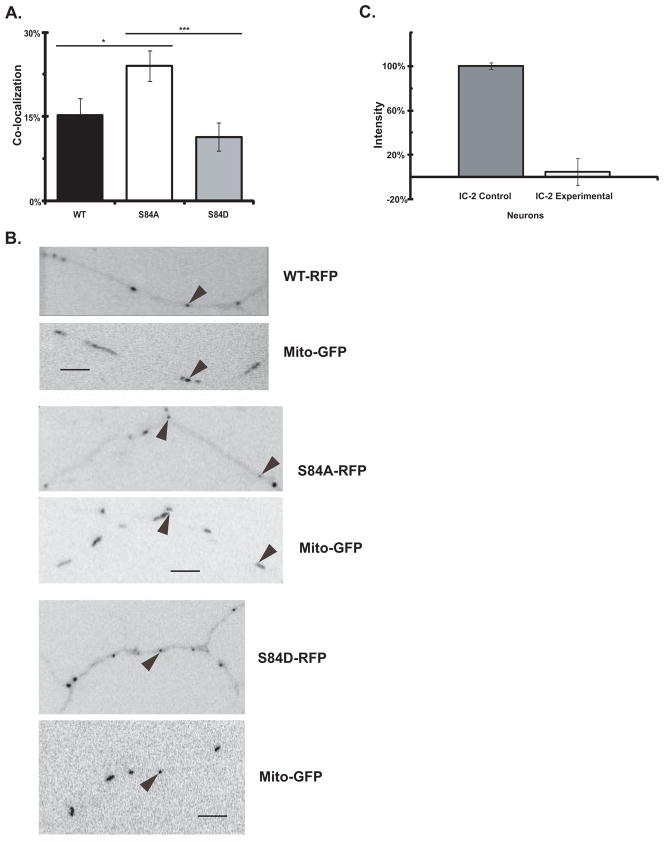
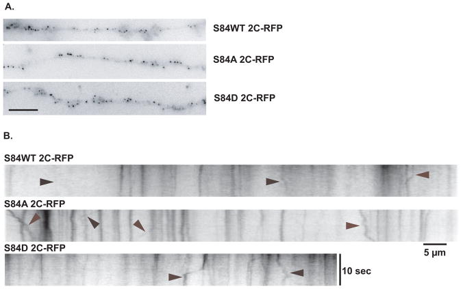
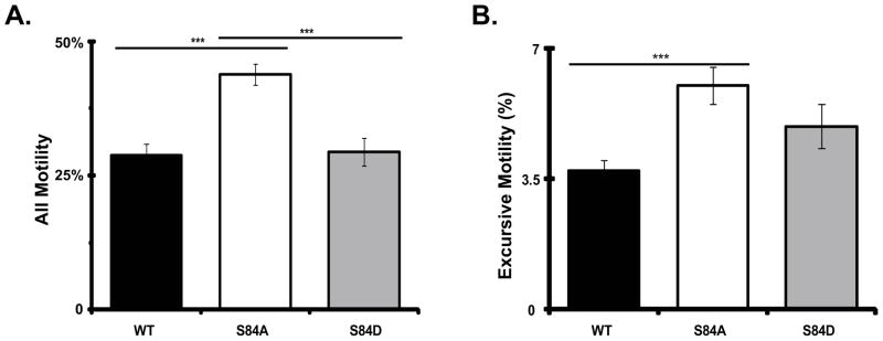
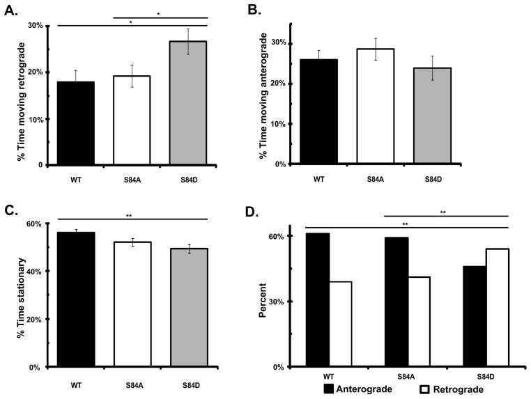
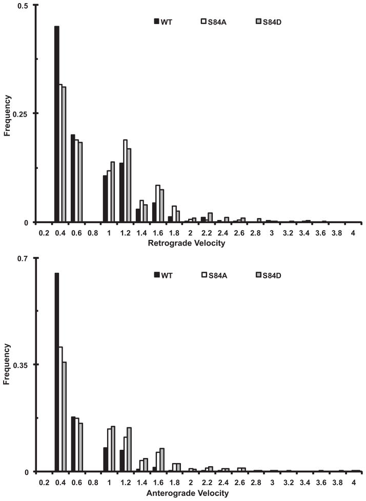
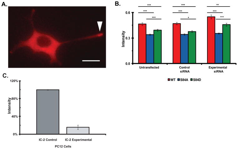
Cytoplasmic dynein is a multisubunit motor protein responsible for intracellular cargo transport toward microtubule minus ends. There are multiple isoforms of the dynein intermediate chain (DYNC1I, IC), which is encoded by two genes. One way to regulate cytoplasmic dynein is by IC phosphorylation. The IC-2C isoform is expressed in all cells, and the functional significance of phosphorylation on IC-2C serine 84 was investigated by using live cell imaging of fluorescent protein-tagged IC-2C wild type (WT) and phospho- and dephosphomimic mutant isoforms in axonal transport model systems. Both mutations modulated dynein functional properties. The dephosphomimic mutant IC-2C S84A had greater colocalization with mitochondria than the IC-2C WT or the phosphomimic mutant IC-2C S84D. The dephosphomimic mutant IC-2C S84A was also more likely to be motile than the phosphomimic mutant IC-2C S84D or the IC-2C WT. In contrast, the phosphomimic mutant IC-2C S84D mutant was more likely to move in the retrograde direction than was the IC-2C S84A mutant. The phosphomimic IC-2C S84D was also as likely as the IC-2C WT to colocalize with mitochondria. Both the S84D phospho- and the S84A dephosphomimic mutants were found to be capable of microtubule minus-end-directed (retrograde) movement in axons. They were also observed to be passively transported in the anterograde direction. These data suggest that the IC-2C S84 has a role in modulating dynein properties.

Keywords: axonal transport; cytoplasmic dynein; cytoskeleton; motor protein.


Figures

Figure 1
Figure 1. Comparison of the co-localization of dynein IC-2C WT and phospho-state IC-2C mutations with mitochondria
Rat embryonic hippocampal neurons were transfected with IC-2 siRNA oligonucleotides by Lonza nucelofection prior to plating and then co-transfected with Mito-GFP and the indicated dynein IC-2C isoform tagged with mRFP on DIV 3 by CaPO4. Axons of living cells were imaged the next day. A. The IC-2C S84A dephospho-mimic mutation enhances dynein co-localization with mitochondria. The amount of co-localization of the indicated isoform with mitochondria was determined by imaging axons of living cells in the two colors simultaneously with a Dualview. The number of puncta in each color and the number that overlap were quantified and the percentage of dynein puncta that co-localized with mitochondria was graphed. WT (black), IC-2C WT, (n = 216 dynein, 346 mitochondria, and 31 co-localizing); S84A (white), IC-2C S84A, (n = 287 dynein, 395 mitochondria, and 64 co-localizing; S84D (gray), IC-2C S84D (n = 310 dynein, 445 mitochondria, annd 36 co-localizing). There were significant differences in co-localization between the WT and S84A isoforms, and between the S84A and S84D isoforms, * P<0.03, *** P<0.001 Student’s t-test. The difference between WT and S84D was not significant. B. Co-localization dynein IC-2C WT and mutant isoforms in cultured hippocampal neurons. Neurons were co-transfected as described and movies of axons of living cells were made with the GFP and mRFP signals collected simultaneously using DualView. The pairs of panels are from single DualView frames with the indicated IC-2C mRFP isoform (top panel of each pair) and Mito-GFP (bottom panel of each pair) signals aligned and the puncta displayed in reverse contrast. Arrows identify puncta that co-localize. Puncta that only showed partial overlap or that did not co-localize in all frames are not identified. In these examples, 15% of the IC-2C WT co-localized with mitochondria, 24% of the IC-2C S84A mutant co-localized with mitochondria, and 11% of the IC-2C S81D co-localized with mitochondria. Scale bar is 5 μm. C. Quantitation of effect of siRNA in reducing the expression level of IC-2 isoforms in neurons. Cultured rat cortical neurons were transfected with siRNA oligonucleotides as described in the Methods and (Mitchell et al. 2012). After 4 days the cells were lysed and polypeptides resolved by SDS-PAGE and Western blotting. The blots were probed with IC-2 specific antibody (Pfister et al. 1996b; Vaughan and Vallee 1995) and the intensity of the endogenous IC-2 bands were quantified using MetaMorph. The intensities of the bands for cells transfected with control scrambled oligonucleotides and experimental siRNA oligonucleotides are graphed (n=2). A greater than 90% reduction in the expression of endogenous IC-2 was achieved.
Figure 2
Figure 2. Live cell imaging of dynein containing IC-2C WT mRFP and the IC-2C S84A and S84D mutations in axons
Mouse catecholaminergic neurons were transfected with either the IC-2C WT mRFP, IC-2C S84A, or IC-2C S84D isoforms with Lipofectamine2000 on the 3rd day of culture and the next day fluorescent dynein was imaged in axons of living cells. A. In axons, all three fluorescent IC isoforms are observed as small puncta. Single frame images from movies of axons of cell expressing the IC-2C or mutant isoform indicated to the right of the image. The WT, S84A and S84D IC-2C isoforms all have the appearance of small puncta in the axons. The scale bar is 5 μm. B. Imaging bi-directional motility of dynein puncta with IC-2C WT mRFP, and the IC-2C S84A and S84D mutants in axons. Kymographs were made from movies of axons expressing the fluorescent IC-2C isoform indicated above the kymographs. Arrowheads point to the traces of dynein puncta with movement in either the anterograde or retrograde directions. The cell body is located to the left for the kymographs of the IC-2C mutants. The time and distance scales are as indicated.
Figure 3
Figure 3. The dephospho-mimic mutant IC-2C S84A dynein puncta have increased motility in axons
Cytoplasmic dynein IC-2C isoforms were separately transfected into neurons and the fluorescent dynein was imaged as for Figure 2. black, IC-2C WT; white, IC-2C S84A; and gray, IC-2C S84D. Number of axons: WT 47, S84A 44, S84D 36; number of puncta: WT 2867, S84A 1795, S84D 2490. A. All Motility. For each of the three IC isoforms, the percentage of moving puncta is graphed. Individual dynein puncta were scored as being motile if they showed any displacement between at least two frames. There were significant differences when the motility of S84A puncta was compared to that of the WT or S84D isoforms, *** P<0.001 Student’s t-test. The difference between dynein puncta containing IC-2C WT and IC-2C S84D was not significant B. Excursive Motility. For each of the three IC isoforms, the percentage of moving puncta exhibiting excursive movement is graphed. Individual dynein puncta were scored as having excursive motility if they showed displacement of at least 1 pixel for four consecutive frames. There was a significant difference when the excursive motility of dynein puncta containing the S84A was compared to that of the WT isoforms, *** P<0.001 Student’s t-test. The differences between S84D and WT or S84A were not significant.
Figure 4
Figure 4. The phospho-mimic mutant IC-2C S84D dynein puncta have increased retrograde motility in axons
Cytoplasmic dynein IC-2C isoforms were separately transfected into neurons and the fluorescent dynein was imaged as for Figure 2. A, B, C. For each of the three IC isoforms the time each excursive puncta in axons spent moving in the retrograde direction, (A), moving in the anterograde direction (B), and stationary (C) was calculated for each axon and the averages are graphed as percent, black, IC-2C WT; white,IC-2C S84A; and gray, IC-2C S84D. There was a significant increase in the amount of time dynein puncta containing the IC-2C S84D, phosphomimic mutant spent moving in the retrograde direction compared to that of IC-2C WT and the IC-2C S84 mutant, *P<0.02 Student’s t-test. There was no significant difference between the time the WT and the S84A isoforms spent in retrograde movement. No significant differences in the time spent moving in the anterograde direction was observed when the three IC isoforms were compared. The IC-2C S84D mutant dynein puncta spent significantly less time stationary (49.3 +/− 0.02−) than did the IC-2C WT dynein isoform (56.0 =/− 0.01%), ** P< 0.01, Student’s t-test. The S84A mutant was stationary 52.1 +/−0.01% of the time and this was not significantly different from that of IC-2C WT or the IC-2C S84D mutant. D. For every excursive puncta, the direction of every movement between two frames was identified and the number in each direction for the three IC isoforms was summed and graphed as percent moving retrograde (white) or anterograde (black), IC-2C WT n = 1,758; IC-2C S84A, n = 1,953; and IC-2C S84D, n= 2017. There were significantly more IC-2C S84D movements in the retrograde direction (and fewer in the anterograde direction) than were observed for IC-2C S84 or IC-2C WT; **, P<0.01, Χ2 test.
Figure 5
Figure 5. Comparison of the velocity distributions of dynein puncta containing wild type and mutant IC-2C isoforms in axons
Cytoplasmic dynein IC-2C isoforms were transfected into neurons and the fluorescent dynein was imaged as for Figure 2. The movements of individual puncta between each frame were tracked manually and analyzed with MetaMorph. A. Comparison of the retrograde interval velocity distributions of the IC-2C WT and mutant isoforms in axons. Velocities (μm/s) of individual retrograde movements of GFP-IC-2C dynein puncta between two frames were plotted against the frequency of their occurrence; black, IC-2C WT dynein (n=684); white IC-2C S84A (n=780), and gray IC-2C S84D dynein puncta (n=1030). B) Comparison of the anterograde interval velocity distributions of the IC-2C WT and mutant isoforms in axons/neurons. Velocities (μm/s) of individual anterograde movements of GFP-IC-2C dynein puncta between two frames were plotted against the frequency of their occurrence; black IC-2C WT (n=1074), white IC-2C S84A (n=1172), and gray IC-2C S84D dynein puncta (n=987).
Figure 6
Figure 6. Mutations of IC-2C at S84 modulate dynein motility
Three PC12 cell lines with the stable expression of mRFP IC-2C WT, and the IC-2C S84A and IC-2C S84D mutations were generated. The cells were differentiated by the addition of NGF and 3–4 days later the cells and the growth cones of neurite processes of the differentiated cells were imaged. A. Dynein distribution in differentiated PC12 cells. Representative image of mRFP IC-2C WT in differentiated PC12 cell showing dynein accumulation in a growth cone of a long process marked with arrowhead, see also Figure 3 in (Myers et al. 2007). The scale bar is 10 μm. B. Mutation of IC-2C S84 changes dynein motility and reduces the accumulation of dynein in growth cone. The fluorescence intensity the mRFP-IC-2C in the three cell lines with stable expression of the tagged IC isoforms per unit growth cone area was measured and corrected for differences in IC-mRFP expression in the different cell bodies; red IC-2C WT, blue IC-2C S84A, and green IC-2C S84D. The IC isoform intensities were measured under three experimental conditions; Untransfected, the three parent cells lines with stable expression of the indicated IC isoform (left set of bars), Control siRNA, the parent cell lines transfected with the control siRNA reagent (middle set of bars), and Experimental siRNA, the cell lines transfected with an IC-2 siRNA reagent to reduce the expression of the endogenous IC-2 (right set of bars). There were significant differences between the fluorescent intensities which measure the accumulation of dynein in growth cones between the IC-2C WT and both mutant cell lines under all three experimental conditions. Significant differences were also observed when the dynein accumulations in cell expressing the S84A & S84D mutations were compared. (Student’s t-test * p < 0.05, ** p < 0.002, and *** P < 0.001). Untrasfected cell lines; IC-2C WT n=84, S84A n=184, S84D n=125. Control siRNA; IC-2C WT n=112, S84A n=169, S84D n=130. Experimental siRNA; IC-2C WT n=119, S84A, n=170, S84D n=47. C. Quantitation of effect of siRNA in reducing the expression level of IC-2 isoforms in PC12 cells. PC12 cells were transfected with siRNA oligonucleotides as described in the Methods and (Ha et al. 2008). After 4 days the cells were lysed and polypeptides resolved by SDS-PAGE and Western blotting. The blots were probed with the 74.1 antibody and the intensities of the bands corresponding to the endogenous IC-2 were quantified using MetaMorph. The intensities of the bands for cells transfected with control scrambled oligonucleotides and experimental siRNA oligonucleotides are graphed (n=3). An ~85% reduction in the expression of endogenous IC-2 was achieved.

Similar articles

Cited by

References

    1. Abe TK, Honda T, Takei K, Mikoshiba K, Hoffman-Kim D, Jay DG, Kuwano R. Dynactin is essential for growth cone advance. Biochemical and biophysical research communications. 2008;372(3):418–422. - PubMed
    1. Addinall SG, Mayr PS, Doyle S, Sheehan JK, Woodman PG, Allan VJ. Phosphorylation by cdc2-CyclinB1 kinase releases cytoplasmic dynein from membranes. The Journal of biological chemistry. 2001;276(19):15939–15944. - PubMed
    1. Akhmanova A, Hammer JA., 3rd Linking molecular motors to membrane cargo. Current opinion in cell biology. 2010;22(4):479–487. - PMC - PubMed
    1. Ally S, Larson AG, Barlan K, Rice SE, Gelfand VI. Opposite-polarity motors activate one another to trigger cargo transport in live cells. J Cell Biol. 2009;187(7):1071–1082. - PMC - PubMed
    1. Baas PW, Deitch JS, Black MM, Banker GA. Polarity orientation of microtubules in hippocampal neurons: uniformity in the axon and nonuniformity in the dendrite. Proceedings of the National Academy of Sciences of the United States of America. 1988;85(21):8335–8339. - PMC - PubMed

MeSH terms

LinkOut - more resources