

Investigating spermatogenesis in Drosophila melanogaster
- PMID: 24798812
- PMCID: PMC4128239
- DOI: 10.1016/j.ymeth.2014.04.020
Investigating spermatogenesis in Drosophila melanogaster
Abstract
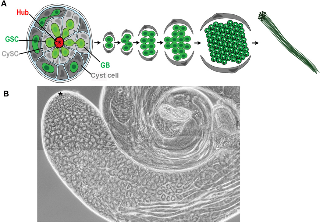
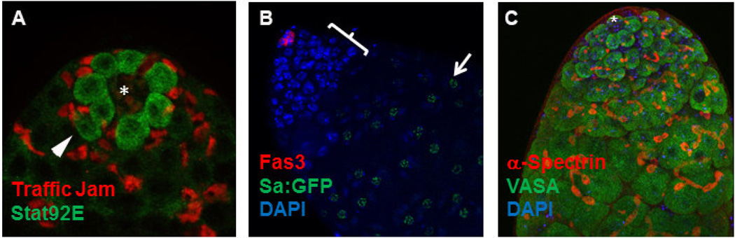
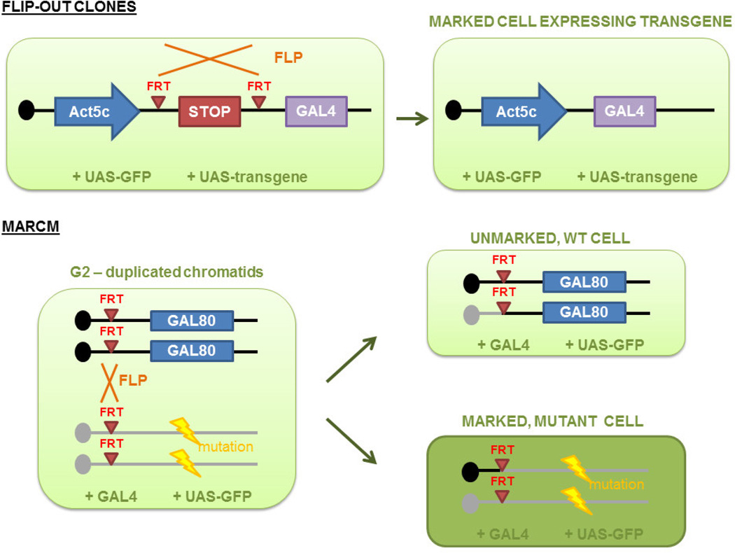
The process of spermatogenesis in Drosophila melanogaster provides a powerful model system to probe a variety of developmental and cell biological questions, such as the characterization of mechanisms that regulate stem cell behavior, cytokinesis, meiosis, and mitochondrial dynamics. Classical genetic approaches, together with binary expression systems, FRT-mediated recombination, and novel imaging systems to capture single cell behavior, are rapidly expanding our knowledge of the molecular mechanisms regulating all aspects of spermatogenesis. This methods chapter provides a detailed description of the system, a review of key questions that have been addressed or remain unanswered thus far, and an introduction to tools and techniques available to probe each stage of spermatogenesis.
Keywords: Cytokinesis; Drosophila; Germ line; Meiosis; Spermatogenesis; Stem cell.
Copyright © 2014 Elsevier Inc. All rights reserved.
Figures
Similar articles
-
The CPEB protein Orb2 has multiple functions during spermatogenesis in Drosophila melanogaster.PLoS Genet. 2012;8(11):e1003079. doi: 10.1371/journal.pgen.1003079. Epub 2012 Nov 29. PLoS Genet. 2012. PMID: 23209437 Free PMC article.
-
Methods for studying oogenesis.Methods. 2014 Jun 15;68(1):207-17. doi: 10.1016/j.ymeth.2014.01.005. Epub 2014 Jan 17. Methods. 2014. PMID: 24440745 Free PMC article. Review.
-
Direct evidence for postmeiotic transcription during Drosophila melanogaster spermatogenesis.Genetics. 2010 Sep;186(1):431-3. doi: 10.1534/genetics.110.118919. Epub 2010 Jul 6. Genetics. 2010. PMID: 20610406 Free PMC article.
-
IV. Tools and methods for studying cell migration and cell rearrangement in tissue and organ development.Methods. 2014 Jun 15;68(1):228-32. doi: 10.1016/j.ymeth.2014.03.004. Epub 2014 Mar 12. Methods. 2014. PMID: 24631575 Review.
-
Unique aspects of transcription regulation in male germ cells.Cold Spring Harb Perspect Biol. 2011 Jul 1;3(7):a002626. doi: 10.1101/cshperspect.a002626. Cold Spring Harb Perspect Biol. 2011. PMID: 21555408 Free PMC article. Review.
Cited by
-
Centrosomal and Non-Centrosomal Microtubule-Organizing Centers (MTOCs) in Drosophila melanogaster.Cells. 2018 Aug 28;7(9):121. doi: 10.3390/cells7090121. Cells. 2018. PMID: 30154378 Free PMC article. Review.
-
scRNA-seq Reveals Novel Genetic Pathways and Sex Chromosome Regulation in Tribolium Spermatogenesis.Genome Biol Evol. 2024 Mar 2;16(3):evae059. doi: 10.1093/gbe/evae059. Genome Biol Evol. 2024. PMID: 38513111 Free PMC article.
-
A postmeiotically bifurcated roadmap of honeybee spermatogenesis marked by phylogenetically restricted genes.PLoS Genet. 2023 Dec 4;19(12):e1011081. doi: 10.1371/journal.pgen.1011081. eCollection 2023 Dec. PLoS Genet. 2023. PMID: 38048317 Free PMC article.
-
Single-cell RNA sequencing reveals cell landscape following antimony exposure during spermatogenesis in Drosophila testes.Cell Death Discov. 2023 Mar 9;9(1):86. doi: 10.1038/s41420-023-01391-4. Cell Death Discov. 2023. PMID: 36894529 Free PMC article.
-
RNA from a simple-tandem repeat is required for sperm maturation and male fertility in Drosophila melanogaster.Elife. 2019 Nov 5;8:e48940. doi: 10.7554/eLife.48940. Elife. 2019. PMID: 31687931 Free PMC article.
References
-
- Fuller MT. Spermatogenesis. In: Bate MaAMA., editor. The Development of Drosophila. Cold Spring Harbor, NY: Cold Spring Harbor Press; 1993. pp. 71–147.
-
- Kimble J. Alterations in cell lineage following laser ablation of cells in the somatic gonad of Caenorhabditis elegans. Dev Biol. 1981;87:286–300. - PubMed
Publication types
MeSH terms
Grants and funding
LinkOut - more resources
Full Text Sources
Other Literature Sources
Molecular Biology Databases
