Review
. 2014 Jun;20(3):199-214.
doi: 10.1089/mdr.2014.0035. Epub 2014 May 5.

Deciphering the metabolism of undecaprenyl-phosphate: the bacterial cell-wall unit carrier at the membrane frontier

Affiliations
Review

Deciphering the metabolism of undecaprenyl-phosphate: the bacterial cell-wall unit carrier at the membrane frontier

Guillaume Manat et al. Microb Drug Resist. 2014 Jun.

Abstract

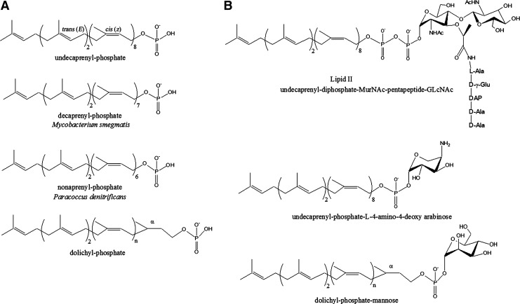
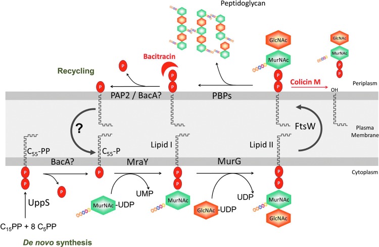
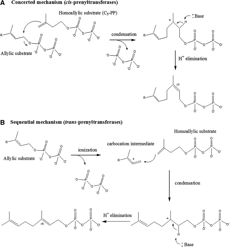
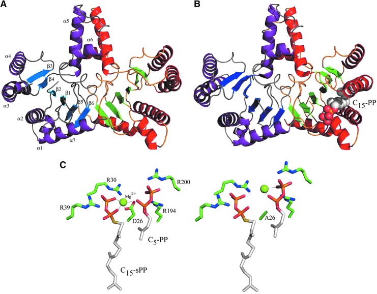
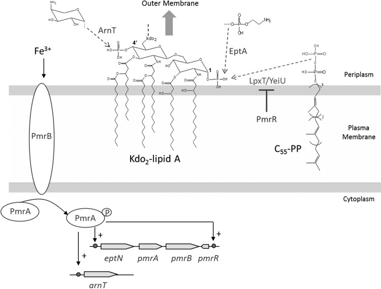
During the biogenesis of bacterial cell-wall polysaccharides, such as peptidoglycan, cytoplasmic synthesized precursors should be trafficked across the plasma membrane. This essential process requires a dedicated lipid, undecaprenyl-phosphate that is used as a glycan lipid carrier. The sugar is linked to the lipid carrier at the inner face of the membrane and is translocated toward the periplasm, where the glycan moiety is transferred to the growing polymer. Undecaprenyl-phosphate originates from the dephosphorylation of its precursor undecaprenyl-diphosphate, with itself generated by de novo synthesis or by recycling after the final glycan transfer. Undecaprenyl-diphosphate is de novo synthesized by the cytosolic cis-prenyltransferase undecaprenyl-diphosphate synthase, which has been structurally and mechanistically characterized in great detail highlighting the condensation process. In contrast, the next step toward the formation of the lipid carrier, the dephosphorylation step, which has been overlooked for many years, has only started revealing surprising features. In contrast to the previous step, two unrelated families of integral membrane proteins exhibit undecaprenyl-diphosphate phosphatase activity: BacA and members of the phosphatidic acid phosphatase type 2 super-family, raising the question of the significance of this multiplicity. Moreover, these enzymes establish an unexpected link between the synthesis of bacterial cell-wall polymers and other biological processes. In the present review, the current knowledge in the field of the bacterial lipid carrier, its mechanism of action, biogenesis, recycling, regulation, and future perspective works are presented.


Figures

<b>FIG. 1.</b>
FIG. 1.
Structures of different polyprenyl-phosphate carrier lipids (A) and lipid intermediates from various glycoconjugate biosynthetic pathways (B).
<b>FIG. 2.</b>
FIG. 2.
C55-P metabolism and membrane steps of peptidoglycan biosynthesis. C55-PP is de novo synthesized by undecaprenyl-diphosphate synthase (UppS) enzyme in the cytoplasm, from which it partitions in the inner side of the plasma membrane. Subsequently, C55-PP should be dephosphorylated to function as a lipid carrier in various polysaccharide biosynthetic pathways, such as the peptidoglycan synthesis. The enzymes MraY and MurG catalyze the successive transfers of the phospho-MurNAc-pentapeptide and GlcNAc motifs from the nucleotide precursors to the lipid carrier C55-P, generating the lipid I and lipid II intermediates, respectively. Lipid II is then translocated by the flippase FtsW to the periplasmic side of the membrane, where polymerization reactions of peptidoglycan that are catalyzed by the penicillin-binding proteins (PBPs) occur. The lipid carrier is released in its pyrophosphate form (C55-PP), which should be dephosphorylated and shuttled back to the inner face of the membrane to be reused. C55-P metabolism is the target of bacitracin that sequestrates C55-PP, thereby inhibiting its dephosphorylation, and of colicin M which cleaves lipid II in dead-end products, C55-OH and 1-pyrophospho-MurNAc(-peptide)-GlcNAc.
<b>FIG. 3.</b>
FIG. 3.
Mechanisms of C5-PP condensation on an allylic substrate by cis-prenyltransferases (A) and trans-prenyltransferases (B).
<b>FIG. 4.</b>
FIG. 4.
Structures of UppS enzyme from Escherichia coli. Overall structure of UppS (1UEH atomic coordinate): one dimer shown with purple α-helices and blue β-strands and the other monomer shown with red α-helices and green β-strands (A); structure of UppS in complex with C15-PP homoallylic substrate (represented with spheres) (1V7U atomic coordinate), highlighting conformational changes occurring in the α3-helix on substrate binding (B). Details of the ternary complex of UppS with Mg2+, C15-sPP, and C5-PP of wild-type enzyme (left) and a D26A mutant (right) (1X06, 1X07, 1X08, and 1X09 atomic coordinates) emphasizing the condensation mechanism and the role played by the aspartate 26 residue in this process (C).
<b>FIG. 5.</b>
FIG. 5.
Phosphorylation of lipid A by LpxT in E. coli, Salmonella enterica, and Salmonella typhimurium. LpxT transfers the distal phosphate group from C55-PP to the 1-phosphate group of nascent lipid A at the periplasmic side of the plasma membrane, releasing lipid A-1-diphosphate and C55-P. The 6′ position of lipid A is linked to two Kdo (3-deoxy-D-manno-octulosonic acid) residues from the core region. Subsequently, the core-lipid A (followed or not by the outermost O-antigen depending on the bacterial species) is exported toward the outer membrane and forms the outer leaflet. In rich media, the modification, whose role is yet to be discovered, occurs in about one third of total lipid A molecules. The activation of the PmrA/PmrB two-component system, by high extracellular concentrations of Fe3+, induces the expression of a small-membrane peptide, PmrR, which inhibits LpxT; along with ArnT and EptA, which catalyze the modification of lipid A with 4-amino-4-deoxy-L-arabinose (to the 4′-phosphate group) and phosphoethanolamine (to the 1-phosphate group), respectively. The latter lipid A decorations confer increased resistance to Fe3+ and cationic antibacterial peptides (CAMPs) and attenuate the recognition of lipid A by the host immune TLR4 system.

References

    1. Amiteye S., Kobayashi K., Imamura D., Hosoya S., Ogasawara N., and Sato T.2003. Bacillus subtilis diacylglycerol kinase (DgkA) enhances efficient Sporulation. J. Bacteriol. 185:5306–5309 - PMC - PubMed
    1. Apfel C.M., Takacs B., Fountoulakis M., Stieger M., and Keck W. 1999. Use of genomics to identify bacterial undecaprenyl pyrophosphate synthetase: cloning, expression, and characterization of the essential uppS Gene. J. Bacteriol. 181:483–492 - PMC - PubMed
    1. Barreteau H., Magnet S., El Ghachi M., Touzé T., Arthur M., Mengin-Lecreulx D., and Blanot D.2009. Quantitative high-performance liquid chromatography analysis of the pool levels of undecaprenyl phosphate and its derivatives in bacterial membranes. J. Chromatogr. B Analyt. Technol. Biomed. Life Sci. 877:213–220 - PubMed
    1. Bernard R., El Ghachi M., Mengin-Lecreulx D., Chippaux M., and Denizot F.2005. BcrC from Bacillus subtilis acts as an undecaprenyl pyrophosphate phosphatase in bacitracin resistance. J. Biol. Chem. 280:28852–28857 - PubMed
    1. Bernard R., Joseph P., Guiseppi A., Chippaux M., and Denizot F.2003. YtscD and YwoA, two independent systems that confer bacitracin resistance to Bacillus subtilis. FEMS Microbiol. Lett. 228:93–97 - PubMed

Publication types

MeSH terms

Substances

LinkOut - more resources