

Urgent bilateral endoscopic marsupialization for respiratory distress due to bilateral dacryocystitis in a newborn
- PMID: 24799096
- PMCID: PMC4025628
- DOI: 10.1097/SCS.0000000000000724
Urgent bilateral endoscopic marsupialization for respiratory distress due to bilateral dacryocystitis in a newborn
Abstract
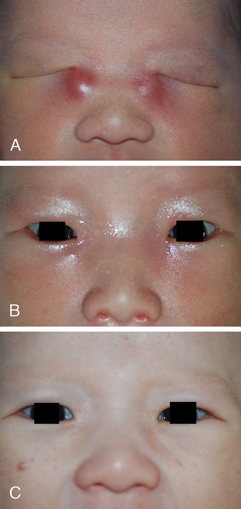
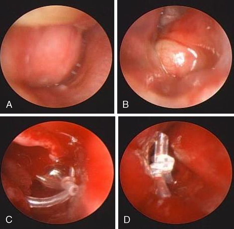
We describe an infant with respiratory distress due to bilateral dacryocystoceles and dacryocystitis who was successfully treated with urgent bilateral endoscopic marsupialization. A male infant was brought to our outpatient department 7 days after birth, with red, acutely inflamed swellings near the medial canthal area of both eyes. From birth, there had been bluish swelling near the medial canthal area, and redness and swellings developed within 3 days. On physical examination, the child was afebrile but showed respiratory distress with coarse breathing sound. That day, the infant was admitted and treated with intravenous cefotaxime 150 mg. After withholding oral intake for appropriate preoperative fasting, urgent bilateral probing with endoscopy was done. On endoscopy, huge bilateral congenital dacryocystoceles were found. Because of its huge size, the inferior surface of the cyst was touching the nasal floor, which made probe unable to perforate the wall of dacryocystocele. Therefore, an endoscopy-assisted marsupialization of dacryocystoceles and bicanalicular silicone intubation were performed. Both swellings and erythema subsided within 48 hours postoperatively, and the patient was discharged after 72 hours from treatment.
Conflict of interest statement
The authors report no conflicts of interest.
Figures
Similar articles
-
Bilateral dacryocystocele with an intranasal cyst as the cause of respiratory distress in a newborn.B-ENT. 2016;12(1):23-7. B-ENT. 2016. PMID: 27097390
-
Endoscopic marsupialization of congenital nasolacrimal duct cyst with dacryocoele.Clin Otolaryngol Allied Sci. 2002 Jun;27(3):167-70. doi: 10.1046/j.1365-2273.2002.00556.x. Clin Otolaryngol Allied Sci. 2002. PMID: 12071990
-
Bilateral dacryocystoceles as a rare cause of neonatal respiratory distress: report of 2 cases.Ear Nose Throat J. 2014 Jan;93(1):E26-8. Ear Nose Throat J. 2014. PMID: 24452900
-
Congenital Dacryocystocele: A Major Review.Ophthalmic Plast Reconstr Surg. 2019 Jul/Aug;35(4):309-317. doi: 10.1097/IOP.0000000000001297. Ophthalmic Plast Reconstr Surg. 2019. PMID: 30601463 Review.
-
Respiratory distress in the neonate. Sequela of a congenital dacryocystocele.Arch Otolaryngol Head Neck Surg. 1995 Dec;121(12):1423-5. doi: 10.1001/archotol.1995.01890120079016. Arch Otolaryngol Head Neck Surg. 1995. PMID: 7488375 Review.
Cited by
-
A rare cause of intermittent respiratory distress and epiphora in the newborn: congenital dacryocystocele.Gland Surg. 2017 Feb;6(1):114-118. doi: 10.21037/gs.2016.06.02. Gland Surg. 2017. PMID: 28210562 Free PMC article.
References
-
- Mansour AM, Cheng KP, Mumma JV, et al. Congenital dacryocele. A collaborative review. Ophthalmology 1991; 98: 1744– 1751 - PubMed
-
- Teixeira CC, Dias RJ, Falcão-Reis FM, et al. Congenital dacryocystocele with intranasal extension. Eur J Ophthalmol 2005; 15: 126– 128 - PubMed
-
- Han BL, Shin HS. Dacryocystocele with congenital unilateral lacrimal puncta agenesis in an adults. J Craniofac Surg 2013; 24: 1242– 1243 - PubMed
-
- Lipton J, Jacobs N, Rosen ES. Bilateral acute dacryocystitis in an infant. Br J Hosp Med 1987; 38: 251. - PubMed
Publication types
MeSH terms
LinkOut - more resources
Full Text Sources
Other Literature Sources
Medical
