Review
. 2014 Oct:30:1-8.
doi: 10.1016/j.ceb.2014.04.002. Epub 2014 May 8.

Drosophila blood cell chemotaxis

Affiliations
Review

Drosophila blood cell chemotaxis

Iwan Robert Evans et al. Curr Opin Cell Biol. 2014 Oct.

Abstract

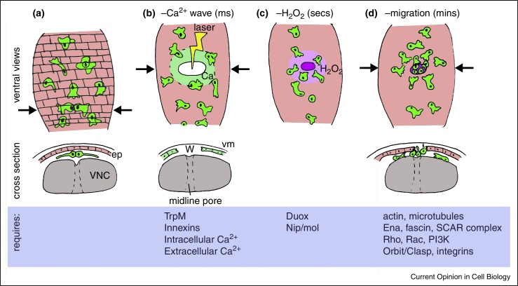
Drosophila melanogaster contains a population of blood cells called hemocytes that represent the functional equivalent of vertebrate macrophages. These cells undergo directed migrations to disperse during development and reach sites of tissue damage or altered self. These chemotactic behaviors are controlled by the expression of PDGF/Vegf-related ligands in developing embryos and local production of hydrogen peroxide at wounds. Recent work reveals that many molecules important in vertebrate cell motility, including integrins, formins, Ena/VASP proteins and the SCAR/WAVE complex, have a conserved function in these innate immune cells. The use of this model organism has elucidated how damage signals are activated by calcium signaling during inflammation and that the steroid hormone ecdysone activates immune competence at key developmental stages.


Figures

Figure 1
Figure 1
Embryonic migration routes and chemoattractant expression. Schematics showing expression of Pvf2 and Pvf3 chemoattractants (pink shading) in the developing Drosophila embryo at stages 11 (a) and 12 (b). Cartoons below embryos correspond to boxed regions and show RhoL-dependent invasion of the germband (gb) towards a source of Pvfs, some of which is expressed by the developing malphigian tubules (mp) (a) and movement along the developing ventral nerve cord (VNC; grey) (b); arrows indicate hemocyte movements at these stages of development. During progression along the VNC hemocytes are tightly confined between the ventral side of the VNC and epithelium (ep) and as they migrate along the VNC in response to the Pvf ligands that are expressed there, the epithelium and VNC separate, creating a channel for hemocyte progression. Hemocytes also migrate along the developing dorsal vessel at this stage (dv); a = anterior, p = posterior, d = dorsal, v = ventral, lat = lateral. Later in development cell–cell repulsion begins to occur and this depends upon the microtubules, which are frequently bundled into an arm-like structure (arrow) that facilitates persistent migration (c). Microtubules labeled via Clip-GFP expression in hemocytes; white line indicates edges of hemocytes, drawn according to mCherry-moesin localization (not shown). After initial dispersal hemocytes migrate at right angles from the ventral midline to the edges of the VNC (purple arrows) to form three lines (white arrows) on the ventral side of the embryo, immediately beneath the epithelium (d). Maximum projection images show GFP and nls-red stinger localization in hemocytes from the ventral side of the embryo; scale bars represent 50 μm; ant = anterior, post = posterior.
Figure 2
Figure 2
Calcium waves direct inflammatory migration of hemocytes. Ventral and cross-sectional views (anterior-posterior position indicated by arrows) showing immune cell recruitment to sites of tissue damage in Drosophila embryos. Hemocytes (green) sit immediately beneath the epidermis (ep, pink) on the ventral nerve cord (VNC, grey) (a). Laser wounding of the epithelium causes an almost instantaneous calcium wave to flood through the epithelium via cell–cell junctions (b); this depends upon functional cell–cell junctions and TrpM. An increase in intracellular calcium activates the NADPH oxidase Duox via its EF hands driving hydrogen peroxide production (c). Hydrogen peroxide is necessary for the recruitment of hemocytes to this point of tissue damage, which is an active, migratory process requiring the function of the actin and microtubule cytoskeletons (d). The relative timescale is indicated in brackets.
Figure 3
Figure 3
Comparison of migration to wounds in larval and embryonic stages of Drosophila development with vertebrate inflammatory responses. Cartoon of macrophage migration to wounds in vertebrates (a). Macrophages (green) form transient adhesions with activated endothelial cells (red) and roll, leading to arrest and extravasation and penetration through the basement membrane (brown) before migrating though tissue largely composed of fibroblasts (fb) and ECM to reach wound sites (W). Larval hemocyte responses (b) consist of an adhesive capture that recapitulates rolling and tethering of vertebrate leukocytes; sessile hemocytes do not respond to wounds. Migration of hemocytes to wounds in the embryo occurs in the context of an environment containing ECM deposited between closely opposed tissues (epithelium and VNC) and requires active migration and resembles movement of vertebrate leukocytes post-extravasation.

References

    1. Barreiro O., Martin P., Gonzalez-Amaro R., Sanchez-Madrid F. Molecular cues guiding inflammatory responses. Cardiovasc Res. 2010;86:174–182. - PubMed
    1. King J.S., Insall R.H. Chemotaxis: finding the way forward with Dictyostelium. Trends Cell Biol. 2009;19:523–530. - PubMed
    1. Wang L., Kounatidis I., Ligoxygakis P. Drosophila as a model to study the role of blood cells in inflammation, innate immunity and cancer. Front Cell Infect Microbiol. 2013;3:113. - PMC - PubMed
    1. Wood W., Jacinto A. Drosophila melanogaster embryonic haemocytes: masters of multitasking. Nat Rev Mol Cell Biol. 2007;8:542–551. - PubMed
    1. Evans I.R., Hu N., Skaer H., Wood W. Interdependence of macrophage migration and ventral nerve cord development in Drosophila embryos. Development. 2010;137:1625–1633. - PMC - PubMed

Publication types

Substances

LinkOut - more resources