

Metabolic shifts toward glutamine regulate tumor growth, invasion and bioenergetics in ovarian cancer
- PMID: 24799285
- PMCID: PMC4188042
- DOI: 10.1002/msb.20134892
Metabolic shifts toward glutamine regulate tumor growth, invasion and bioenergetics in ovarian cancer
Abstract
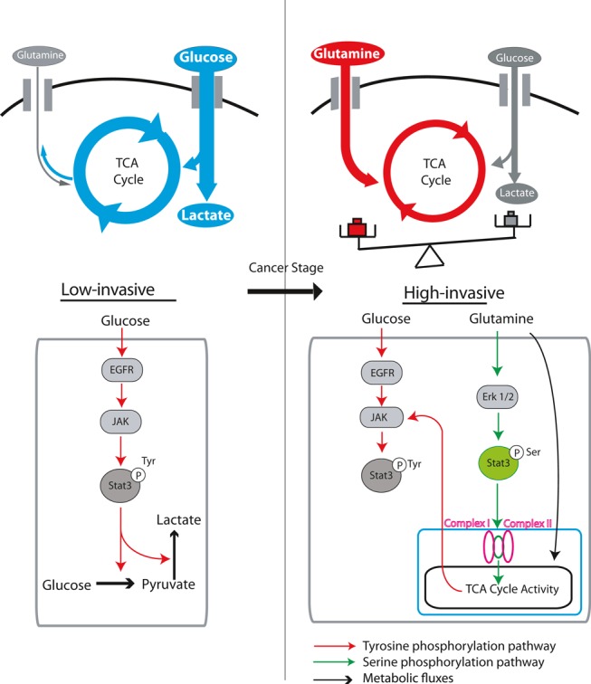
Glutamine can play a critical role in cellular growth in multiple cancers. Glutamine-addicted cancer cells are dependent on glutamine for viability, and their metabolism is reprogrammed for glutamine utilization through the tricarboxylic acid (TCA) cycle. Here, we have uncovered a missing link between cancer invasiveness and glutamine dependence. Using isotope tracer and bioenergetic analysis, we found that low-invasive ovarian cancer (OVCA) cells are glutamine independent, whereas high-invasive OVCA cells are markedly glutamine dependent. Consistent with our findings, OVCA patients' microarray data suggest that glutaminolysis correlates with poor survival. Notably, the ratio of gene expression associated with glutamine anabolism versus catabolism has emerged as a novel biomarker for patient prognosis. Significantly, we found that glutamine regulates the activation of STAT3, a mediator of signaling pathways which regulates cancer hallmarks in invasive OVCA cells. Our findings suggest that a combined approach of targeting high-invasive OVCA cells by blocking glutamine's entry into the TCA cycle, along with targeting low-invasive OVCA cells by inhibiting glutamine synthesis and STAT3 may lead to potential therapeutic approaches for treating OVCAs.
Figures
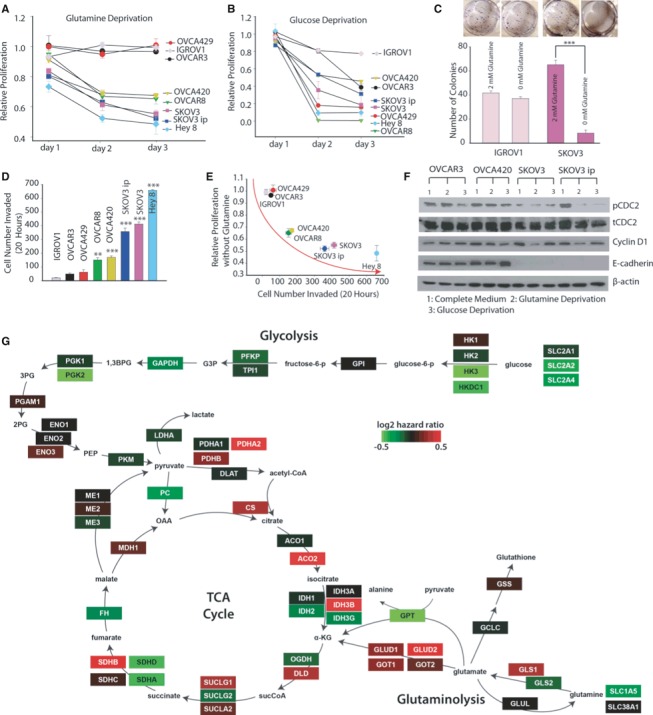
Gln deprivation effect on proliferation rate of a panel of
OVCA cells for 24, 48 and 72 h.OVCAR 3,IGROV 1,OVCA 429 cells wereG ln independent;OVCA 420 andOVCAR 8 cells were moderatelyG ln dependent; andSKOV 3,SKOV 3ip andH ey 8 cells wereG ln dependent. n ≥ 15.Glucose deprivation effect on proliferation rate of
OVCA cell lines for 24, 48, 72 h. n ≥ 15.Gln deprivation effect on clonogenic formation in
IGROV 1 andSKOV 3. n = 6.Matrigel invasion assay was conducted to characterize invasiveness of
OVCA cell lines.OVCAR 3,IGROV 1 andOVCA 429 cells were noninvasive,OVCA 420 andOVCAR 8 cells were moderately invasive, whileSKOV 3,SKOV 3ip and Hey 8 were highly invasiveOVCA cell lines. n ≥ 6.Correlation between proliferation rate at 72 h of
OVCA cell lines under Gln‐depleted conditions with their corresponding number of invaded cells.Western blot analysis of cell cycle proteins linked with growth rate (
CDC 2, CyclinD 1) and protein linked with metastatic potential (E ‐cadherin) inOVCA cell lines. β‐actin was used as the loading control.Genes in the glutaminolysis and tricarboxylic acid cycle metabolic pathways are associated with higher risk in
OVCA patients. Shown is a metabolic network and the genes (rectangles) that produce the enzyme catalyzing the reactions in the network. The genes in the network are colored by their correlation withOVCA patient survival based on calculating theirC ox hazard values (color key). The gene expression and clinical data were fitted with a Cox proportional hazard model to determine the hazard ratio for each gene inG ln/glucose metabolic network. A higher hazard ratio (poor outcome) was observed in gene products that catalyze reactions inG ln catabolism and higher expression of glycolytic genes culminated in better patient survival (low hazard ratio).
- A
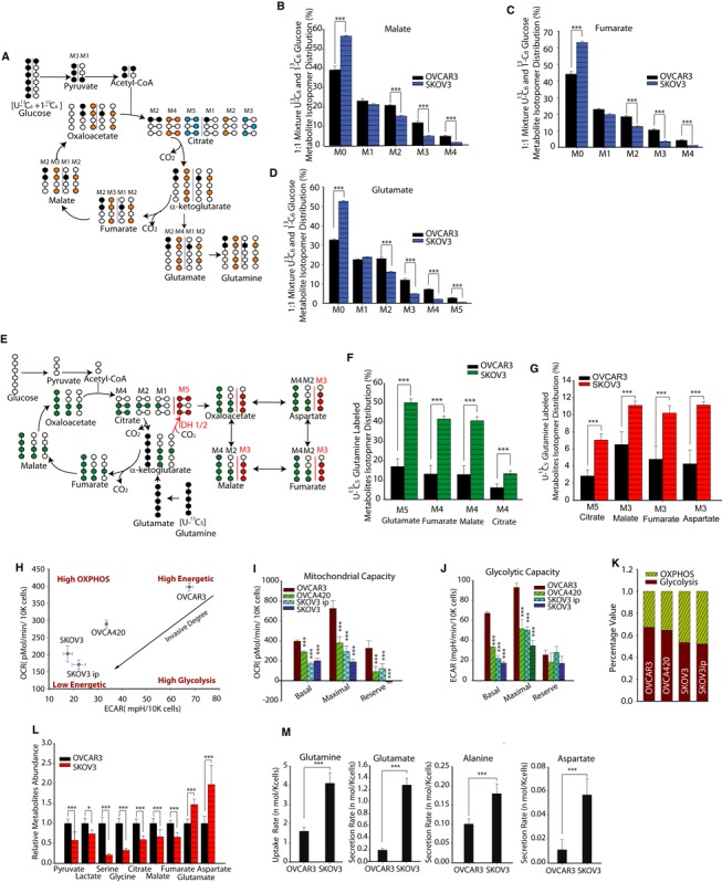
Schematic of carbon atom transitions using 1:1 mixture of 13C6 glucose and 1‐13C‐labeled glucose. This allows estimations of the contribution of glucose toward
TCA cycle metabolites and synthesis of glutamate and Gln. Black color represents labeled carbon on 1stTCA cycle, yellow color represents labeled carbon on 2ndTCA cycle, and blue color represents labeled carbon on 3rdTCA cycle. - B–D
Glucose's contribution to
TCA metabolites and glutamate pool. Comparison of mass isotopomer distribution (MID ) of malate (B), fumarate (C), glutamate (D) in high‐invasive (SKOV 3) and low‐invasive (OVCAR 3) cells cultured with a 1:1 mixture of 13C6 glucose and 1‐13C‐labeled glucose. - E
Schematic of carbon atom transitions using 13C5‐labeled Gln. Black color represents labeled carbon of Gln before its entry into TCA cycle. Green color represents Gln's direct effect on canonical
TCA cycle, red color represents Gln's effect onTCA cycle through reductive carboxylation. - F
Gln's contribution to
TCA cycle metabolites pool. Comparisons ofMID ofM 5 glutamate,M 4 fumarate,M 4 malate,M 4 citrate inOVCAR 3 andSKOV 3 cultured with 13C5‐labeledG ln. - G
Mass isotopomer analysis of isotopomers linked with reductive carboxylation. M5 citrate,
M 3 malate,M 3 fumarate andM 3 aspartate analysis indicates significantly higher reductive carboxylation fluxes in high‐invasive than in low‐invasiveOVCA cells. - H
Basal oxygen consumption rate (
OCR ) and extracellular acidification rate (ECAR ) were measured forOVCAR 3,OVCA 420,SKOV 3 andSKOV 3ip cell lines. BasalOCR is a measure ofOXPHOS activity, and basalECAR is a measure of glycolysis activity. - I
Using oligomycin,
FCCP and antimycin, we estimated mitochondrial functional state inOVCA cells through maximal and reserve mitochondrial capacities. - J
Using 2
DG , we estimated glycolytic functional state inOVCA cells through maximal and reserve glycolytic capacities. - K
Percentage of
OXPHOS and glycolysis contribution to cancer cell's energetic demand. - L
Relative metabolite abundances were measured using
GC ‐MS inOVCAR 3 andSKOV 3 cells. - M
Extracellular uptake/secretion fluxes of amino acids involved in glutaminolysis (Glutamine:
G ln; Glutamate:G lu; Alanine: Ala; Aspartate:A sp) were measured using ultra‐high‐performance liquid chromatography.
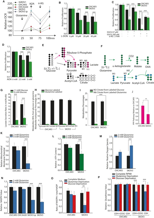
Oxygen consumption rate (
OCR ) was measured in high‐invasive (SKOV 3 andSKOV 3ip), low‐invasive (IGROV 1 andOVCAR 3) and moderately invasive (OVCA 420) cells in media containing Gln, aminooxyacetate (AOA ) and α‐ketoglutarate (α‐KG ).OCR was normalized with value before injections.Analysis of proliferation of
OVCA cell lines with range of concentrations of 6‐diazo‐5‐oxo‐L‐norleucine (L‐DON ), a Gln analog which inhibitsG ln's conversion to glutamate. Data were normalized with complete media conditions withoutL ‐DON .Analysis of proliferation of
OVCA cell lines with range of concentrations of epigallocatechin gallate (EGCG , an inhibitor of glutamate dehydrogenase, which converts glutamate into α‐KG ) used to inhibit glutamate entering the tricarboxylic acid (TCA ) cycle. α‐KG was used to rescue reduction of cell proliferation byEGCG . Data were normalized with complete media conditions withoutEGCG .Analysis of proliferation of
OVCA cell lines when glutaminolysis was inhibited with range of concentrations ofAOA . Data were normalized with complete media conditions withoutAOA .Lactate and citrate generation from glucose through direct glycolysis or through both oxidative pentose phosphate pathway (
PPP ) and glycolysis. Black circles represent labeled carbons, empty circles represent unlabeled carbons, pink circles represent unlabeled carbons in oxidative PPP.Lactate and citrate generation from glutaminolysis. Blue circles represent labeled carbons from malic enzymes, and green circles are labeled carbons from canonical
TCA cycle.Glucose deprivation effect on lactate secretion rate.
Comparison of
M 3 pyruvate andM 3 lactate derived from either glucose (1:1 mixture ofU ‐13C 6 glucose and 1‐13C 1 glucose) orG ln (U ‐13C 5G ln) in high‐ and low‐invasiveOVCA cells. Since all conditions have complete media, their total pyruvate and lactate content should be the same.Comparison of
M 2 citrate labeling from glucose and M6 citrate labeling from Gln inOVCAR 3 andSKOV 3 cells. Total pyruvate and lactate content in all conditions should be equal as explained for (H).Oxidative pentose phosphate pathway fluxes estimated using the mathematical relation (percentage of unlabeled M0 lactate—percentage of labeled
M 1 lactate) to represent relative oxidativePPP fluxes inOVCAR 3 andSKOV 3 cells cultured with 1:1 mixture of 13C6 glucose and 1‐13C‐labeled glucose.Role of
G ln in maintaining mitochondrial membrane potential (MMP ) levels in low‐ and high‐invasive cells.Gln maintains
ATP content selectively in high‐invasiveOVCA cells.Gln reduces reactive oxygen species induced by
H 2O 2 in high‐invasive cells, but not in low‐invasive cells.Gln maintains
NADPH level in high‐invasiveOVCA cells.Gln increases ratio of
NADPH /totalNADPH ratio selectively in high‐invasiveOVCA cells. Glucose is unable to provide enough reducing equivalents in high‐invasiveOVCA cells.Gln/glucose deprivation's effect on total glutathione and reduced glutathione level in cancer cells.
- A
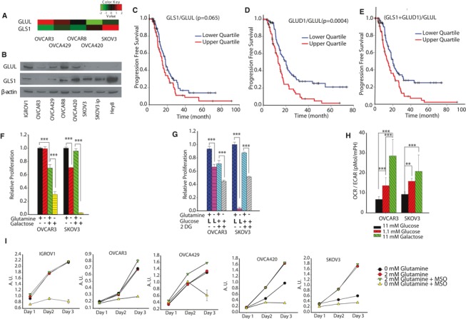
Gene expression levels of
G ln synthetase (GLUL ) and glutaminase (GLS1 ) were determined using KyotoOv38, a database for gene expression of ovarian cancer cell lines. - B
Western blot of the
GLUL andGLS 1 protein expression levels forOVCA cell lines. - C–E
Progression free survival rate for
OVCA patients, categorized according toGLS 1/GLUL (C),GLUD 1/GLUL (D), (GLS 1 +GLUD 1)/GLUL (E). The survival analysis is executed from comparison of upper quartile and lower quartile patients. (total patients n = 539). - F, G
Converting Gln‐independent
OVCA cells into Gln dependent by replacing glucose with galactose (F) or low glucose concentrations (L glucose) and 2‐DG (10 mM) (G). - H
Galactose and low glucose's effect on oxygen consumption rate (
OCR )/extracellular acidification rate. - I
Combined approach of targeting high‐invasive
OVCA cells by blocking glutamine's entry into theTCA cycle along with targeting low‐invasiveOVCA cells by inhibiting glutamine synthesis leads to pronounced reduction of cell growth. TargetingGLUL usingMSO under Gln deprivation decreasesOVCA cell growth. Arbitrary Units (AU ) is proportional to cell number (n ≥ 10).
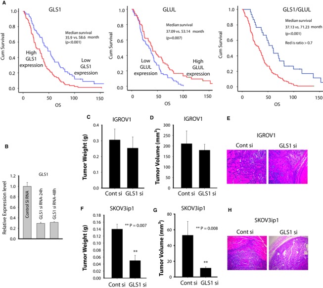
- A
Kaplan–Meier curves of disease‐specific survival for patients with epithelial
OVCA (n = 139) based onGLS 1 andG ln synthetase protein expression. The log‐rank test (two sided) was used to compare difference between groups. - B
Following transfection with either
GLS 1 siRNA or control siRNA ,mRNA levels were assessed withqRT ‐PCR . - C–H
Therapeutic efficacy of si
RNA ‐mediatedGLS downregulation: (C) tumor nodule (D) volume (E) pattern of invasion of low‐invasiveOVCA cell line (IGROV 1); (F) tumor nodule (G) volume and (H) pattern of invasion ofSKOV 3ip1 cells in nude mouse models. Following subcutaneous injection of nude mice with 2.0 × 10⁶IGROV 1 orSKOV 3ip1 cells, mice were randomly allocated to one of the following groups: control siRNA ‐DOPC orGLS 1 siRNA ‐DOPC . Treatment was started 3 days after tumor cell injection, and siRNA ‐liposomes were administered twice weekly at a dose of 150 μg/kg body weight and continued for 2 weeks. At the time of sacrifice, mouse weight, tumor weight and tumor volume were recorded. Statistical analysis for tumor weights was performed by Student's t‐test. **P = 0.007 and **P = 0.008 compared with control siRNA ‐DOPC .
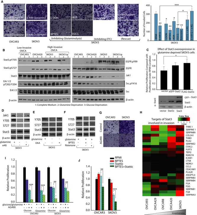
Comparison of
OVCAR 3 invasive capacity inG ln depleted and complete media conditions. Invasive capacity ofSKOV 3 is measured under complete media, Gln depleted media, and drugs inhibitingG ln's entry into tricarboxylic acid (TCA ) cycle. L‐DON ,BPTES ,EGCG ,AOA , and Rotenone decreaseSKOV 3's Matrigel invasive capacity. α‐Ketoglutarate (α‐KG ) addition under glutamine deprivation orEGCG conditions rescuesSKOV 3's invasive capacity. Data are expressed as mean ± SEM, *P < 0.05, **P < 0.01, ***P < 0.001, n ≥ 4.Activation of Stat3 through tyrosine‐705 phosphorylation (Stat3.
pY 705) is elevated in high‐invasive ovarian cancer (OVCA ) cells. The phosphorylation level ofEGFR andE rk 1/2 increases with increasing degree of invasiveness. Tyrosine kinase signaling pathway activities can be affected by metabolic stress and in high‐invasive cells that are glutamine and glucose deprived. Stat3.pY 705 is reduced along with total Jak levels. Stat3.pY 705 levels are only reduced in theOVCA 420 cell lines upon glucose deprivation, while the regulation ofS tat3 phosphorylation by metabolic stress is absent in the low‐invasive cell line,OVCAR 3. Stat3 serine‐727 phosphorylation (S tat3.pS 727) is also reduced along with phosphorylation level ofE rk 1/2 in the high‐invasive cell lines and only in response to glutamine deprivation. β‐actin as loading control.Cell proliferation in high‐invasive
OVCA cell line is reduced when glutamine starved. Proliferation can be partially rescued by overexpression of transgenic Stat3 and a constitutively active mutant of Stat3, Stat3c, mean ±SD , n = 3, *P < 0.05. β‐actin as loading control.α‐
KG addition rescuesSTAT 3 tyrosine phosphorylation and Jak1, but cannot rescue Stat3 serine‐727 phosphorylation. β‐actin as loading control. Western blot figures are cut from same gel, but lanes are rearranged to show complete media conditions in first column, glutamine deprivation in second and α‐KG addition in the third column. The full Western blot images of the gel and the detailed description are included in the source file.OAA addition rescuesSTAT 3 tyrosine phosphorylation. β‐actin as loading control. Western blot figures are cut from same gel, but lanes are rearranged to include only the relevant conditions. The full Western blot images of the gel and the detailed description are included in the source file.BPTES and rotenone inhibitSTAT 3 phosphorylation at tyrosine 705 (Y 705). β‐actin as loading control. Western blot figures are cut from same gel, but lanes are arranged to show only the relevant conditions. The full Western blot images of the gel and the detailed description are included in the source file.Inhibition of Stat3 decreases
OVCA metastasis. Treatment ofSKOV 3 cells withAG 490 results in reduced invasion.Gene expression levels of targets of
STAT 3 involved in invasion were determined using KyotoOv38 in fiveOVCA cells. High‐invasiveOVCA cells had higher gene expression of invasive genes.The addition of
AG 490, a Stat3 inhibitor enhances the effect of glucose and glutamine deprivation on proliferation inOVCA cell lines.The addition of stattic, inhibitor of
STAT 3, has a combinational cytotoxic effect on both cell lines.
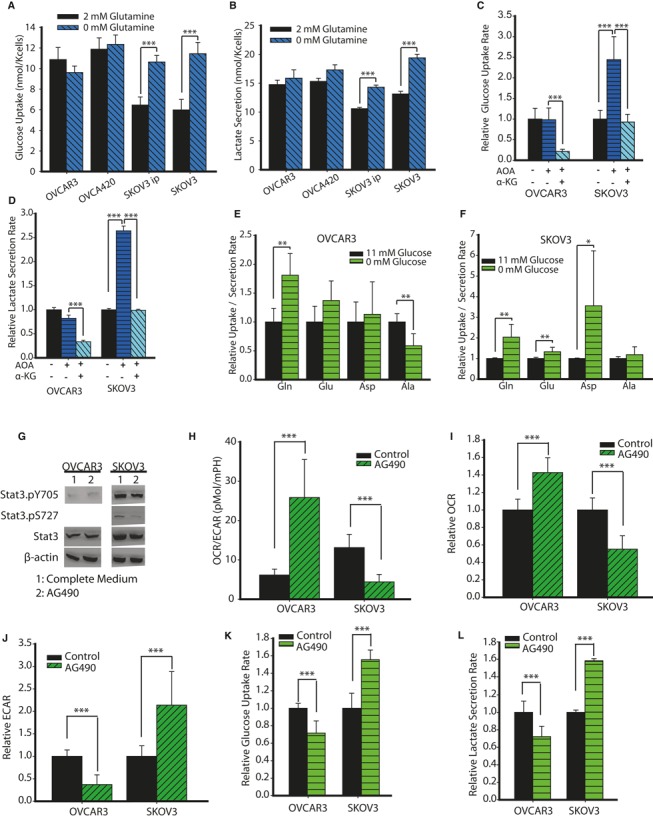
- A, B
Effect of Gln deprivation on glucose uptake (A) and lactate secretion (B) fluxes in
OVCAR 3,OVCA 420,SKOV 3,SKOV 3ip cells. - C, D
Influence of
AOA , α‐ketoglutarate (α‐KG ) on glucose uptake (C) and lactate secretion (D) fluxes inOVCA cells. - E, F
Effect of glucose deprivation on Gln, glutamate, aspartate and alanine uptake/secretion fluxes in high‐invasive (F) and low‐invasive (E)
OVCA cells. - G
AG 490's effect on tyrosine and serine phosphorylation ofSTAT 3 in low‐invasive (OVCAR 3) and high‐invasive (SKOV 3)OVCA cells. - H
AG 490 shifts low‐invasiveOVCA cells from glycolysis toOXPHOS , whereas reverse was true for high‐invasive cells. - I
AG 490 increases oxygen consumption rate (OCR ) forOVCAR 3 and decreasesOCR forSKOV 3 cells. - J
AG 490 decreases extracellular acidification rate (ECAR ) forOVCAR 3 and increasesECAR forSKOV 3 cells. - K, L
AG 490 decreases glucose uptake (K) and lactate secretion rate (L) forOVCAR 3 and increases glucose uptake and lactate secretion rate forSKOV 3.
References
-
- Agus DB, Alexander JF, Arap W, Ashili S, Aslan JE, Austin RH, Backman V, Bethel KJ, Bonneau R, Chen WC, Chen‐Tanyolac C, Choi NC, Curley SA, Dallas M, Damania D, Davies PC, Decuzzi P, Dickinson L, Estevez‐Salmeron L, Estrella V et al (2013) A physical sciences network characterization of non‐tumorigenic and metastatic cells. Sci Rep 3: 1449. - PMC - PubMed
-
- Armaiz‐Pena GN, Allen JK, Cruz A, Stone RL, Nick AM, Lin YG, Han LY, Mangala LS, Villares GJ, Vivas‐Mejia P, Rodriguez‐Aguayo C, Nagaraja AS, Gharpure KM, Wu Z, English RD, Soman KV, Shazhad MM, Zigler M, Deavers MT, Zien A et al (2013) Src activation by beta‐adrenoreceptors is a key switch for tumour metastasis. Nat Commun 4: 1403. - PMC - PubMed
Publication types
MeSH terms
Substances
Grants and funding
LinkOut - more resources
Full Text Sources
Other Literature Sources
Medical
Molecular Biology Databases
Miscellaneous
