

DPP and DSP are Necessary for Maintaining TGF-β1 Activity in Dentin
- PMID: 24799420
- PMCID: PMC4107551
- DOI: 10.1177/0022034514534690
DPP and DSP are Necessary for Maintaining TGF-β1 Activity in Dentin
Abstract
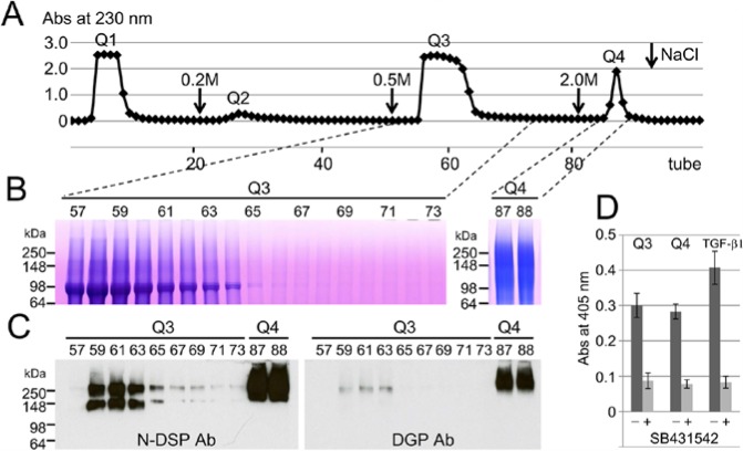
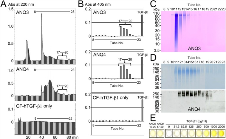
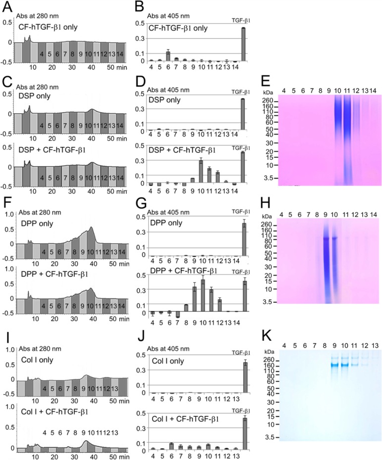
Porcine dentin sialophosphoprotein (DSPP) is the most abundant non-collagenous protein in dentin. It is processed by proteases into 3 independent proteins: dentin sialoprotein (DSP), dentin glycoprotein (DGP), and dentin phosphoprotein (DPP). We fractionated DPP and DSP along with TGF-β activity by ion exchange (IE) chromatography from developing pig molars and measured their alkaline phosphatase (ALP)-stimulating activity in human periodontal (HPDL) cells with or without TGF-β receptor inhibitor. We then purified TGF-β-unbound or -bound DPP and DSP by reverse-phase high-performance liquid chromatography (RP-HPLC) using the ALP-HPDL system. The TGF-β isoform bound to DPP and DSP was identified as being TGF-β1 by both ELISA and LC-MS/MS analysis. We incubated carrier-free human recombinant TGF-β1 (CF-hTGF-β1) with TGF-β-unbound DPP or DSP and characterized the binding on IE-HPLC using the ALP-HPDL system. When only CF-hTGF-β1 was incubated, approximately 3.6% of the ALP-stimulating activity remained. DPP and DSP rescued the loss of TGF-β1 activity. Approximately 19% and 10% of the ALP stimulating activities were retained by the binding of TGF-β to DPP and DSP, respectively. The type I collagen infrequently bound to CF-hTGF-β1. We conclude that both DPP and DSP help retain TGF-β1 activity in porcine dentin.
Keywords: HPLC; cell culture; extracellular matrix (ECM); isolation and purification; phosphophoryn; tooth.
© International & American Associations for Dental Research.
Conflict of interest statement
The authors declare no potential conflicts of interest with respect to the authorship and/or publication of this article.
Figures
References
-
- Baker SM, Sugars RV, Wendel M, Smith AJ, Waddington RJ, Cooper PR, et al. (2009). TGF-beta/extracellular matrix interactions in dentin matrix: a role in regulating sequestration and protection of bioactivity. Calcif Tissue Int 85:66-74. - PubMed
-
- Bègue-Kirn C, Smith AJ, Ruch JV, Wozney JM, Purchio A, Hartmann D, et al. (1992). Effects of dentin proteins, transforming growth factor beta 1 (TGF beta 1) and bone morphogenetic protein 2 (BMP2) on the differentiation of odontoblast in vitro. Int J Dev Biol 36:491-503. - PubMed
-
- Butler WT, Bhown M, Brunn JC, D’Souza RN, Farach-Carson MC, Happonen RP, et al. (1992). Isolation, characterization and immunolocalization of a 53-kDal dentin sialoprotein (DSP). Matrix 12:343-351. - PubMed
-
- Cam Y, Lesot H, Colosetti P, Ruch JV. (1997). Distribution of transforming growth factor beta1-binding proteins and low-affinity receptors during odontoblast differentiation in the mouse. Arch Oral Biol 42:385-391. - PubMed
-
- Cassidy N, Fahey M, Prime SS, Smith AJ. (1997). Comparative analysis of transforming growth factor-beta isoforms 1-3 in human and rabbit dentine matrices. Arch Oral Biol 42:219-223. - PubMed
Publication types
MeSH terms
Substances
Grants and funding
LinkOut - more resources
Full Text Sources
Other Literature Sources
Miscellaneous
