. 2014 Jun 2;211(6):1231-42.
doi: 10.1084/jem.20131853. Epub 2014 May 5.

Siglec-5 and Siglec-14 are polymorphic paired receptors that modulate neutrophil and amnion signaling responses to group B Streptococcus

Affiliations

Siglec-5 and Siglec-14 are polymorphic paired receptors that modulate neutrophil and amnion signaling responses to group B Streptococcus

Syed Raza Ali et al. J Exp Med. .

Abstract

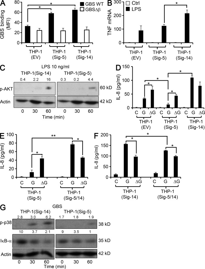
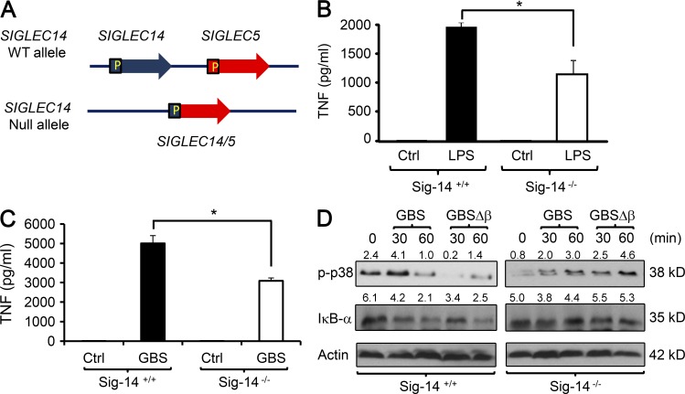
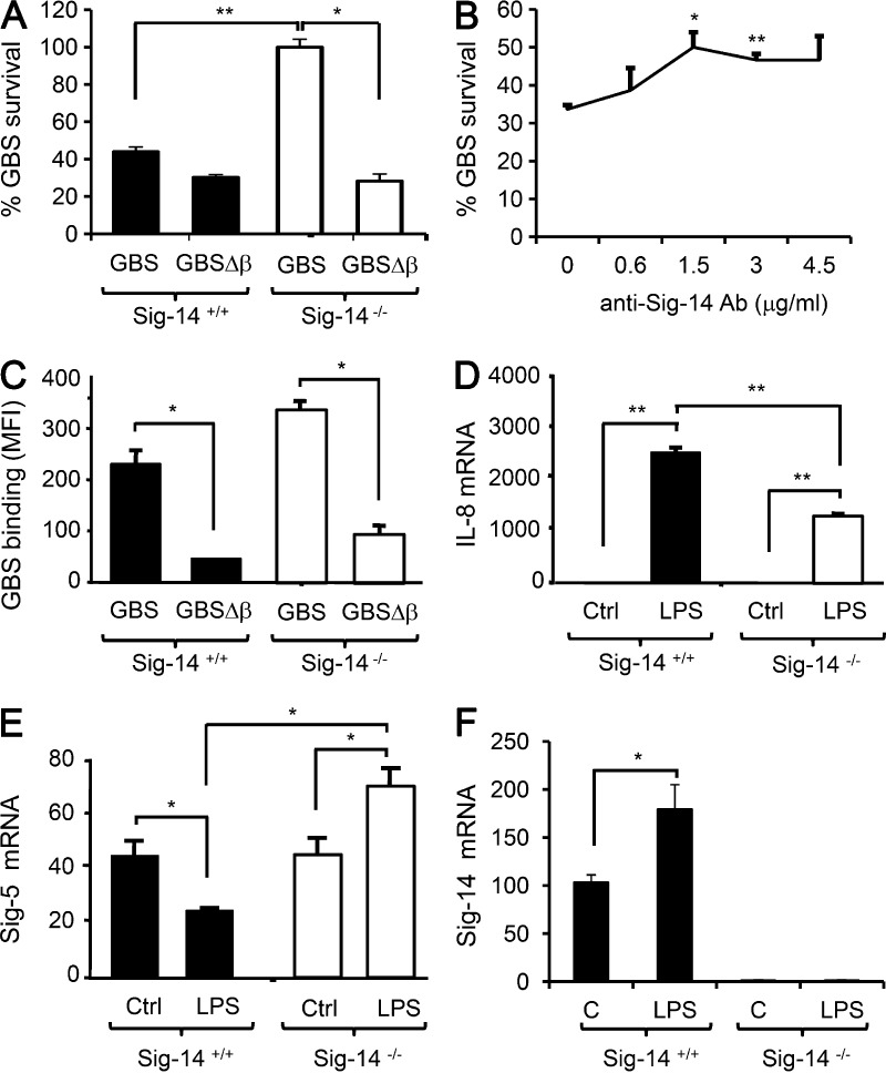
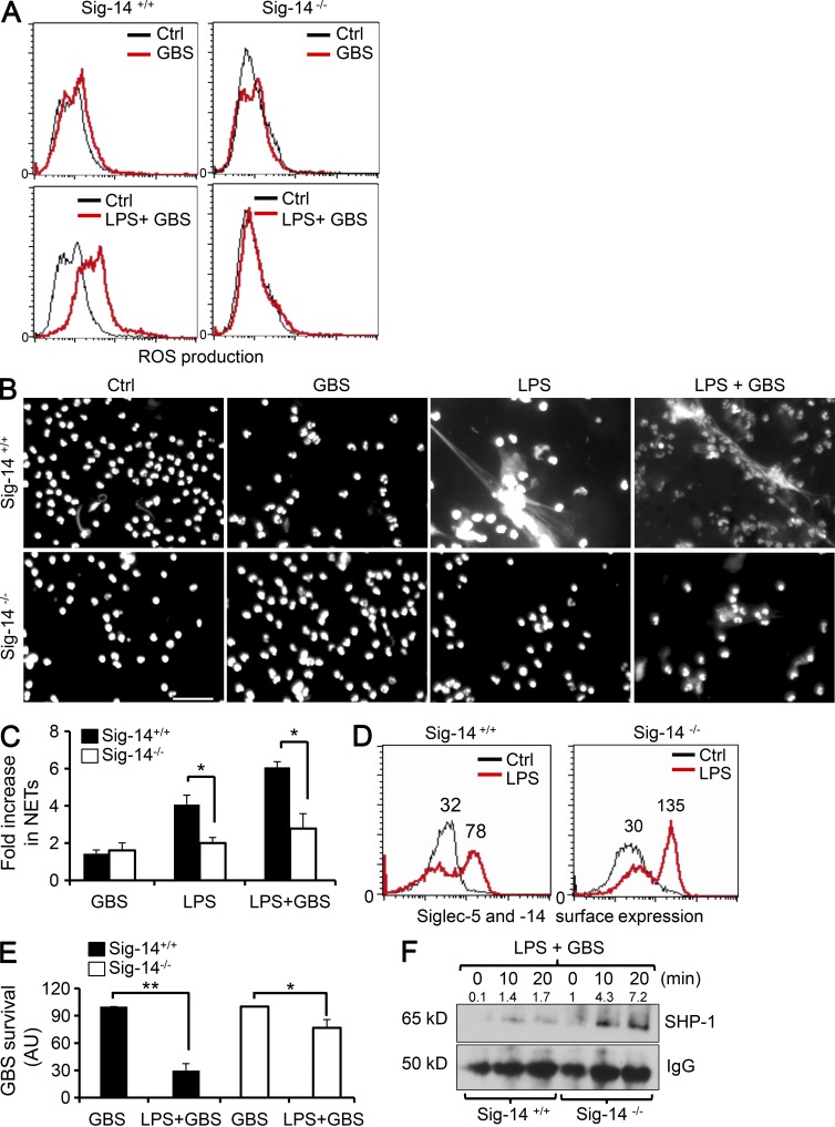
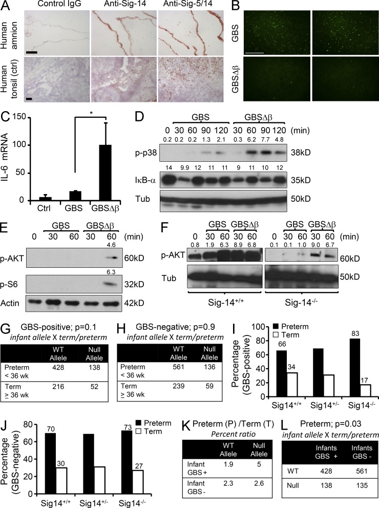
Group B Streptococcus (GBS) causes invasive infections in human newborns. We recently showed that the GBS β-protein attenuates innate immune responses by binding to sialic acid-binding immunoglobulin-like lectin 5 (Siglec-5), an inhibitory receptor on phagocytes. Interestingly, neutrophils and monocytes also express Siglec-14, which has a ligand-binding domain almost identical to Siglec-5 but signals via an activating motif, raising the possibility that these are paired Siglec receptors that balance immune responses to pathogens. Here we show that β-protein-expressing GBS binds to both Siglec-5 and Siglec-14 on neutrophils and that the latter engagement counteracts pathogen-induced host immune suppression by activating p38 mitogen-activated protein kinase (MAPK) and AKT signaling pathways. Siglec-14 is absent from some humans because of a SIGLEC14-null polymorphism, and homozygous SIGLEC14-null neutrophils are more susceptible to GBS immune subversion. Finally, we report an unexpected human-specific expression of Siglec-5 and Siglec-14 on amniotic epithelium, the site of initial contact of invading GBS with the fetus. GBS amnion immune activation was likewise influenced by the SIGLEC14-null polymorphism. We provide initial evidence that the polymorphism could influence the risk of prematurity among human fetuses of mothers colonized with GBS. This first functionally proven example of a paired receptor system in the Siglec family has multiple implications for regulation of host immunity.


Figures

Figure 1.
Figure 1.
Expression of Siglec-14 on THP-1 monocytes increases responsiveness to LPS and GBS. (A) THP-1 cells expressing Siglec-5 (THP-1–Sig-5), Siglec-14 (THP-1–Sig-14), or empty vector (THP-1–EV) were infected with FITC-labeled GBS or with GBSΔβ at 4oC, and then bacterial binding to neutrophils at the 20-min time point was analyzed by FACS. The graph depicts MFI of GFP fluorescence on neutrophils. (B) The indicated THP-1 cell variants were stimulated with 10 ng/ml LPS or media control for 2 h, and TNF mRNA was measured by Q-RT-PCR and normalized to GAPDH mRNA. (C) The indicated THP-1 cell variants were stimulated with or without 10 ng/ml LPS for the indicated times, lysed, and analyzed for AKT phosphorylation by immunoblot. The p-AKT/actin (×10) densitometry value is shown on the immunoblot. (D–F) IL-8 protein was measured in the supernatant of uninfected THP-1 cell variants and those infected with GBS or GBSΔβ (MOI = 10) for 6 h. (G) The indicated THP-1 cell variants were infected or not with GBS, and cell lysates were prepared and analyzed for p38 phosphorylation and IκB-α degradation by immunoblotting. The p-p38/actin and IκB-α/actin (×10) densitometry value is shown on corresponding blots. All data are representative of two to three independent experiments. Results are means ± SD; *, P < 0.05, **, P < 0.01.
Figure 2.
Figure 2.
The Siglec-5/14 genotype influences primary human monocyte responses to LPS and GBS. (A) Pictorial representation of SIGLEC-14/5 polymorphism in humans. Highly similar regions of SIGLEC14 and SIGLEC5 resulted in the generation of a SIGLEC14/5 fusion gene, leading to deletion of SIGLEC14 and expression of SIGLEC5 under the SIGLEC14 promoter. (B) Human blood monocytes of the indicated genotypes were left unstimulated (Ctrl) or stimulated with 10 ng/ml LPS, and TNF protein release was measured at 6 h by ELISA. (C) Human blood monocytes of the indicated genotypes were left uninfected (Ctrl) or infected with GBS at MOI = 10, and TNF protein release was measured at 6 h by ELISA. (D) Human monocytes of the indicated genotypes were left uninfected or infected with GBS or GBSΔβ for the indicated times and then lysed and analyzed for phosphorylation of p38 MAPK and IκB-α degradation by immunoblot. The p-p38/actin and IκB-α/actin (×10) densitometry values are shown on corresponding blots. Data in B–D are representative of two to four independent experiments with one to three different donors per genotype in each experiment. Results are means ± SD; *, P < 0.05.
Figure 3.
Figure 3.
The Siglec-5/14 genotype influences human neutrophil responses to GBS. (A) Human neutrophils of the indicated genotypes were left uninfected or infected with GBS or GBSΔβ (MOI = 5), and bacterial survival at 40 min was assessed by CFU enumeration. (B) Sig-14+/+ neutrophils were pretreated with the indicated concentrations of anti–Siglec-14 antibody (Ab) for 15 min, followed by GBS infection as in A; after 40 min, bacterial survival was assayed by CFU enumeration. Data are representative of three independent experiments with one donor in each experiment. (C) Human neutrophils of the indicated genotypes were infected with FITC-labeled GBS or with GBSΔβ at 4°C, and then bacterial binding to neutrophils at the 20-min time point was analyzed by FACS. The graph depicts MFI of GFP fluorescence on neutrophils. (D–F) Human neutrophils of the indicated genotypes were left unstimulated or stimulated with LPS, and IL-8 (D), Siglec-5 (E), and Siglec-14 (F) mRNA were analyzed by Q-RT-PCR at 2 h. Results were normalized to the amount of GAPDH mRNA. Data in this figure are representative of two to four independent experiments with one to three different donors per genotype in each experiment. Results are means ± SD; *, P < 0.05, **, P < 0.01.
Figure 4.
Figure 4.
TLR priming increases GBS suppression of Sig-14−/− but not Sig-14+/+ neutrophil responses. (A) Human neutrophils of the indicated genotypes were left unstimulated or stimulated with LPS for 6 h, incubated with H2DFCA for 30 min, and then infected with GBS (MOI = 10); ROS production was measured 20 min after infection by FACS. The histogram shows fluorescence of ROS indicator H2DCFDA. (B) Human neutrophils of the indicated genotypes were left unstimulated or stimulated with LPS for 6 h and infected with GBS (MOI = 10), and NET formation was visualized by myeloperoxidase and DAPI staining 30 min after infection. Bar, 50 µm. (C) Quantification of NETs shown in B. (D) Human neutrophils of the indicated genotypes were left unstimulated or stimulated with LPS for 8 h; expression of Siglec-5 and Siglec-14 was analyzed by FACS using antibody recognizing both human Siglec-5 and Siglec-14. The histogram depicts combined surface expression of Siglec-5 and Siglec-14. (E) Human neutrophils of the indicated genotypes were stimulated or not with LPS for 6 h and infected with GBS (MOI = 10), and bacterial killing was assayed 20 min after infection. (F) Human neutrophils of the indicated genotypes were stimulated with LPS for 6 h and infected with GBS (MOI = 10), and cell lysates prepared at the indicated times were immunoprecipitated with Siglec-5– and Siglec-14–recognizing antibody. SHP-1 recruitment and total IgG were analyzed by immunoblot. The SHP-1/IgG (×10) densitometry value is shown on the immunoblot. Data in this figure are representative of two to four independent experiments with one to three different donors per genotype in each experiment. Results are means ± SD; *, P < 0.05, **, P < 0.01.
Figure 5.
Figure 5.
The unusual ectopic expression of Siglec-5 and Siglec-14 on human AMs influences inflammatory responses to GBS. (A) Human AMs were stained for Siglec-5 and Siglec-14 expression using a human Siglec-5– and Siglec-14–recognizing antibody and Siglec-14 antibody by immunohistochemistry. (B) Human AMs were incubated either with FITC-labeled GBS or FITC-labeled GBSΔβ, and bacterial binding to the membrane was analyzed by fluorescence microscopy. (A and B) Bars, 50 µm. (C) Sig-14−/− AMs were cut into small pieces of similar size and infected either with GBS or GBSΔβ; IL-6 mRNA was analyzed by Q-RT-PCR after 2 h of infection. Results were normalized to GAPDH mRNA. Results are means ± SD; *, P < 0.05. (D and E) Sig-14−/− AMs were infected either with GBS or GBSΔβ as above. At the indicated times, cell lysates were prepared and analyzed for p38 phosphorylation and IκB-α degradation (D) and phosphorylation of AKT and S6 (E) by immunoblotting. (F) AMs of the indicated genotypes were infected either with GBS or GBSΔβ. At the indicated times, cell lysates were prepared and analyzed for phosphorylation of AKT protein by immunoblotting. The p-AKT/tubulin (×10) densitometry ratio is shown on the blot. p-AKT/tubulin, IκB-α/tubulin, and p-S6/tubulin (×10) densitometry values are shown on corresponding blots. Data in A–F are representative of two to five independent experiments with two to three different amnions per genotype in each experiment. (G–L) Human amnion genotyping: Association of the SIGLEC14-null allele with preterm birth in infants of GBS-positive pregnancies. (G and H) Tables showing distribution of WT and null alleles in term and preterm groups in infants of GBS+ (G) and GBS (H) pregnancies. Fisher’s exact test was performed using data computing infant allele × term/preterm. (I and J) Percentage of infants with various genotypes (Sig-14+/+, Sig-14+/−, or Sig-14−/−) in preterm and term group; infants from GBS-positive and -negative pregnancies were analyzed in I and J, respectively. (K) Table showing the ratio of preterm to term genotype percentage obtained from I and J. (L) Association of SIGLEC14/5 alleles with GBS colonization in preterm infants. The table shows the distribution of WT and null alleles in GBS-positive and -negative infants in the preterm group only. Fisher’s exact test was performed using data computing infant allele × term/preterm.

Similar articles

Cited by

References

    1. Abi-Rached L., Parham P. 2005. Natural selection drives recurrent formation of activating killer cell immunoglobulin-like receptor and Ly49 from inhibitory homologues. J. Exp. Med. 201:1319–1332 10.1084/jem.20042558 - DOI - PMC - PubMed
    1. Akkaya M., Barclay A.N. 2013. How do pathogens drive the evolution of paired receptors? Eur. J. Immunol. 43:303–313 10.1002/eji.201242896 - DOI - PubMed
    1. Ananth C.V., Vintzileos A.M. 2008. Medically indicated preterm birth: recognizing the importance of the problem. Clin. Perinatol. 35:53–67 10.1016/j.clp.2007.11.001 - DOI - PubMed
    1. Angata T., Hayakawa T., Yamanaka M., Varki A., Nakamura M. 2006. Discovery of Siglec-14, a novel sialic acid receptor undergoing concerted evolution with Siglec-5 in primates. FASEB J. 20:1964–1973 10.1096/fj.06-5800com - DOI - PubMed
    1. Angata T., Ishii T., Motegi T., Oka R., Taylor R.E., Soto P.C., Chang Y.C., Secundino I., Gao C.X., Ohtsubo K., et al. 2013. Loss of Siglec-14 reduces the risk of chronic obstructive pulmonary disease exacerbation. Cell. Mol. Life Sci. 70:3199–3210 10.1007/s00018-013-1311-7 - DOI - PMC - PubMed

Publication types

MeSH terms