. 2014 Jun 15;192(12):5933-42.
doi: 10.4049/jimmunol.1303470. Epub 2014 May 5.

Simian virus 40 large T antigen induces IFN-stimulated genes through ATR kinase

Affiliations

Simian virus 40 large T antigen induces IFN-stimulated genes through ATR kinase

Adriana Forero et al. J Immunol. .

Abstract

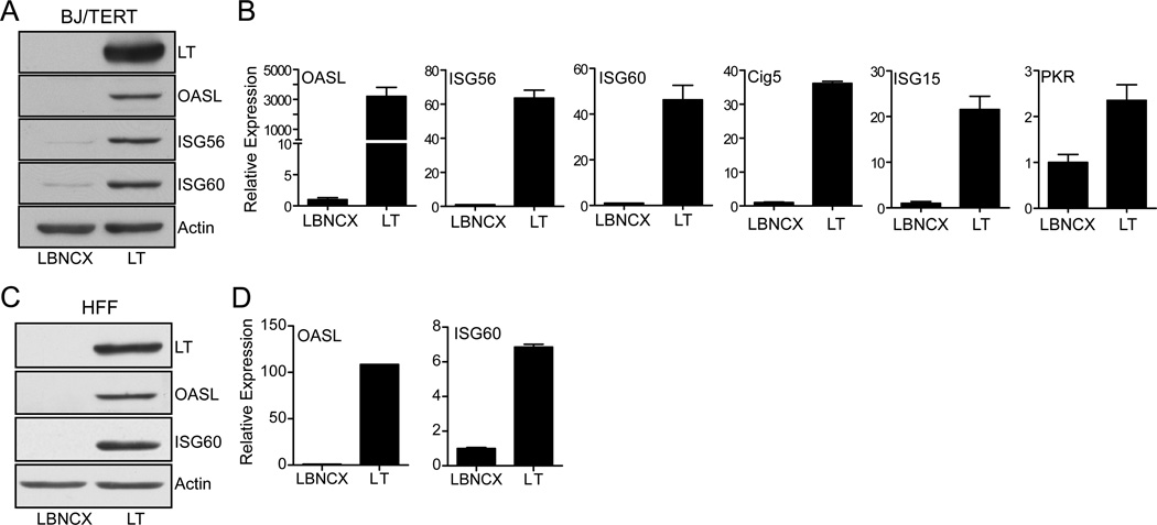
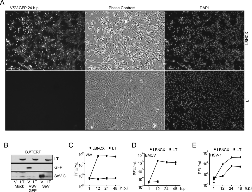
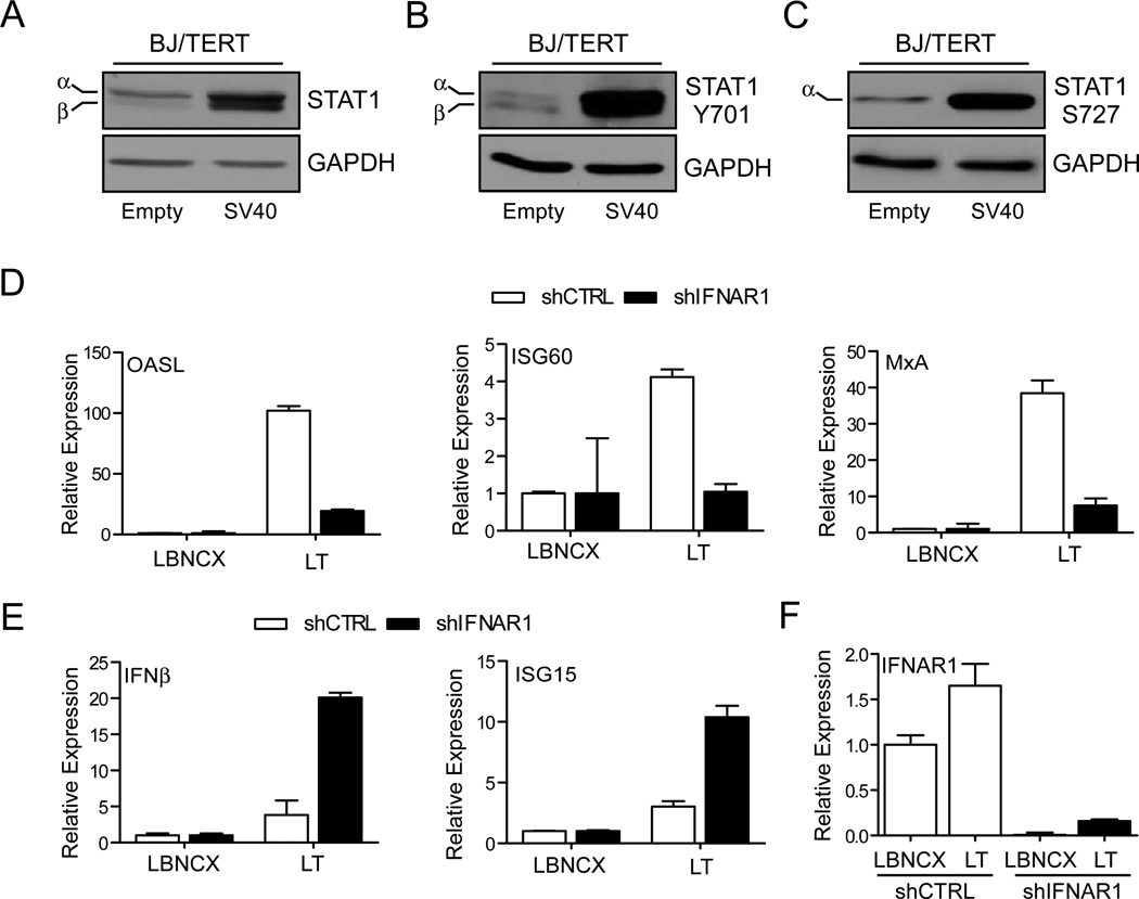
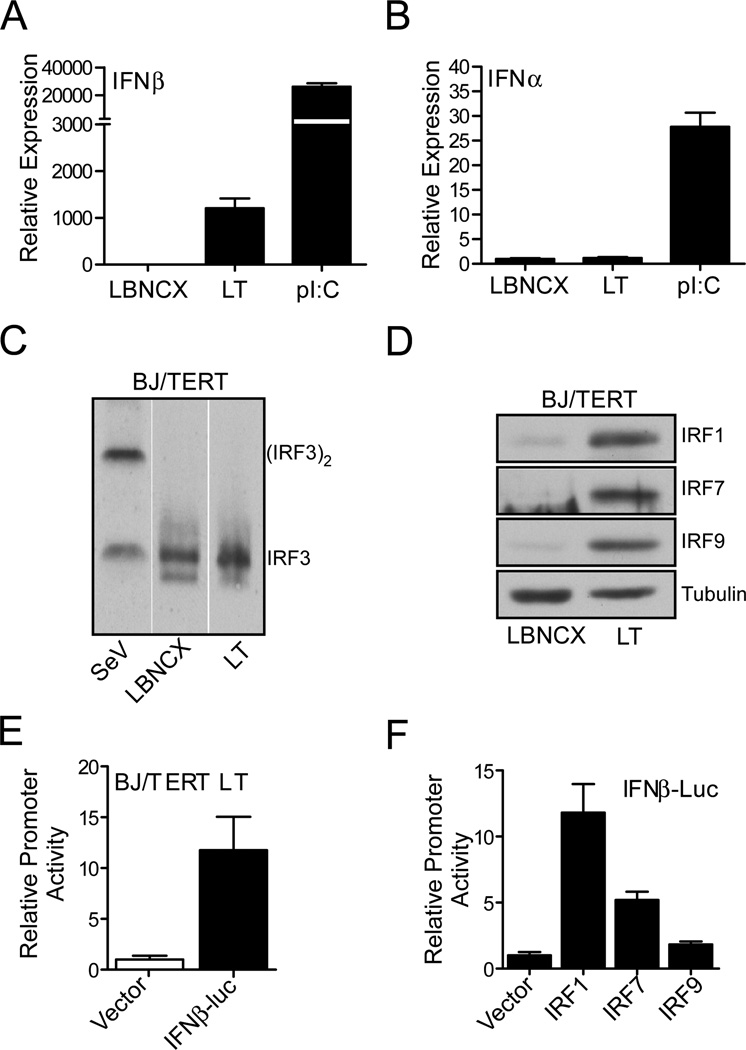
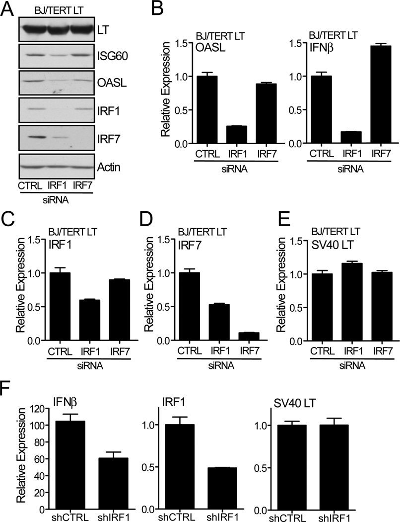
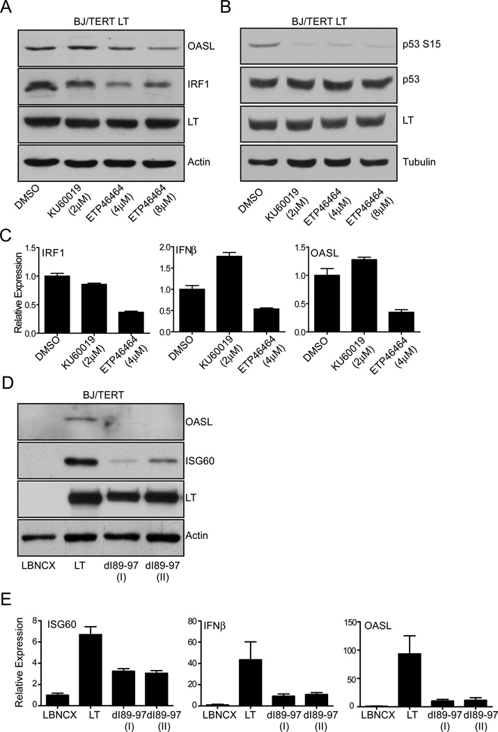
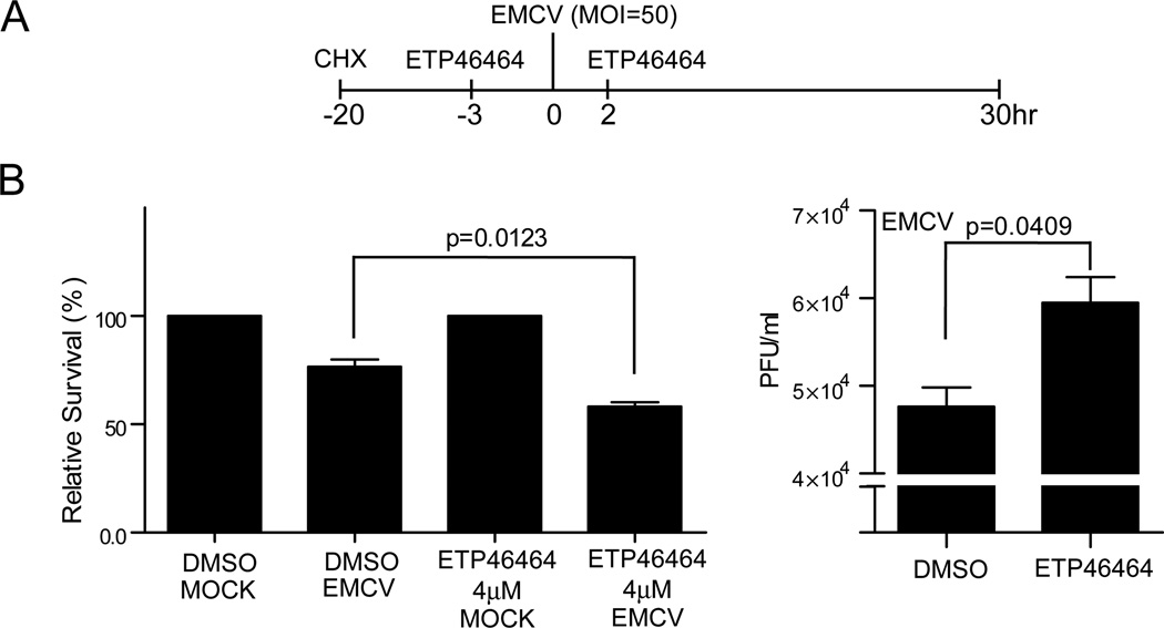
Polyomaviruses encode a large T Ag (LT), a multifunctional protein essential for the regulation of both viral and host cell gene expression and productive viral infection. Previously, we have shown that stable expression of LT protein results in upregulation of genes involved in the IFN induction and signaling pathway. In this study, we focus on the cellular signaling mechanism that leads to the induction of IFN responses by LT. Our results show that ectopic expression of SV40 LT results in the induction of IFN-stimulated genes (ISGs) in human fibroblasts and confers an antiviral state. We describe a LT-initiated DNA damage response (DDR) that activates IFN regulatory factor 1, causing IFN-β production and consequent ISG expression in human cells. This IFN-β and ISG induction is dependent on ataxia-telangiectasia mutated and Rad3-related (ATR) kinase, but independent of ATM. ATR kinase inhibition using a selective kinase inhibitor (ETP-46464) caused a decrease in IFN regulatory factor 1 stabilization and ISG expression. Furthermore, expression of a mutant LT that does not induce DDR also does not induce IFN-β and ISGs. These results show that, in the absence of viral infection, LT-initiated activation of ATR-dependent DDR is sufficient for the induction of an IFN-β-mediated innate immune response in human cells. Thus, we have uncovered a novel and critical role for ATR as a mediator of antiviral responses utilizing LT.


Conflict of interest statement

CONFLICT OF INTEREST

The authors declare that they have no conflict of interest.

Figures

Fig. 1
Fig. 1. SV40 Large T antigen induces ISGs
(A) Induction of ISG protein expression in human fibroblasts. Lysates were prepared from BJ/TERT cells transduced by retrovirus infection with SV40 Large T antigen (LT) cDNA or empty vector (LBNCX) and probed with antibodies against LT, OASL, ISG56, IS60, and Actin. (B) Transcriptional induction of ISG in human fibroblasts. Total RNA was extracted from BJ/TERT cells stably expressing SV40 LT cDNA or empty vector (described above). OASL, ISG56, and IS60, Cig5, ISG15, and PKR mRNA were assessed by quantitative RT-PCR. Expression of mRNA was normalized to the housekeeping gene RPL32 and expressed as fold change compared to empty vector expressing cells (value 1). (C) Induction of ISG protein expression in primary human fibroblasts (HFF). Lysates were prepared from primary HFF cells transduced by retrovirus infection with SV40 Large T antigen (LT) cDNA or empty vector and probed with antibodies against LT, OASL, IS60, and Actin. (D) Transcriptional induction of ISG60 and OASL in primary human fibroblasts. Total RNA was extracted from primary HFF cells stably expressing SV40 LT cDNA or empty vector. ISG60 and OASL mRNA was measured by qRT-PCR. Expression of mRNA was normalized to RPL32 and expressed as fold change compared to empty vector expressing cells (value 1).
Fig. 2
Fig. 2. Expression of LT induces an antiviral state
(A) GFP expression in VSV-GFP infected BJ/TERT cells. BJ/TERT cells expressing SV40 LT cDNA or vector control were infected with VSV-GFP (m.o.i 1) for 24hrs. GFP expression was detected by fluorescence microscopy (left), cell morphology was examined by phase contrast microscopy (middle). Nuclei were stained with DAPI (right). (B) Analysis of viral protein expression in infected BJ/TERT cells. BJ/TERT cells expressing SV40 LT cDNA or vector control were infected with VSV-GFP (m.o.i. 5) or SeV (200 HAU/ml) for 24 hrs. Lysates were prepared and probed with antibodies against LT, GFP, and SeV C-protein. (C) Analysis of VSV growth in BJ/TERT cells. BJ/TERT cells expressing SV40 LT cDNA or vector control were infected with VSV-GFP (m.o.i. 5). Supernatants were harvested at the indicated time-points and virus replication was measured by plaque assay on BHK21 cells. Virus growth is expressed as plaque forming units (PFU)/ml. (D) Analysis of EMCV growth in BJ/TERT cells. BJ/TERT cells expressing SV40 LT cDNA or vector control were infected with EMCV (m.o.i. 5). Supernatants were harvested at the indicated time-points and virus replication was measured by plaque assay on Vero cells. Virus growth is expressed as PFU/ml. (E) Analysis of HSV-1 growth in BJ/TERT cells. BJ/TERT cells expressing SV40 LT or vector control were infected with HSV-1 (K26-GFP) (m.o.i. 5). Supernatants were harvested at the indicated time-points and virus replication was measured by plaque assay on Vero cells. Virus growth is expressed as PFU/ml.
Fig. 3
Fig. 3. IFNAR-dependent and -independent induction of ISGs
(A–C) STAT1 activation in human fibroblasts by LT expression. BJ/TERT cells were transfected with SV40 early region or vector and placed under selection with Puromycin (1µg/ml) for 7 days. Whole cell extracts were prepared from antibiotic resistant cells. Lysates were probed with antibodies against total STAT1 (A), STAT1 Y701 (B), STAT1 S727 (C), and GAPDH. (D) Analysis of ISG induction in IFNAR1 knockdown cells. BJ/TERT cells were infected with lentivirus encoding a short hairpin targeting IFNAR1 or scrambled control. Cells were then selected with puromycin (1 µg/ml). Puromycin resistant cells were then infected with a retrovirus encoding the SV40 LT cDNA or empty control (Vector) and selected for resistance to blasticidin (5 µg/ml). Total RNA was harvested and expression of OASL, ISG60, and MxA mRNA was analyzed by qRT-PCR. Samples were normalized to RPL32 and expressed as fold change with respect to vector control cells (value 1). (E) Analysis of IFNβ and ISG15 induction in IFNAR1 knockdown cells. RNA was harvested from BJ/TERT cells expressing either empty vector or SV40 LT cDNA as well as a short hairpin targeting IFNAR1 (described above). Expression of IFNβ and ISG15 mRNA was analyzed by qRT-PCR. Samples were normalized to RPL32 and expressed as fold change with respect to vector control cells (value 1). (F) Validation of IFNAR1 knockdown in BJ/TERT cells. RNA was harvested from BJ/TERT cells expressing either empty vector or SV40 LT cDNA as well as a short hairpin targeting IFNAR1. Expression of IFNAR1 mRNA was analyzed by qRT-PCR. Samples were normalized to RPL32 and expressed as fold change with respect to vector control cells expressing the non-targeting shRNA (value 1).
Fig. 4
Fig. 4. SV40 Large T antigen induces IFNβ expression
(A and B) SV40LT induction of type I IFN mRNA. BJ/TERT cells were stimulated with 100 mg/ml poly(I):(C) for 24hrs or stably transduced with empty vector or SV40 LT cDNA by retroviral infection and blasticidin selection. Total RNA was extracted from 1×10⁷ cells and IFNβ (A) and Pan-IFNα (B) mRNA levels were analysed by qRT-PCR. mRNA expression was normalized to RPL32 and expressed as fold change relative to vector control expressing cells (value 1). (C) IRF3 dimerization assay. Lysates were prepared from BJ/TERT cells stably expressing vector control, SV40 LT cDNA, or cells infected with 300 HAU/ml for 8hrs. Protein was resolved by Native PAGE and probed with anti-IRF3 antibodies. (D) Modulation of IRF gene expression by SV40 LT. Lysates were prepared from BJ/TERT cells stably expressing vector control or SV40 LT cDNA and probed with antibodies against the antiviral mediators IRF1, IRF7, and IRF9 as well as Tubulin. (E) SV40 LT expression induces the IFNβ promoter activity. BJ/TERT LT cells (4×10⁵) were transiently transfected with 1 µg of IFNβ luciferase reporter construct or pGL3 basic vector control using Lipofectamine 2000 at a 1:2 DNA to transfection reagent ratio. Six hours post transfection, fresh culture medium was added to cells and cells were incubated for 48hrs prior to measurement of luciferase activity. Firefly luciferase was measured by Dual-luciferase assay (Promega) and normalized to empty vector expressing cells (value 1). (F) Induction of IFNβ promoter activity by IRF proteins. HEK293T (1.5×10⁵) cells were co-trasnfected with pcDNA, IRF1, IRF7, or IRF9 cDNA (250ng), IFNβ125-luc (400ng) and pRL-null (50ng). 24hrs post transfection cells were collected by trypsin-EDTA transfection and seeded in white-walled 96-well plates. Twenty four hours later, firefly and renilla luciferase activity was measure by Dual-luciferase assay (Promega). Firefly luciferase activity was normalized to renilla luciferase activity and promoter activity was expressed as fold change relative to vector control transfected cells (value 1).
Fig. 5
Fig. 5. Induction of IRF1 transactivates IFNβ
(A) Requirement for IRF1 in the induction of ISG protein synthesis. Briefly, BJ/TERT LT cells were plated in 60 mm dishes and transfected with 160 pmoles of siRNAs pools targeting IRF1 (target sequences; 5’-CGTGTGGATCTTGCCACATTT -3’ and 5’-CCTCTGTCTATGGAGACTTTA -3’), IRF7 (M-011810-02-0005, Thermo Fisher, Pittsburgh, PA) or control (D-001220-01-05, Thermo Fisher) using 6 µl of Lipofectamine RNAiMAX (Invitrogen). After 48 hrs, lysates were prepared and probed with antibodies against OASL, ISG60, SV40 LT, IRF1, IRF7, and Actin. (B–E) IRF1 and IRF7 requirement for the transcriptional induction of IFNβ and OASL. IFNβ (B), IRF1 (C), IRF7 (D), and SV40 LT (E) mRNA levels were assessed by qRT-PCR. BJ/TERT LT cells were transfected with siRNA against IRF1 and IRF7 as described above. Total RNA was harvested and cDNA was synthezised. Expression of target genes was determined by qRT-PCR analysis and normalized to RPL32 and control siRNA transfected cells (value 1). (F) Analysis of IFNβ mRNA expression in IRF1 knockdown cells. BJ/TERT cells were stably transduced with an shRNA targeting IRF1 or a non-targeting shRNA. Cells were then stably transduced with vector control or LT encoding retrovirus. Total RNA was harvested and mRNA expression was determined by qRT-PCR. Expression of target genes was normalized to RPL32 and reported relative to the enhancement observed over empty vector expressing cells (value 1).
Fig. 6
Fig. 6. ATR kinase activity is necessary for the induction of IFNβ and subsequent ISG expression
(A) Analysis of ISG induction in ATM/ATR kinase inhibitor treated BJ/TERT LT cells. Lysates were prepared from BJ/TERT LT cells stimulated for 24hrs with the indicated doses of KU60019 or ETP46464. OASL, IRF1, LT, and Actin proteins were detected by immunoblot analysis. (B) Analysis of p53 phosphorylation in ATM/ATR kinase inhibitor treated BJ/TERT LT cells. Cells were treated as described above. Lysates were prepared, and probed with antibodies against phosphorylated p53 (Serine 15), total p53, and Actin. (C) Analysis of IFNβ mRNA in ATM/ATR kinase inhibitor treated BJ/TERT LT cells. Total RNA was prepared from BJ/TERT cells stimulated for 24hrs with 2 µM KU60019 or 4 µM ETP-46464. Expression of IFNβ, IRF1, and OASL mRNA expression was determined by qRT-PCR. Expression of target genes was normalized to the housekeeping gene RPL32 and reported relative to the enhancement observed over empty vector expressing cells (value 1). (D) Analysis of induction of ISGs by LT mutants defective in DDR induiction. Lysates were prepared from BJ/TERT cells stably expressing either the wt LT or LT dI89-97. Expression of OASL, ISG60, LT, and Actin was detected by immunoblot analysis. (E) Analysis of type I IFN and ISG mRNA induction by LT mutant. Total RNA was harvested from BJ/TERT cells expressing LT, dI89-97, or empty vector. ISG60, IFNβ, and OASL mRNA expression was determined by qRT-PCR. Expression of target genes was normalized to the housekeeping gene RPL32 and reported relative to the enhancement observed over empty vector expressing cells (value 1).
Fig. 7
Fig. 7. ATR kinase activity is necessary to generate and antiviral state
(A and B) EMCV growth in BJ/TERT LT cells treated with ETP-46464. In brief, BJ/TERT LT cells were plated in 12-well plates (80% confluency). Cells were treated with CHX (50 ng/ml) for 17hrs prior to viral infection. Three hours prior to EMCV infection, cells were treated with 5 µM of ETP-46464. Cells were then infected with EMCV (m.o.i 50) (A). Infected cells were stained with crystal violet to determine cell survival relative to mock-infected cells. Briefly, Infected cells were stained with 0.1% crystal violet (10% ethanol) overnight. Plates were washed to remove excess stain with distilled water and left to air-dry overnight. The retained crystal violet was solubilized in 600 µl 2% SDS solution in PBS for 30 minutes at room temperature with constant shaking. Absorbance at 550 nm was then used to determine crystal violet retention (B, left). Supernatants were harvested 30hrs post infection and viral growth in DMSO or ATR kinase inhibitor treated infected cells was determined by plaque assay on Vero cells (B, right).

Similar articles

Cited by

References

    1. Zurhein G, Chou SM. Particles Resembling Papova Viruses in Human Cerebral Demyelinating Disease. Science. 1965;148:1477–1479. - PubMed
    1. Padgett BL, Walker DL, ZuRhein GM, Eckroade RJ, Dessel BH. Cultivation of papova-like virus from human brain with progressive multifocal leucoencephalopathy. Lancet. 1971;1:1257–1260. - PubMed
    1. Bennett SM, Broekema NM, Imperiale MJ. BK polyomavirus: emerging pathogen. Microbes Infect. 2012;14:672–683. - PMC - PubMed
    1. Pipas JM. SV40: Cell transformation and tumorigenesis. Virology. 2009;384:294–303. - PubMed
    1. Marechal A, Zou L. DNA damage sensing by the ATM and ATR kinases. Cold Spring Harb Perspect Biol. 2013;5 - PMC - PubMed

Publication types

MeSH terms

Substances