. 2014 Jul;82(7):3002-14.
doi: 10.1128/IAI.01664-14. Epub 2014 May 5.

Bile acids and bicarbonate inversely regulate intracellular cyclic di-GMP in Vibrio cholerae

Affiliations

Bile acids and bicarbonate inversely regulate intracellular cyclic di-GMP in Vibrio cholerae

Benjamin J Koestler et al. Infect Immun. 2014 Jul.

Abstract

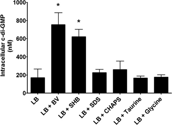
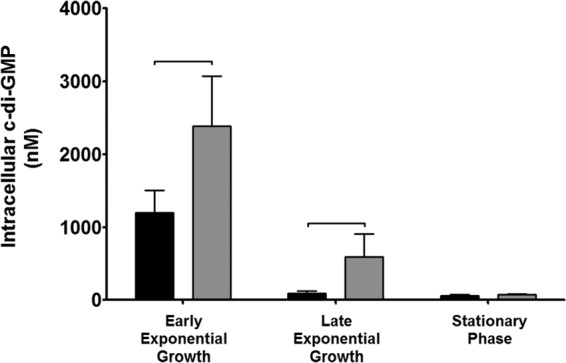
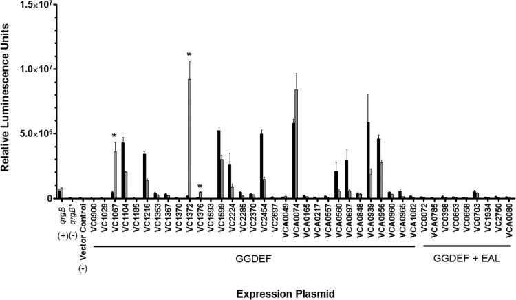
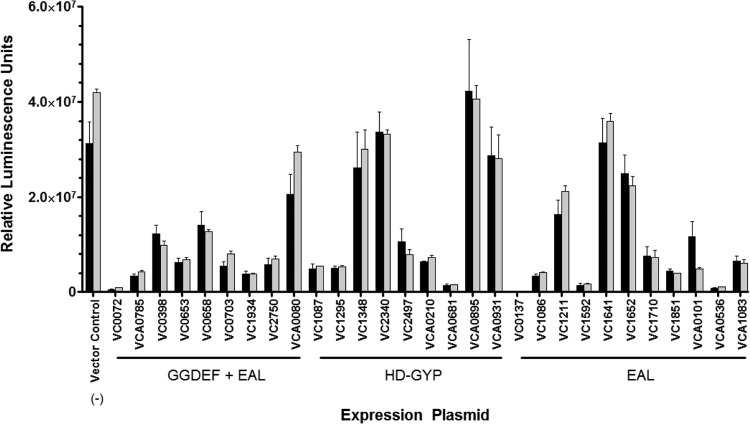
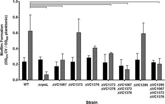
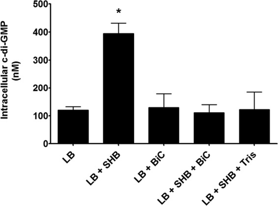
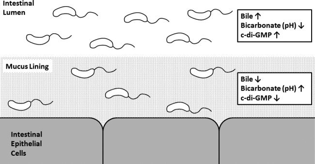
Vibrio cholerae is a Gram-negative bacterium that persists in aquatic reservoirs and causes the diarrheal disease cholera upon entry into a human host. V. cholerae employs the second messenger molecule 3',5'-cyclic diguanylic acid (c-di-GMP) to transition between these two distinct lifestyles. c-di-GMP is synthesized by diguanylate cyclase (DGC) enzymes and hydrolyzed by phosphodiesterase (PDE) enzymes. Bacteria typically encode many different DGCs and PDEs within their genomes. Presumably, each enzyme senses and responds to cognate environmental cues by alteration of enzymatic activity. c-di-GMP represses the expression of virulence factors in V. cholerae, and it is predicted that the intracellular concentration of c-di-GMP is low during infection. Contrary to this model, we found that bile acids, a prevalent constituent of the human proximal small intestine, increase intracellular c-di-GMP in V. cholerae. We identified four c-di-GMP turnover enzymes that contribute to increased intracellular c-di-GMP in the presence of bile acids, and deletion of these enzymes eliminates the bile induction of c-di-GMP and biofilm formation. Furthermore, this bile-mediated increase in c-di-GMP is quenched by bicarbonate, the intestinal pH buffer secreted by intestinal epithelial cells. Our results lead us to propose that V. cholerae senses distinct microenvironments within the small intestine using bile and bicarbonate as chemical cues and responds by modulating the intracellular concentration of c-di-GMP.


Figures

FIG 1
FIG 1
V. cholerae was grown in LB medium and LB medium with bovine bile (BV), synthetic human bile (SHB), the detergents SDS and CHAPS, and the amino acids taurine and glycine. Intracellular c-di-GMP was measured using LC-MS/MS. The reported values indicate the means. Error bars indicate standard deviations, and asterisks indicate statistical significance compared to LB medium as determined by a Student two-tailed t test (n = 3, P < 0.05).
FIG 2
FIG 2
The intracellular c-di-GMP concentration of wild-type V. cholerae was quantified over the course of growth. All intracellular c-di-GMP concentrations were determined using LC-MS/MS. Black bars indicate strains grown in LB medium, and gray bars indicate strains grown in LB medium with SHB. The reported values indicate the means, and the error bars indicate the standard deviations. Brackets indicate statistical significance as determined by a one-tailed Student t test (n = 3, P > 0.05).
FIG 3
FIG 3
V. cholerae strains containing a DGC expression plasmid and the 6:C9-lux reporter plasmid were measured for luminescence in LB medium (black bars) and LB medium with SHB (gray bars). Expression plasmids carrying qrgB and its mutant allele counterpart, qrgB*, were included as positive and negative controls, respectively. The vector control indicates expression of 6:C9-lux in the absence of protein induction. The error bars indicate standard deviations. Statistical significance (*) was determined for cultures exhibiting a positive fold change (LB medium + SHB/LB medium) greater than 2 as determined by a Student one-tailed t test (n = 3, P < 0.01).
FIG 4
FIG 4
V. cholerae strains containing a PDE expression plasmid, the 6:C9-lux reporter plasmid, and the qrgB expression plasmid were measured for luminescence in LB medium (black bars) and LB medium with SHB (gray bars). The vector control indicates expression of 6:C9-lux with qrgB induction and without PDE expression. The error bars indicate standard deviations (n = 3).
FIG 5
FIG 5
The predicted hydrophobicity of the amino acid sequence of VC1067 (A), VC1372 (B), and VC1376 (C) was used to predict transmembrane domains using the toppred program (left). The dotted line indicates the cutoff value for a potential transmembrane domain. A depiction of the potential N-terminal signaling domains and transmembrane domains (black bars) and the C-terminal GGDEF domain of each DGC is shown (right). The images were created using the SMART database (57).
FIG 6
FIG 6
Intracellular levels of c-di-GMP in V. cholerae expressing an empty vector or DGC VC1067, VC1372, or VC1376 quantified with LC-MS/MS. The intracellular c-di-GMP levels of strains expressing alleles containing mutations in the active site motif of each DGC were also quantified. The black bars indicate strains grown in LB medium, while the gray bars indicate strains grown in LB medium with SHB. Error bars indicate standard deviations. Brackets indicate statistical significance, which was determined using a Student one-tailed t test (n = 3, P < 0.05).
FIG 7
FIG 7
V. cholerae strains containing a transcriptional fusion of each HD-GYP promoter to luciferase were grown in LB medium (black) or LB medium with SHB (gray). Luminescence was quantified after 6 h of growth (n = 4) under each environmental condition. Each culture was normalized to an OD600 reading. Error bars indicate standard deviations. The asterisk indicates a statistically significant differences from the LB medium condition, determined by a one-tailed Student t test (P < 0.05).
FIG 8
FIG 8
The intracellular c-di-GMP concentrations of wild-type V. cholerae and the DGC and PDE mutant strains were quantified after growth in LB medium (black bars) or LB medium with SHB (gray bars) using LC-MS/MS. The reported values indicate the means, and the error bars indicate standard deviations. Brackets indicate statistical significance as determined by a one-tailed Student t test (n = 9 to 10, P < 0.05).
FIG 9
FIG 9
The biofilm formation of wild-type V. cholerae and V. cholerae DGC and PDE mutants was quantified in test tubes using crystal violet (CV). A strain containing a mutation in the vpsL gene was included as a negative control. The CV value was normalized by the OD600 value of the planktonic culture to account for differences in growth. The reported values indicate the means, and error bars indicate standard deviations from the means. Brackets indicate statistical significance as determined by a Student one-tailed t test (n = 3, P < 0.05).
FIG 10
FIG 10
The intracellular c-di-GMP concentration of wild-type V. cholerae was quantified in LB medium, LB medium with bicarbonate (BiC), or LB medium with SHB and different supplements using LC-MS/MS. The reported values indicate the means, and the error bars indicate standard deviations. The asterisk indicates statistical significance compared to LB medium as determined by a one-tailed Student t test (n = 3, P > 0.05).
FIG 11
FIG 11
Proposed model of V. cholerae c-di-GMP regulation in the human small intestine. c-di-GMP is elevated in the lumen, where the concentration of bile is elevated and the concentrations of bicarbonate and pH are low. Upon entry into the mucosal layer, where bile is low and the bicarbonate concentration and pH are elevated, c-di-GMP is repressed.

References

    1. Halpern M, Broza YB, Mittler S, Arakawa E, Broza M. 2004. Chironomid egg masses as a natural reservoir of Vibrio cholerae non-O1 and non-O139 in freshwater habitats. Microb. Ecol. 47:341–349. 10.1007/s00248-003-2007-6 - DOI - PubMed
    1. Huq A, Small EB, West PA, Huq MI, Rahman R, Colwell RR. 1983. Ecological relationships between Vibrio cholerae and planktonic crustacean copepods. Appl. Environ. Microbiol. 45:275–283 - PMC - PubMed
    1. Tamplin ML, Gauzens AL, Huq A, Sack DA, Colwell RR. 1990. Attachment of Vibrio cholerae serogroup-O1 to zooplankton and phytoplankton of Bangladesh waters. Appl. Environ. Microbiol. 56:1977–1980 - PMC - PubMed
    1. Tischler AD, Camilli A. 2004. Cyclic diguanylate (c-di-GMP) regulates Vibrio cholerae biofilm formation. Mol. Microbiol. 53:857–869. 10.1111/j.1365-2958.2004.04155.x - DOI - PMC - PubMed
    1. Simm R, Morr M, Kader A, Nimtz M, Romling U. 2004. GGDEF and EAL domains inversely regulate cyclic di-GMP levels and transition from sessility to motility. Mol. Microbiol. 53:1123–1134. 10.1111/j.1365-2958.2004.04206.x - DOI - PubMed

Publication types

MeSH terms

LinkOut - more resources