. 2014 May 20;111(20):7385-90.
doi: 10.1073/pnas.1403252111. Epub 2014 May 5.

Caspase-8 mediates caspase-1 processing and innate immune defense in response to bacterial blockade of NF-κB and MAPK signaling

Affiliations

Caspase-8 mediates caspase-1 processing and innate immune defense in response to bacterial blockade of NF-κB and MAPK signaling

Naomi H Philip et al. Proc Natl Acad Sci U S A. .

Abstract

Toll-like receptor signaling and subsequent activation of NF-κB- and MAPK-dependent genes during infection play an important role in antimicrobial host defense. The YopJ protein of pathogenic Yersinia species inhibits NF-κB and MAPK signaling, resulting in blockade of NF-κB-dependent cytokine production and target cell death. Nevertheless, Yersinia infection induces inflammatory responses in vivo. Moreover, increasing the extent of YopJ-dependent cytotoxicity induced by Yersinia pestis and Yersinia pseudotuberculosis paradoxically leads to decreased virulence in vivo, suggesting that cell death promotes anti-Yersinia host defense. However, the specific pathways responsible for YopJ-induced cell death and how this cell death mediates immune defense against Yersinia remain poorly defined. YopJ activity induces processing of multiple caspases, including caspase-1, independently of inflammasome components or the adaptor protein ASC. Unexpectedly, caspase-1 activation in response to the activity of YopJ required caspase-8, receptor-interacting serine/threonine kinase 1 (RIPK1), and Fas-associated death domain (FADD), but not RIPK3. Furthermore, whereas RIPK3 deficiency did not affect YopJ-induced cell death or caspase-1 activation, deficiency of both RIPK3 and caspase-8 or FADD completely abrogated Yersinia-induced cell death and caspase-1 activation. Mice lacking RIPK3 and caspase-8 in their hematopoietic compartment showed extreme susceptibility to Yersinia and were deficient in monocyte and neutrophil-derived production of proinflammatory cytokines. Our data demonstrate for the first time to our knowledge that RIPK1, FADD, and caspase-8 are required for YopJ-induced cell death and caspase-1 activation and suggest that caspase-8-mediated cell death overrides blockade of immune signaling by YopJ to promote anti-Yersinia immune defense.

Keywords: apoptosis; innate immunity; macrophage; programmed necrosis; pyroptosis.


Conflict of interest statement

The authors declare no conflict of interest.

Figures
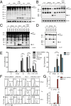
Fig. 1.
Fig. 1.
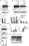
RIPK1 is required for Yersinia-induced caspase-1 processing and cell death. (A and B) B6 BMDMs were either left uninfected (UI), LPS-primed (50 ng/mL) for 3 h followed by ATP (2.5 mM) for 1 h (LPS+ATP), or infected with WT Yersinia (Yp) or YopJ-deficient Yp (ΔYopJ) for 2 h or Salmonella (STm) for 1 h. Cell lysates were probed for caspase-1 or -8 processing by Western analysis. (C and D) Ripk1+/+ and Ripk1−/− fetal liver-derived macrophages (FLDMs) were infected as in A and B. (E) Percent cytotoxicity was measured by LDH release from B6 BMDMs (Left) and Ripk1+/+ or Ripk1−/− FLDMs (Right) that were uninfected or infected with ΔYopJ, Yp, or LPS+ATP for 4 h, or STm for 1 h. (F) Flow cytometry for PI uptake (percent PI+ cells) by cells as treated in E. (G) IL-18 assayed by ELISA and LDH release on cells treated as in E; 30 μM Nec-1 and 100 μM zVAD-fmk were used 3 h before infection where indicated. Grey peaks indicate uninfected cells. Error bars indicate mean ± SEM of triplicates and are representative of three or more independent experiments. ***P < 0.0001, **P < 0.001, *P < 0.01.
Fig. 2.
Fig. 2.
Caspase-1 activation and IL-18 secretion in response to Yersinia infection require caspase-8. (A and B) Lysates from BMDMs left uninfected (UI), infected with Yp for 2 h, STm for 1 h, or LPS-primed for 3 h and ATP for 1 h (LPS+ATP) were probed for caspase-1 processing by Western analysis. (C) Percent cytotoxicity (LDH release) of BMDMs infected with Yp for 4 h, STm for 1 h, or treated with LPS+ATP as in A. (D) BMDMs treated with 4-hydroxytamoxifen (4-OHT) (50 nM) and infected as in A. (E) IL-18 assayed by ELISA from BMDMs left uninfected (UI), infected with isogenic Yp 32777, YopJ C172A, or STm, or LPS-primed for 4 h. (F and G) iR3−/−C8−/− BMDMs were reconstituted with empty vector, WT caspase-8 (WT C8), or noncleavable mutant caspase-8 (D3A C8). (F) LDH release 4 h postinfection. (G) Caspase-1 processing by Western analysis, MOI of 50:1; 30 μM Nec-1 and 100 μM zVAD-fmk were used 3 h before infection where indicated. N.D., not detected. Error bars indicate mean ± SEM of triplicates and are representative of three or more independent experiments. ***P < 0.0001.
Fig. 3.
Fig. 3.
FADD is required for Yersinia-induced caspase-1 processing and cell death in the absence of RIPK3. (A and B) BMDM cell lysates were probed for caspase-1 by Western analysis after infection with ΔYopJ or Yp for 2 h, STm for 1 h, treated with LPS for 3 h and ATP for 1 h (LPS+ATP), or left uninfected (UI). (C) Percent cytotoxicity (LDH release) of BMDMs left uninfected, infected with ΔYopJ or Yp for 4 h, STm for 1 h, or LPS+ATP as in A. N.D., not detected. Error bars indicate mean ± SEM of triplicates and are representative of three or more independent experiments. ***P < 0.0001.
Fig. 4.
Fig. 4.
RIPK3/caspase-8–deficient mice are highly susceptible to Yersinia infection and have dysregulated cytokine production. Lethally irradiated B6.SJL mice were reconstituted with B6, Ripk3+/−Casp8+/−, Ripk3−/−Casp8+/−, Ripk3−/−, or Ripk3−/−Casp8−/− BM and orally infected with 8–10 × 107 cfu Yersinia (Yp) per mouse. Mice were assayed for (A) survival, (B) serum IFN-γ, IL-6, IL-1β, and MCP-1 by Luminex, (C) bacterial loads per gram of tissue, and (D) percent TNF+ inflammatory monocytes in mLNs by flow cytometry. (E–H) IFN-γ production from splenic NK cells (NK1.1+) was assessed by flow cytometry. (E) Representative plots, (F) total numbers, and (G) mean fluorescence intensity (MFI). (B–G) Day 6 postinfection. Dagger denotes 13 dead or moribund mice not harvested for cfus. Dotted lines represent limit of detection. Solid lines represent means (ELISA) or geometric means (cfu). Flow cytometry plots in D were gated on live CD45.2+, CD11c, CD11bhi, Ly6G, and Ly6Chi cells and in E–G on live CD45.2+ CD3 cells. R3C8, Ripk3Casp8. Statistical analysis in F and G was performed using the Mann–Whitney U test. Data representative of two or more independent experiments.
Fig. 5.
Fig. 5.
RIPK3/caspase-8–deficient mice survive ΔYopJ infection and partially recover intracellular cytokine production. Lethally irradiated B6.SJL mice were reconstituted with B6, Ripk3−/−, or Ripk3−/−Casp8−/− BM and orally infected with 8–10 × 107 cfu Yersinia (Yp) or ΔYopJ Yersinia (ΔYopJ) per mouse. Mice were assayed for (A) survival, (B) bacterial loads per gram of tissue, and (C) percent TNF+ inflammatory monocytes in mLNs by flow cytometry. (D and E) Representative images of H&E-stained (D) spleen and (E) liver sections in naïve and infected chimeric mice, showing lesions of necrosuppurative inflammation (dashed white lines) and extracellular bacterial colonies (arrows) in infected mice. (Scale bars, 50 μm.) (F and G) Quantification of number and extent of lesions. (B) Day 5. (C) Representative plots from pooled data on days 5 and 6 (% ± SEM). (D–G) Day 6. Dagger denotes five dead or moribund mice not harvested for cfus. Dotted lines represent limit of detection. Solid lines represent means (histology) or geometric means (cfu). Flow cytometry plots were gated on live CD45.2+, CD11c, CD11bhi, Ly6G, Ly6Chi cells. **P < 0.01, WT vs. ΔYopJ infected Ripk3−/−Casp8−/− monocytes. R3C8, Ripk3Casp8. Data representative of two or more independent experiments.

References

    1. Medzhitov R. Recognition of microorganisms and activation of the immune response. Nature. 2007;449(7164):819–826. - PubMed
    1. Broz P, Monack DM. Newly described pattern recognition receptors team up against intracellular pathogens. Nat Rev Immunol. 2013;13(8):551–565. - PubMed
    1. Mukherjee S, et al. Yersinia YopJ acetylates and inhibits kinase activation by blocking phosphorylation. Science. 2006;312(5777):1211–1214. - PubMed
    1. Orth K, et al. Inhibition of the mitogen-activated protein kinase kinase superfamily by a Yersinia effector. Science. 1999;285(5435):1920–1923. - PubMed
    1. Palmer LE, Hobbie S, Galán JE, Bliska JB. YopJ of Yersinia pseudotuberculosis is required for the inhibition of macrophage TNF-alpha production and downregulation of the MAP kinases p38 and JNK. Mol Microbiol. 1998;27(5):953–965. - PubMed

Publication types

LinkOut - more resources