

Postsurgical pathologies associated with intradural electrical stimulation in the central nervous system: design implications for a new clinical device
- PMID: 24800260
- PMCID: PMC3988712
- DOI: 10.1155/2014/989175
Postsurgical pathologies associated with intradural electrical stimulation in the central nervous system: design implications for a new clinical device
Abstract
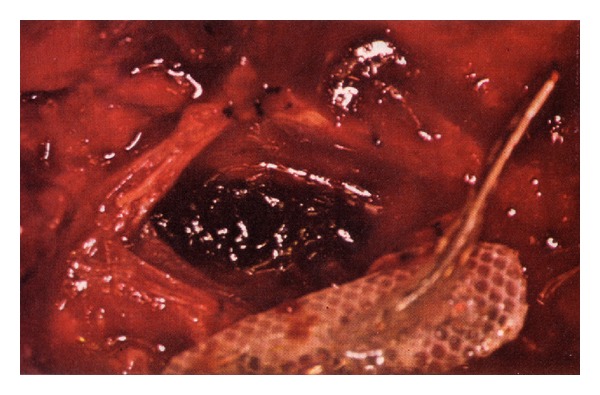
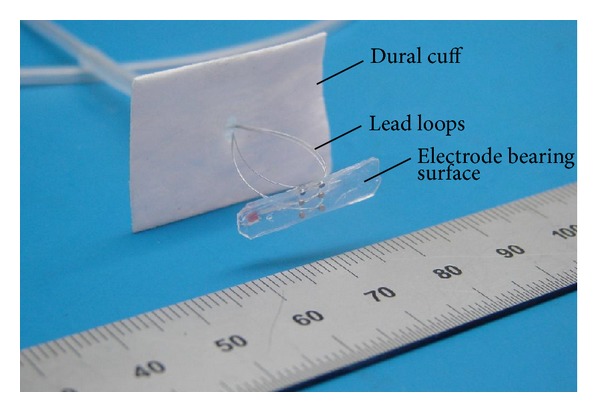
Spinal cord stimulation has been utilized for decades in the treatment of numerous conditions such as failed back surgery and phantom limb syndromes, arachnoiditis, cancer pain, and others. The placement of the stimulating electrode array was originally subdural but, to minimize surgical complexity and reduce the risk of certain postsurgical complications, it became exclusively epidural eventually. Here we review the relevant clinical and experimental pathologic findings, including spinal cord compression, infection, hematoma formation, cerebrospinal fluid leakage, chronic fibrosis, and stimulation-induced neurotoxicity, associated with the early approaches to subdural electrical stimulation of the central nervous system, and the spinal cord in particular. These findings may help optimize the safety and efficacy of a new approach to subdural spinal cord stimulation now under development.
Figures
Similar articles
-
Cervical cord compression due to delayed scarring around epidural electrodes used in spinal cord stimulation.J Neurosurg Spine. 2010 Apr;12(4):409-12. doi: 10.3171/2009.10.SPINE09193. J Neurosurg Spine. 2010. PMID: 20367377
-
Spinal cord stimulation: uses and applications.Neuroimaging Clin N Am. 2010 May;20(2):243-54. doi: 10.1016/j.nic.2010.02.012. Neuroimaging Clin N Am. 2010. PMID: 20439020
-
Spinal cord stimulators: typical positioning and postsurgical complications.AJR Am J Roentgenol. 2011 Feb;196(2):437-45. doi: 10.2214/AJR.10.4789. AJR Am J Roentgenol. 2011. PMID: 21257898
-
Stimulation of the Dorsal Root Ganglion.Prog Neurol Surg. 2015;29:213-24. doi: 10.1159/000434673. Epub 2015 Sep 4. Prog Neurol Surg. 2015. PMID: 26394301 Review.
-
[Treatment of pain syndromes by the technic of chronic electrostimulation of the posterior columns of the spinal cord].Zh Vopr Neirokhir Im N N Burdenko. 1986 Mar-Apr;(2):41-7. Zh Vopr Neirokhir Im N N Burdenko. 1986. PMID: 2939674 Review. Russian. No abstract available.
Cited by
-
Ovine model of neuropathic pain for assessing mechanisms of spinal cord stimulation therapy via dorsal horn recordings, von Frey filaments, and gait analysis.J Pain Res. 2018 Jun 15;11:1147-1162. doi: 10.2147/JPR.S139843. eCollection 2018. J Pain Res. 2018. PMID: 29942150 Free PMC article.
-
Spinal dura mater: biophysical characteristics relevant to medical device development.J Med Eng Technol. 2018 Feb;42(2):128-139. doi: 10.1080/03091902.2018.1435745. Epub 2018 Mar 23. J Med Eng Technol. 2018. PMID: 29569970 Free PMC article. Review.
-
Chronic tissue response to untethered microelectrode implants in the rat brain and spinal cord.J Neural Eng. 2015 Feb;12(1):016019. doi: 10.1088/1741-2560/12/1/016019. Epub 2015 Jan 21. J Neural Eng. 2015. PMID: 25605679 Free PMC article.
-
Developing and Evaluating a Flexible Wireless Microcoil Array Based Integrated Interface for Epidural Cortical Stimulation.Int J Mol Sci. 2017 Feb 5;18(2):335. doi: 10.3390/ijms18020335. Int J Mol Sci. 2017. PMID: 28165427 Free PMC article.
-
Intradural Spinal Cord Stimulation: Performance Modeling of a New Modality.Front Neurosci. 2019 Mar 19;13:253. doi: 10.3389/fnins.2019.00253. eCollection 2019. Front Neurosci. 2019. PMID: 30941012 Free PMC article.
References
-
- Howard MA, III, Utz M, Brennan TJ, et al. Intradural approach to selective stimulation in the spinal cord for treatment of intractable pain: design principles and wireless protocol. Journal of Applied Physics. 2011;110(4)044702
-
- Holsheimer J, Barolat G, Struijk JJ, He J. Significance of the spinal cord position in spinal cord stimulation. Acta Neurochirurgica. 1995;64:119–124. - PubMed
-
- Flouty O, Oya H, Kawasaki H, et al. A new device concept for directly modulating spinal cord pathways: initial in vivo experimental results. Physiological Measurement. 2012;33:2003–2015. - PubMed
-
- Eldabe S, Kumar K, Buchser E, Taylor RS. An analysis of the components of pain, function, and health-related quality of life in patients with failed back surgery syndrome treated with spinal cord stimulation or conventional medical management. Neuromodulation. 2010;13(3):201–209. - PubMed
Publication types
MeSH terms
LinkOut - more resources
Full Text Sources
Other Literature Sources
Medical
