. 2014 May 6;9(5):e96502.
doi: 10.1371/journal.pone.0096502. eCollection 2014.

Immunosuppressive activity of daphnetin, one of coumarin derivatives, is mediated through suppression of NF-κB and NFAT signaling pathways in mouse T cells

Affiliations

Immunosuppressive activity of daphnetin, one of coumarin derivatives, is mediated through suppression of NF-κB and NFAT signaling pathways in mouse T cells

Bocui Song et al. PLoS One. .

Abstract

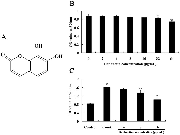
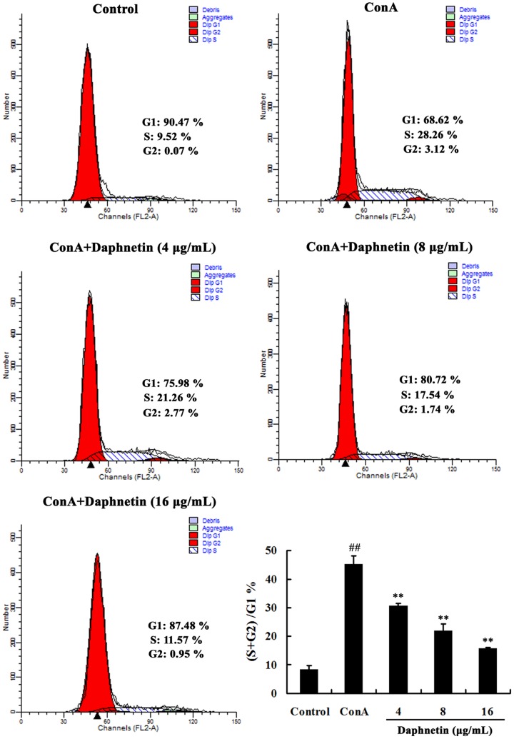
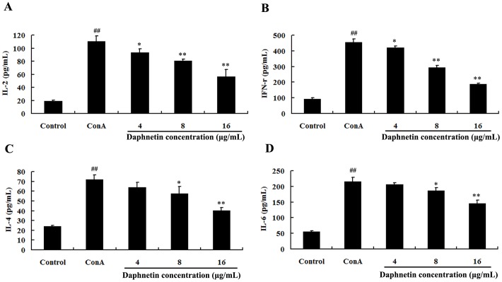
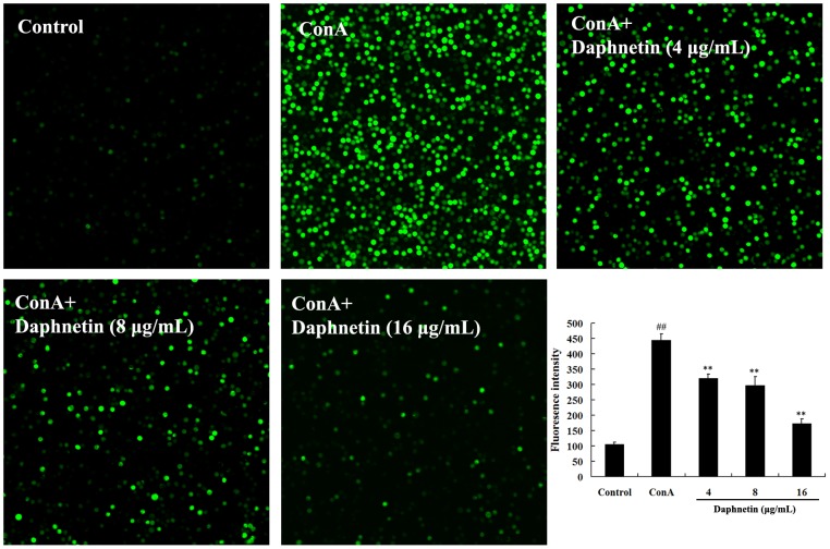
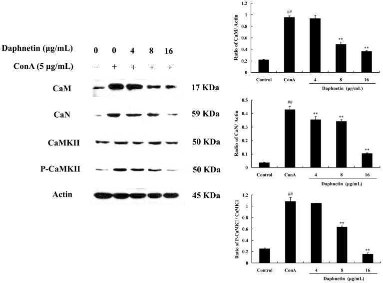
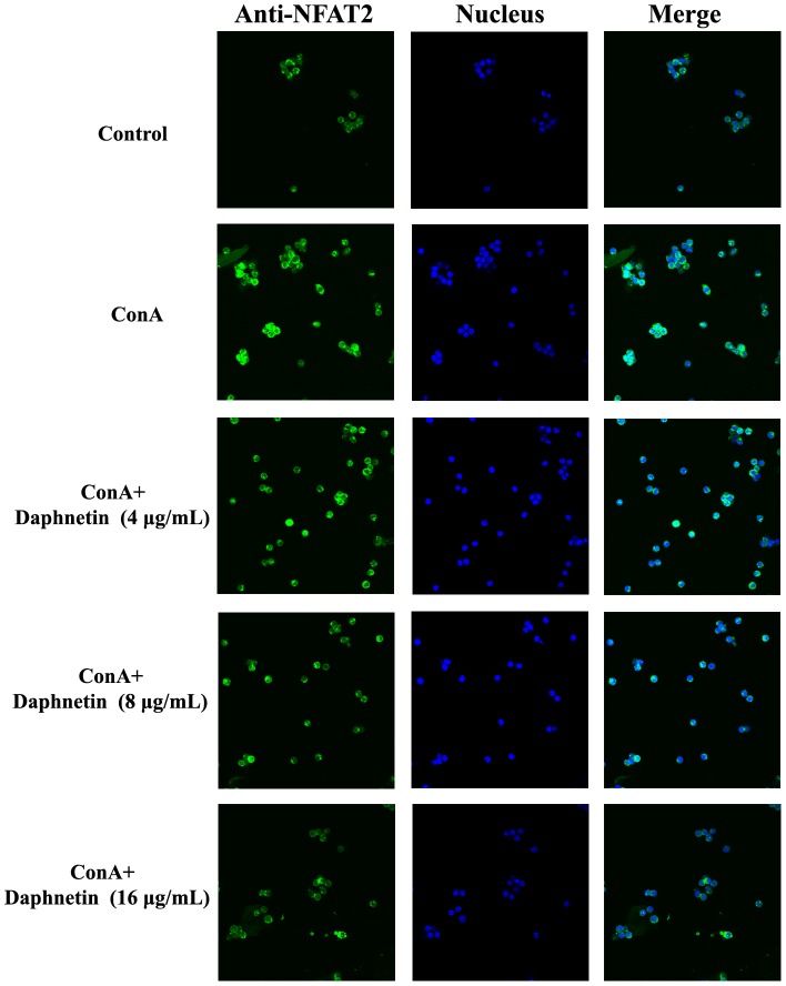
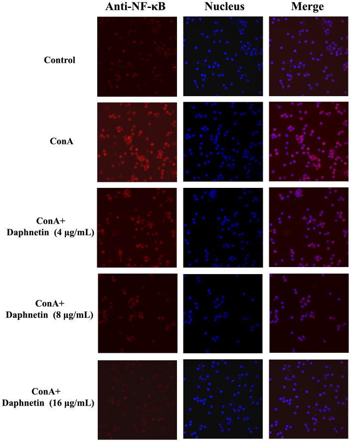
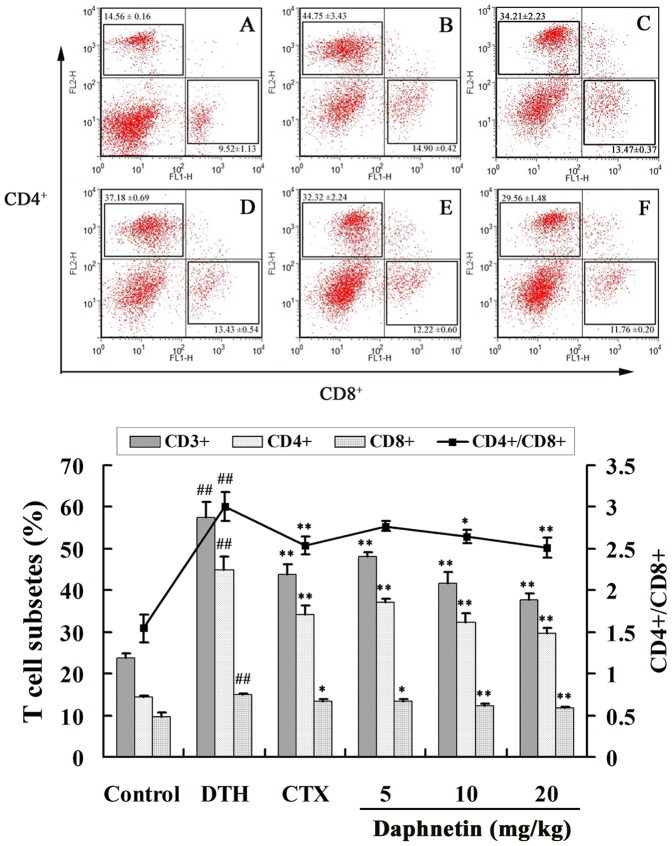
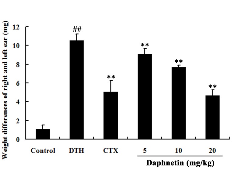
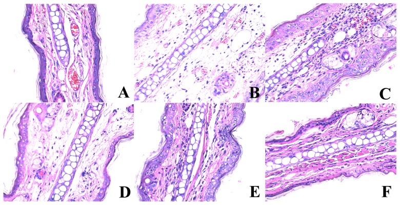
Daphnetin, a plant-derived dihydroxylated derivative of coumarin, is an effective compound extracted from a plant called Daphne Korean Nakai. Coumarin derivates were known for their antithrombotic, anti-inflammatory, and antioxidant activities. The present study was aimed to determine the immunosuppressive effects and the underlying mechanisms of daphnetin on concanavalin A (ConA) induced T lymphocytes in mice. We showed that, in vitro, daphnetin suppressed ConA-induced splenocyte proliferation, influenced production of the cytokines and inhibited cell cycle progression through the G0/G1 transition. The data also revealed that daphnetin could down-regulate activation of ConA induced NF-κB and NFAT signal transduction pathways in mouse T lymphocyte. In vivo, daphnetin treatment significantly inhibited the 2, 4- dinitrofluorobenzene (DNFB) -induced delayed type hypersensitivity (DTH) reactions in mice. Collectively, daphnetin had strong immunosuppressive activity both in vitro and in vivo, suggesting a potential role for daphnetin as an immunosuppressive agent, and established the groundwork for further research on daphnetin.


Conflict of interest statement

Competing Interests: The authors have declared that no competing interests exist.

Figures

Figure 1
Figure 1. Effect of daphnetin on mouse splenocytes viability and proliferation.
(A) Chemical structure of daphnetin used in the study. (B) Effect of daphnetin on the viability of mouse splenocytes. The cells were treated with daphnetin (0–64 µg/mL) for 48 h. The cell viability was determined by MTT assay. (C) Effect of daphnetin on the ConA induced mouse splenocytes proliferation. Splenocytes cultured with fisetin (4, 8, 16 µg/mL) combined with ConA (5 µg/mL) for 24 h. Cell proliferation was assessed by MTT assay. Data are presented as means ± SD of three independent experiments. Significant differences from control group were indicated by *P<0.05 and **P<0.01.
Figure 2
Figure 2. Cell cycle arrest of splenocytes lymphocytes by daphnetin.
Cells were harvested and DNA contents were stained with propidium iodide for flow cytometric analysis. The results were from three independent experiments and presented as mean ± SD. ## P<0.01 vs. Control group; **P<0.01 vs. ConA group.
Figure 3
Figure 3. Effect of daphnetin on cytokines secretion in cell culture supernatant.
The levels of cytokines IL-2 (A), IFN-γ (B), IL-4 (C) and IL-6 (D) were analyzed after culturing the splenocytes in the presence of different concentration daphnetin and ConA (5 µg/mL) for 24 h. The cytokine levels were quantified by ELISA. The experiments were performed in triplicates and the data were present as means ± SD. ##P<0.01 vs. Control group. *P<0.05 or **P<0.01 vs. ConA group.
Figure 4
Figure 4. Effects of different concentration of daphnetin on the [Ca2+]i in mouse T cells.
T Cells were pretreated with 20 µM Fluo-3-AM and incubated in the presence of daphnetin (4, 8, 16 µg/mL) for 30 min at 37°C and measured at 37°C using a Confocal Laser Microscope. The fluorescence intensity in ConA group was increased compared with the control group, and daphnetin could diminish the fluorescence intensity. The results were from three independent experiments and presented as mean ± SD. ## P<0.01 vs. Control group; **P<0.01 vs. ConA group.
Figure 5
Figure 5. Effect of daphnetin on [Ca2+]i -related signaling pathway in mouse T cells.
Mouse CD3 T cells were treated with daphnetin (4, 8, 16 µg/mL) for 0.5 h and then stimulated with ConA (5 µg/mL) for 0.5 h. Representative western blots showed protein expression of CaM, CaN, CaMKII and P-CaMKII. Data were presented as means ± SD of three independent experiments. ## P<0.01 vs. Control group; **P<0.01 vs. ConA group.
Figure 6
Figure 6. Effect of daphnetin treatment on nuclear translocation of NFAT induced by ConA.
Mouse CD3 T cells were pretreated with different concentrations of daphnetin (4, 8, 16 µg/mL) for 1 h and then stimulated with ConA for another 30 min. NFAT localization was analyzed by immunocytochemistry and confocal microscopy.
Figure 7
Figure 7. Influence of daphnetin on NF-κB translocation in mouse T cells.
Mouse CD3 T cells were treated with 4, 8 and 16 µg/mL of daphnetin for 1 h and then stimulated with ConA for 30 min. Control group: NF-κB was localized to the cytoplasm; ConA group: Cells treated with ConA showed nuclear distribution of NF-κB; Daphnetin group: The localization of NF-κB was mainly cytoplasmic, demonstrating a reduction in the ability to translocate NF-κB. The fluorescence images were recorded with an Olympus Confocal Laser Scanning Biological Microscope.
Figure 8
Figure 8. Flowcytometric evaluation of the effect of daphnetin on CD4 and CD8.
(A) Control group, (B) DTH group, (C) CTX group, (D) Daphnetin 5 mg/kg group, (E) Daphnetin 10 mg/kg group, (F) Daphnetin 20 mg/kg group. The results were from three independent experiments and presented as mean ± SD. ## P<0.01 vs. Control group. *P<0.05 or **P<0.01 vs. ConA group.
Figure 9
Figure 9. Effects of daphnetin on cell immunity of DNFB-treated mice evaluated by DTH.
The DTH reaction was evaluated by the increase in the ear patch weight (8-mm punches) between the left and right ear was measured 24 h after the second challenge. ## P<0.01 vs. Control group. **P<0.01 vs. DTH group.
Figure 10
Figure 10. Daphnetin reduced ear swelling and leukocytes infiltration.
Histological changes in right ear of mice at 24 h following elicitation with DNFB. (A) Control group: microphotograph showed normal structure of the ear; (B) DTH group: microphotograph showed histopathologic changes (edema, infiltration of inflammatory cells) of the ear; (C) CTX group; Daphnetin group (D: 5 mg/kg; E; 10 mg/kg; F: 20 mg/kg): microphotograph showing decreased histopathologic changes of the ear.

References

    1. Abbas AK, Murphy KM, Sher A (1996) Functional diversity of helper T lymphocytes. Nature 383: 787–793. - PubMed
    1. Snell GI, Westall GP, Paraskeva MA (2013) Immunosuppression and allograft rejection following lung transplantation: evidence to date. Drugs 73: 1793–1813. - PubMed
    1. van Sandwijk MS, Bemelman FJ, Ten Berge IJ (2013) Immunosuppressive drugs after solid organ transplantation. Neth J Med 71: 281–289. - PubMed
    1. Pearce EL (2010) Metabolism in T cell activation and differentiation. Curr Opin Immunol 22: 314–320. - PMC - PubMed
    1. Pfeifhofer C, Kofler K, Gruber T, Tabrizi NG, Lutz C, et al. (2003) Protein kinase C theta affects Ca2+ mobilization and NFAT cell activation in primary mouse T cells. J Exp Med 197: 1525–1535. - PMC - PubMed

Publication types

MeSH terms

LinkOut - more resources