Comparative Study
. 2014 Aug;56(8):609-20.
doi: 10.1007/s00234-014-1364-9. Epub 2014 May 7.

Computed tomographic angiography criteria in the diagnosis of brain death-comparison of sensitivity and interobserver reliability of different evaluation scales

Affiliations
Comparative Study

Computed tomographic angiography criteria in the diagnosis of brain death-comparison of sensitivity and interobserver reliability of different evaluation scales

Marcin Sawicki et al. Neuroradiology. 2014 Aug.

Abstract

Introduction: The standardized diagnostic criteria for computed tomographic angiography (CTA) in diagnosis of brain death (BD) are not yet established. The aim of the study was to compare the sensitivity and interobserver agreement of the three previously used scales of CTA for the diagnosis of BD.

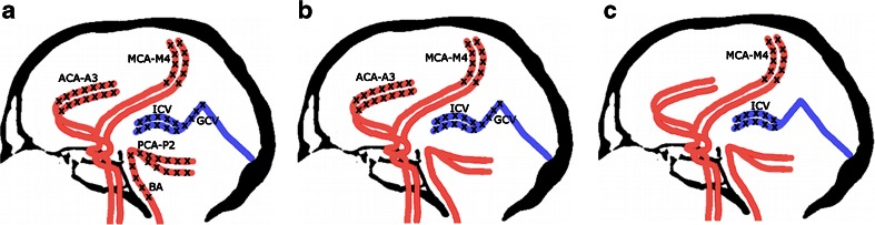
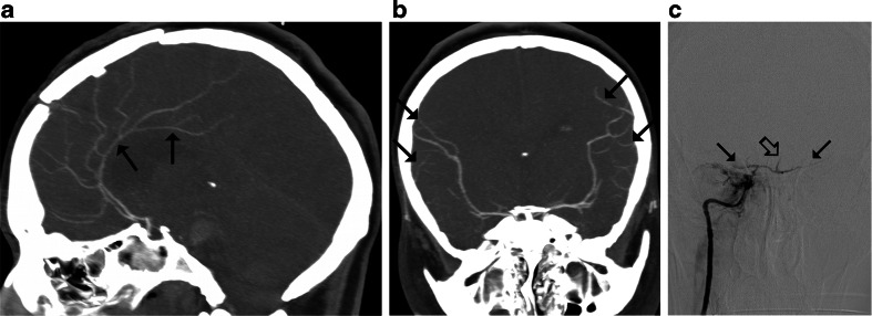
Methods: Eighty-two clinically brain-dead patients underwent CTA with a delay of 40 s after contrast injection. Catheter angiography was used as the reference standard. CTA results were assessed by two radiologists, and the diagnosis of BD was established according to 10-, 7-, and 4-point scales.

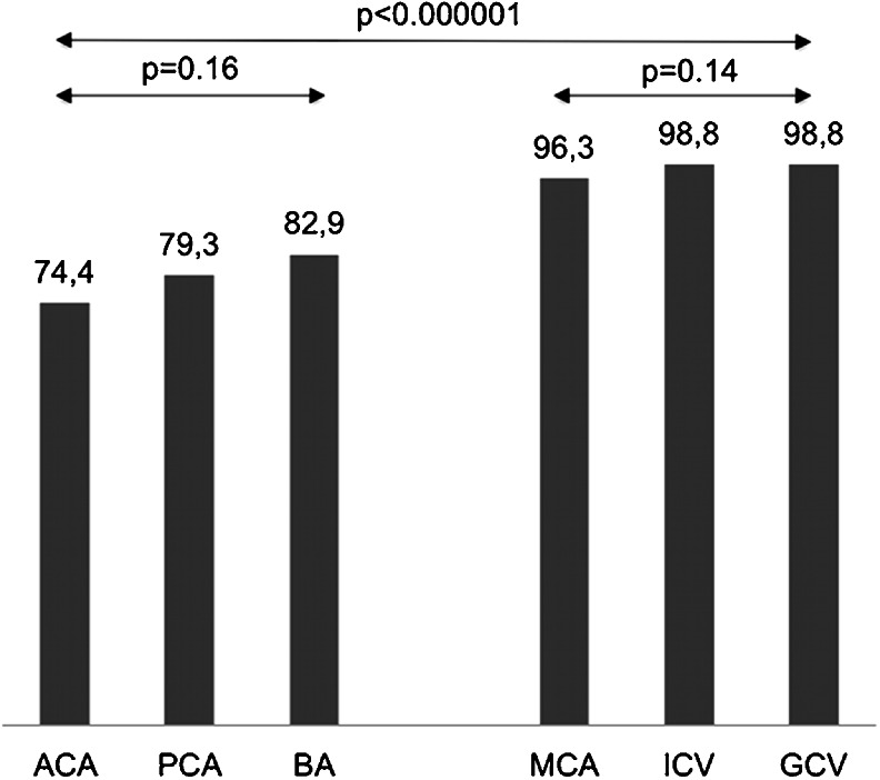
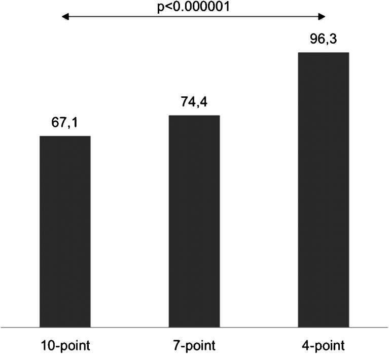
Results: Catheter angiography confirmed the diagnosis of BD in all cases. Opacification of certain cerebral vessels as indicator of BD was highly sensitive: cortical segments of the middle cerebral artery (96.3 %), the internal cerebral vein (98.8 %), and the great cerebral vein (98.8 %). Other vessels were less sensitive: the pericallosal artery (74.4 %), cortical segments of the posterior cerebral artery (79.3 %), and the basilar artery (82.9 %). The sensitivities of the 10-, 7-, and 4-point scales were 67.1, 74.4, and 96.3 %, respectively (p<0.001). Percentage interobserver agreement in diagnosis of BD reached 93 % for the 10-point scale, 89 % for the 7-point scale, and 95 % for the 4-point scale (p=0.37).

Conclusions: In the application of CTA to the diagnosis of BD, reducing the assessment of vascular opacification scale from a 10- to a 4-point scale significantly increases the sensitivity and maintains high interobserver reliability.


Figures

Fig. 1
Fig. 1
Different criteria for the diagnosis of BD by CTA: a Positive result in the 10-point scale (score = 10) confirming the diagnosis of BD was recorded when the following vessels were not opacified: the bilateral PCA-P2, the BA, the bilateral ACA-A3, the bilateral MCA-M4, the bilateral ICV, and the GCV. Scores from 0 to 9 were classified as negative results excluding the diagnosis of BD; b In the 7-point scale, positive result (score = 7) was recorded with a lack of opacification of the bilateral ACA-A3, the bilateral MCA-M4, the bilateral ICV, and the GCV. Scores from 0 to 6 were classified as negative results; c Positive result in the 4-point scale (score = 4) was recorded when the bilateral MCA-M4 and the bilateral ICV were not opacified. Scores from 0 to 3 were classified as negative results
Fig. 2
Fig. 2
Sensitivity of cerebral vessels as indicators of cerebral circulatory arrest in CTA. All vessels were classified into the high sensitivity group (cortical segments of the MCA, ICV, and GCV) and the low sensitivity group (the pericallosal artery and cortical segments of the PCA and BA)
Fig. 3
Fig. 3
CTA findings in a 50-year-old man (patient no. 45) with traumatic brain injury (epidural hematoma in the right parietal region, massive intracerebral, and subarachnoid and intraventricular hemorrhage) and right sided craniectomy presented with signs of BD on clinical examination: a Ten millimeter maximum intensity projection (MIP) in sagittal plane. CTA shows opacification of the BA (thin arrow) and a trace of contrast in A2 segments of the ACAs (thick arrow). b Ten millimeter MIP in coronal plane. CTA shows opacification of the M1 segment of the left MCA (thin arrow) and the A1 segments of the ACAs (thick arrow); these findings exclude the diagnosis of BD according to the 10-point scale but confirm BD according to the 7- and 4-point scales. c Catheter angiography of the right VA performed 0.5 h later revealed delayed, residual filling of the BA (arrow) that occurred 21 s after injection. This result was interpreted as stasis filling consistent with the diagnosis of BD
Fig. 4
Fig. 4
CTA findings in a 22-year-old woman (patient no. 42) with a brain stem ischemic stroke and a right sided craniectomy who presented with signs of BD on clinical examination. a Ten millimeter MIP in sagittal plane. CTA shows opacification of the right pericallosal artery (thin arrows); b Ten millimeter MIP in coronal plane. CTA shows opacification of the M1 segments of the MCAs (thin arrows). These findings exclude the diagnosis of BD according to the 10- and 7-point scales but confirm BD according to the 4-point scale. c Catheter angiography from the aortic arch performed 1 h later revealed delayed, residual filling of the M1 segment of the right MCA (thick arrow) and A2 segment of the right ACA (thin arrow) that occurred 32 s after injection. This result was interpreted as stasis filling consistent with the diagnosis of BD
Fig. 5
Fig. 5
CTA findings in a 34-year-old woman (patient no. 44) with brain stem hematoma and frontal craniotomy presenting signs of BD on clinical examination. a Ten millimeter MIP in sagittal plane. CTA shows opacification of both pericallosal arteries (thin arrows); b Ten millimeter MIP in coronal plane. CTA shows opacification of the cortical segments of the MCAs (thin arrows); these findings exclude the diagnosis of BD according to the 10-, 7-, and 4-point scales. c Catheter angiography of the right ICA performed 3 h later revealed delayed, residual filling of the M1 segments of the MCAs (thin arrows) and the A2 segments of the ACAs (thick arrow) that occurred 18 s after injection. This result was interpreted as stasis filling consistent with the diagnosis of BD
Fig. 6
Fig. 6
Sensitivity of three CTA scales for the diagnosis of BD. The sensitivities differed significantly (p < 0.001, Cochran’s Q test)

References

    1. Young GB, Shemie SD, Doig CJ, Teitelbaum J. Brief review: the role of ancillary tests in the neurological determination of death. Can J Anaesth=J Can Anesth. 2006;53(6):620–627. doi: 10.1007/BF03021855. - DOI - PubMed
    1. Wijdicks EF. The case against confirmatory tests for determining brain death in adults. Neurology. 2010;75(1):77–83. doi: 10.1212/WNL.0b013e3181e62194. - DOI - PubMed
    1. Roberts DJ, MacCulloch KA, Versnick EJ, Hall RI. Should ancillary brain blood flow analyses play a larger role in the neurological determination of death? Can J Anaesth=J Can Anesth. 2010;57(10):927–935. doi: 10.1007/s12630-010-9359-4. - DOI - PubMed
    1. Aichner F, Felber S, Birbamer G, Luz G, Judmaier W, Schmutzhard E. Magnetic resonance: a noninvasive approach to metabolism, circulation, and morphology in human brain death. Ann Neurol. 1992;32(4):507–511. doi: 10.1002/ana.410320405. - DOI - PubMed
    1. Ishii K, Onuma T, Kinoshita T, Shiina G, Kameyama M, Shimosegawa Y. Brain death: MR and MR angiography. AJNR Am J Neuroradiol. 1996;17(4):731–735. - PMC - PubMed

Publication types