. 2014 May 6;9(5):e96576.
doi: 10.1371/journal.pone.0096576. eCollection 2014.

Haploinsufficiency of Def activates p53-dependent TGFβ signalling and causes scar formation after partial hepatectomy

Affiliations

Haploinsufficiency of Def activates p53-dependent TGFβ signalling and causes scar formation after partial hepatectomy

Zhihui Zhu et al. PLoS One. .

Abstract

The metazoan liver exhibits a remarkable capacity to regenerate lost liver mass without leaving a scar following partial hepatectomy (PH). Whilst previous studies have identified components of several different signaling pathways that are essential for activation of hepatocyte proliferation during liver regeneration, the mechanisms that enable such regeneration to occur without accompanying scar formation remain poorly understood. Here we use the adult zebrafish liver, which can regenerate within two weeks following PH, as a new genetic model to address this important question. We focus on the role of Digestive-organ-expansion-factor (Def), a nucleolar protein which has recently been shown to complex with calpain3 (Capn3) to mediate p53 degradation specifically in the nucleolus, in liver regeneration. Firstly, we show that Def expression is up-regulated in the wild-type liver following amputation, and that the defhi429/+ heteroozygous mutant (def+/-) suffers from haploinsufficiency of Def in the liver. We then show that the expression of pro-inflammatory cytokines is up-regulated in the def+/- liver, which leads to distortion of the migration and the clearance of leukocytes after PH. Transforming growth factor β (TGFβ) signalling is thus activated in the wound epidermis in def+/- due to a prolonged inflammatory response, which leads to fibrosis at the amputation site. Fibrotic scar formation in def+/- is blocked by the over-expression of Def, by the loss-of-function of p53, and by treatment with anti-inflammation drug dexamethasone or TGFβ-signalling inhibitor SB431542. We finally show that the Def- p53 pathway suppresses fibrotic scar formation, at least in part, through the regulation of the expression of the pro-inflammatory factor, high-mobility group box 1. We conclude that the novel Def- p53 nucleolar pathway functions specifically to prevent a scar formation at the amputation site in a normal amputated liver.


Conflict of interest statement

Competing Interests: The authors have declared that no competing interests exist.

Figures

Figure 1
Figure 1. def+/− liver suffers from haploinsufficiency of Def.
(A) qPCR analysis of def transcripts in adult wild-type and def+/− liver. The expression of def was normalised against elf1a and is shown as the fold change in expression, with the value from def+/− being set at 1. The values plotted represent the means ± standard errors of the mean. (B) Western blotting analysis of the Def protein. Bhmt: loading control. Densitometry was performed using Photoshop. The value of Def was obtained by normalising against Bhmt expression. The relative value of Def protein in def+/− was set at 1. (C) Representative images of immunostaining of Def (red) in wild-type and def+/− liver 2 days after PH. lv, liver tissue. Scale bar: 100 µm (C).
Figure 2
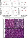
Figure 2. The liver-to-body ratio of the regenerated livers did not differ significantly between the wild-type and def+/− mutant fish after PH.
(A) Liver-to-body ratio of the wild-type sham control and amputated wild-type liver at 3, 7 and 36 days after PH (n≥5). (B) Liver-to-body ratios of the sham controls (n≥5) for the wild-type and def+/− mutant fish. (C–F) Comparison of the liver-to-body ratios of the amputated wild-type and def+/− livers 3 days after PH (C), 7 days after PH (D), 14 days after PH (E) and 30 days after PH (F) as indicated (n≥5 in each case). NS: not significant; *: p<0.05. (G) Representative images of haematoxylin-eosin staining of sections across the wild-type or def+/− hepatic parenchyma without performing PH or 9 months after PH. Scale bar: 50 µm.
Figure 3
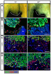
Figure 3. Def haploinsufficiency results in a failure in the remodelling of the wound epidermis to liver tissue.
(A) Ventral view of the gross morphology of an amputated adult wild-type and def+/−liver 7 days after PH. The dotted white line indicates the amputation site. 1: ventral lobe, 2: left dorsal lobe, 3: right dorsal lobe. (B–D) Representative images of co-immunostaining of PCNA and Bhmt to compare the process of epithelialisation of the amputation site 2 days after PH (B), wound epidermis formation 3 days after PH (C), and wound epidermis remodelling 5.5 days after PH (D) between the wild-type and the def+/− mutant. Nuclei were stained with 4',6-diamidino-2-phenylindole (DAPI) (blue). In each case or at each time-point, more than 10 sections from at least three wild-type or def+/− mutant fish were examined. bc, blood clot; lv, liver tissue; we, wound epidermis. Scale bar: 5 mm (A) and 75 µm (B–D).
Figure 4
Figure 4. Def haploinsufficiency results in accumulation of ColI at the amputation site after PH.
(A,B) Representative images of immunostaining of keratin 18 (green) (A) and co-immunostaining of ColI (red) and Bhmt (green) (B) at the amputation site in wild-type and def+/− strains 3 days after PH. Nuclei were stained with DAPI (blue). At each time-point, more than 10 sections from at least three wild-type or def+/− mutant fish were examined. lv, liver tissue; we, wound epidermis. Scale bars: 75 µm (A,B) and 100 µm (C).
Figure 5
Figure 5. Def haploinsufficiency results in the formation of a fibrotic scar at the amputation site after PH.
Masson staining of sections across the amputation site 3 (A), 5.5 (B) and 30 (C) days after PH (dPH), respectively. Fibrotic tissue was stained in blue (highlighted with an asterisk), cytoplasm in light red and nuclei in dark brown. Green, dashed lines outline the amputation site. Scale bar: 100 µm.
Figure 6
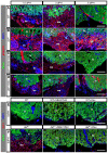
Figure 6. Constitutive activation of TGFβ signalling promotes ColI deposition at the amputation site in def+/− fish after PH.
(A–D) Representative images of immunostaining of pSmad2 (red) and Bhmt (green) in wound epidermis in wild-type (A), def+/− (B), def+/− treated with SB431542 (C) or def+/−treated with Dex (D) 1.5, 3 and 5 days after PH, respectively. (E,F) Representative images of immunostaining of ColI (red) and Bhmt (green) in the wound epidermis in wild-type and def+/−fish treated or not treated with SB431542 (E) or Dex (F) 5 days after PH, respectively. Nuclei were stained with DAPI (blue). In each case, more than 10 sections from at least three wild-type or def+/− mutant fish were examined. bc, blood clot; lv, liver tissue; we, wound epidermis. Scale bar:75 µm (A–F).
Figure 7
Figure 7. def+/− mutant liver exhibited a prolonged inflammatory reaction at the amputation site after PH.
(A) qPCR analysis of cytokine genes TNFα, IL-1β, IL-6 and IL-8 in adult wild-type, def+/−, fabp10a:def+/− and def+/− p53M214K /M214K livers prior to the surgical procedure. Gene expression is expressed as the fold change after normalisation against β-actin. The value for each gene in the wild-type was set at 1. The p value represents the statistical differences between def+/− and another corresponding group. *: p<0.05, **: p<0.01, NS: not significant. (B–D) Adult fish of Tg(zlyz:EGFP) in a wild-type and def+/− background were subjected to PH, and collected 6 h after PH (B), 12 h after PH (C) and 36 h after PH (D) for EGFP fluorescence imaging. Images were taken from the region outlined in white in the corresponding bright field picture shown on the left. The region above the dashed magenta lines is the liver tissue adjacent to the amputation site. Black asterisks highlight the EGFP signal yielded possibly from neutrophils in the intestine. A representative image is shown for each time-point. bc, blood clot; in, intestine; lv, liver tissue. Scale bar: 5 mm (bright field) and 2 mm (GFP field).
Figure 8
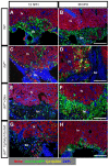
Figure 8. Def haploinsufficiency caused an altered migration behaviour of leukocytes in def+/− fish that was rescued by treatment with Dex or ectopic expression of Def in hepatocytes.
(A–H) Representative images of co-immunostaining of EGFP (green, label macrophage), dsRed (yellow, label neutrophil) and Bhmt (red) in the wound epidermis in wild-type (A,B), def+/− (C,D), def+/− treated with Dex (E,F) and def+/−Tg(fabp10a:def) progeny (G,H) 12 and 36 h after PH, respectively. All are in the background of Tg(coro1a:eGFP;lyz:dsRED). Nuclei were stained with DAPI (blue). In each case, more than 10 sections from at least three wild-type or def+/− mutant fish were examined. bc, blood clot; lv, liver tissue. Scale bars: 150 µm (A–H).
Figure 9
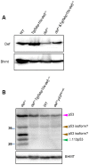
Figure 9. Def expression in the def+/− hepatocytes was restored in the def+/− Tg(fabp10a:def) fish and Def haploinsufficiency up-regulates p53 and Δ113p53 expression in the def+/− liver.
(A) Western blot of Def using an antibody against zebrafish Def to compare its expression levels in the wild-type, Tg(fabp10a:def), def+/− and def+/−Tg(fabp10a:def) adult livers. Bhmt: loading control. (B) Western blot of p53 and Δ113p53 in the wild-type, Tg(fabp10a:def), def+/− and def+/−p53M214K/M214K adult livers using an antibody against zebrafish p53. Two uncharacterised p53 isoforms are highlighted by a brown arrow head. Bhmt: loading control.
Figure 10
Figure 10. Formation of the fibrotic scar in def+/− fish after PH was mediated by the activation of the p53-pathway.
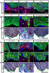
(A) Immunostaining of pSmad2 (red) and Bhmt (green) in the wound dermis in def+/−Tg(fabp10:def)+/− progeny 1.5, 3 and 5 days after PH, respectively. (B) Immunostaining of ColI (red) and Bhmt (green) in the wild-type, def+/− and def+/−Tg(fabp10:def)+/− progeny 5 days after PH. (C) Masson staining of the amputation site (indicated by a green dashed line) in the wild-type, def+/− and def+/− Tg(fabp10:def)+/− progeny 14 days after PH. (D,E) Immunostaining of pSmad2 (red) and Bhmt (green) (D) and of ColI (red) and Bhmt (green) (E) in the wound epidermis in the wild-type, def+/− and def+/−p53M214K/M214K 5 days after PH. (F) Masson staining of the amputation site in the wild-type, def+/− and def+/−p53M214K/M214K progeny 14 days after PH. Fibrotic tissue in the def+/− fish is highlighted by a red asterisk. In (A,B,D,E), nuclei were stained with DAPI (blue). In each case, more than 10 sections from at least three wild-type or def+/− mutant fish were examined. Representative images are shown here. bc, blood clot; lv, liver tissue; we, wound epidermis. Scale bars: 150 µm (C,F) and 75 µm (A,B,D,E).
Figure 11
Figure 11. A model to explain how Def haploinsufficiency activates the p53-dependent inflammation-mediated TGFβ signalling and causes fibrotic scar formation in def+/− after PH.
(A) Western blot of HMGB1 in the adult liver from wild-type, def+/−, def+/−Tg(fabp10:def)+/− and def+/−p53M214K/M214K fish. Loading control: Bhmt. (B) Immunostaining of ColI and Bhmt in the wound epidermis in wild-type, def+/− and def+/−fish treated with 100 µM of glycyrrhizic acid ammonium 5 days after PH. lv, liver; we, wound epidermis. (C) In a wild-type liver, Def complexes with Capn3 to mediate p53 degradation in the nucleoli to mitigate the inflammatory response after acute injury. In def+/− fish, the activity of the Def-Capn3 protein degradation pathway is compromised and p53 protein is thus stabilised, which in turn activates the p53 pathway. Constant activation of the p53 pathway up-regulates the expression of the pro-inflammatory factor HMGB1 that up-regulates the expression of cytokines (e.g., TNFα, IL-1β, IL-6 and IL-8) and other signalling pathways that lead to a prolonged inflammatory response in def+/-. The prolonged inflammatory response activates TGFβ signalling and causes the over-production of fibrotic molecules (e.g., collagens, elastin and fibronectin) in the wound epidermis that finally forms a fibrotic scar at the amputation site in def+/−. Fibrotic scar formation in def+/− can be blocked by over-expressing Def specifically in the hepatocytes, by the loss of function of p53, and by treatment with Dex (anti-inflammatory drug), SB431542 (inhibitor of TGFβ signalling) or glycyrrhizic acid ammonium (inhibitor of HMGB1 function).

Similar articles

Cited by

References

    1. Taub R (2004) Liver regeneration: from myth to mechanism. Nat Rev Mol Cell Biol 5: 836–847. - PubMed
    1. Fausto N, Campbell JS, Riehle KJ (2006) Liver regeneration. Hepatology 43: S45–S53. - PubMed
    1. Michalopoulos GK (2010) Liver regeneration after partial hepatectomy: critical analysis of mechanistic dilemmas. Am J Pathol 176: 2–13. - PMC - PubMed
    1. Costa RH, Kalinichenko VV, Holterman AX, Wang X (2003) Transcription factors in liver development, differentiation, and regeneration. Hepatology 38: 1331–1347. - PubMed
    1. Schachtrup C, Le MN, Passino MA, Akassoglou K (2011) Hepatic stellate cells and astrocytes: Stars of scar formation and tissue repair. Cell Cycle 10: 1764–1771. - PMC - PubMed

Publication types

MeSH terms

Substances

LinkOut - more resources