. 2014;13(13):2046-55.
doi: 10.4161/cc.29079. Epub 2014 May 6.

Loss of Notch1-dependent p21(Waf1/Cip1) expression influences the Notch1 outcome in tumorigenesis

Affiliations

Loss of Notch1-dependent p21(Waf1/Cip1) expression influences the Notch1 outcome in tumorigenesis

Samantha Cialfi et al. Cell Cycle. 2014.

Abstract

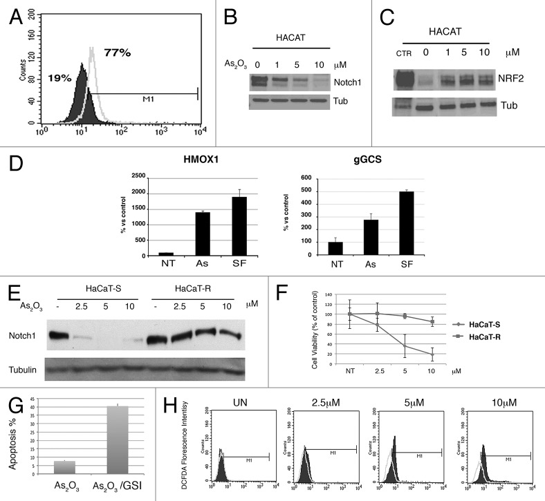
Notch signaling plays a complex role in carcinogenesis, and its signaling pathway has both tumor-suppressor and oncogenic components. In this study we investigated the effects of reactive oxygen species (ROS) on Notch1 signaling outcome in keratinocyte biology. We demonstrate that Notch1 function contributes to the arsenic-induced keratinocyte transformation. We found that acute exposure to arsenite increases oxidative stress and inhibits proliferation of keratinocyte cells by upregulation of p21(waf1/Cip1). The necessity of p21(waf1/Cip1) for arsenite-induced cell death was demonstrated by targeted downregulation of p21(waf1/Cip1) by using RNA interference. We further demonstrated that on acute exposure to arsenite, p21(waf1/Cip1) is upregulated and Notch1 downmodulated, whereas on chronic exposure to arsenite, malignant progression of arsenite-treated keratinocytes cells was accompanied by regained expression and activity of Notch1. Notch1 activity in arsenite-transformed keratinocytes inhibits arsenite-induced upregulation of p21(waf1/Cip1) by sustaining c-myc expression. We further demonstrated that c-myc collaborates with Nrf2, a key regulator for the maintenance of redox homeostasis, to promote metabolic activities that support cell proliferation and cytoprotection. Therefore, Notch1-mediated repression of p21(waf1/Cip1) expression results in the inhibition of cell death and keratinocytes transformation. Our results not only demonstrate that sustained Notch1 expression is at least one key event implicated in the arsenite human skin carcinogenic effect, but also may provide mechanistic insights into the molecular aspects that determine whether Notch signaling will be either oncogenic or tumor suppressive.

Keywords: Notch; Nrf2; metabolism; p21.


Figures

None
Figure 1. Effect of As2O3 in immortalized and transformed keratinocytes. (A) Flow cytometric analysis of the amount of ROS by DCFDA staining in the untreated and As2O3-treated keratinocytes. The percentage of positive cells is shown. (A) Overlay of FACS profile of keratinocytes untreated (black diagram) onto the profile of keratinocytes treated with As2O3 (empty diagram). (B and C) Effects of As2O3 on expression of Notch1 (B) and Nrf2 (C); Proteins were isolated and western blot analysis was performed with antibodies against Notch1 and Nrf2. Equal loading and transfer were verified by reprobing the membranes for Tubulin. (D) Immortalized HaCaT cells were treated with various concentrations of As2O3 (As) and Sulforaphane (SF) for 24 h. RNA was isolated and RT-PCR analysis was performed with primers against heme oxygenase-1 (HMOX1) gene and g-glutamylcysteine synthetase (gGCS). (E) Effects of As2O3 on Notch1 expression in immortalized and transformed keratinocytes. Immortalized and As2O3-transformed HaCaT cells were treated with various concentrations of As2O3 for 24 h. Proteins were isolated and western blot analysis was performed with antibodies against Notch1. Equal loading and transfer were verified by reproving the membranes for tubulin. (F) Viability of immortalized (square) and As2O3-transformed keratinocytes cells (circle); Cells were treated with various concentrations of As2O3 and the proliferation was measured by MTT assay. The percentage of viable cells was determined as the ratio of treated cells to untreated controls (100%). Results are expressed as mean ± SEM of triplicate experiments. (G) Apoptosis of As2O3-transformed keratinocytes cells treated with GSI 72 h. Flow cytometric analysis of the cell death phenotype—by PI staining, the % of apoptotic cells is indicated in As2O3 (5 μΜ) and As2O3/GSI (5 μΜ) treated cells. Results are expressed as mean ± SEM of triplicate experiments. (H) Flow cytometric analysis of the amount of ROS by DCFDA staining in the immortalized (dark area) and As2O3-transformed keratinocytes (non-filled area) after treatment with increased amount of As2O3.
<b>Figure 2</b>.
Figure 2.
Increased miR-223 levels in As2O3 transformed keratinocytes. (A) HaCaT cells were untreated or treated with As2O3. Twenty-four hours (24 h) post-treatment, cells were treated with MG132 for 5 h before collection; immunoblotting was performed with the indicated antibodies. (B) Immortalized and As2O3-transformed keratinocyte cells were treated with As2O3 at the indicated concentrations for 24 h. The cells were processed and the proteins were isolated. Aliquots of proteins were immunoblotted with specific primary antibodies against Notch1, Fbxw7, and tubulin. (C) qRT-PCR assay of miRNA levels in immortalized and As2O3-transformed keratinocyte cells. (D) As2O3-transformed keratinocytes were subjected with scramble antagomir (CTR) or miR-223 antagomir (mir-223) treatment. Proteins were isolated and analyzed by immunoblot with antibodies against Notch1, Fbxw7, and Tubulin. Protein levels were evaluated through densitometry and expressed as the fold change of the antagomir-223 over the antagomir-CTR after normalization to the loading control, tubulin.
None
Figure 3. Notch1-mediated repression of p21waf1/cip1 expression enhances cell survival in As2O3-transfromed HaCaT cells. (A) Cell cycle distribution in immortalized and As2O3-transformed keratinocytes with and without As2O3 treatment. Cells stained with propidium iodide were subjected to flow cytometry analysis to determine the cell cycle distribution. (B) Effects As2O3 treatment for 24 h on the expression of cell cycle-related proteins were measured by western blot analysis. (C) Immunoblot for Notch1-Val1744, Notch1, p21waf1/cip1 in HaCaT-R cells treated with the indicated amount of GSI. (D) Immunoblot for Notch1 and p21waf1/cip1 expression in HaCaT-R cells treated with either anti-Notch1 inhibitory antibodies or control IgG. (E) Immortalized HaCaT cells (HaCaT-S) were transfected with 100 nM control (siRNA-CTR) or p21waf1/cip1-specific (siRNA-p21) siRNA oligonucleotides; 24 h later, cells were treated with As2O3 (5 μΜ) for 24 h and analyzed by immunoblot with the indicated antibodies. (F) Apoptosis of HaCaT-S cells treated as in (E) were analyzed by flow cytometry by PI staining. The percentage of viable cells was determined as the ratio of siRNA-p21waf1/cip1-treated cells to siRNA-CTR controls (100%). Results are expressed as mean ± SEM of triplicate experiments.
None
Figure 4. As2O3 promotes metabolic adaptation in transformed cells. (A) Immortalized HaCaT keratinocytes (HaCaT-S) and As2-O3-transformed HaCaT cells (HaCaT-R) were treated with 8 mM 2-DG for 24 h. Apoptosis was detected using PI-staining analyzed by flow cytometry and the fold inductions over untreated cells (CTR) are indicated. (B) Basal, untreated and 2-DG induced ROS levels were analyzed by flow cytometry 24 h after treatment with the indicated amount of 2-DG. (C) Quantification of basal metabolic rate in As2O3-transformed cells. SOD1, G6PD, LDH, enzymatic activity, and O2 /glucose consumption were quantified in untreated immortalized HaCaT keratinocytes (HaCaT-S) and As2-O3-transformed HaCaT cells (HaCaT-R).
None
Figure 5. Altered metabolism in As2O3-transformed cells. (A) Quantification of metabolic intermediates in pentose phosphate pathway. All samples were analyzed in triplicate. Error bars indicate SDs. Quantifications were expressed as fold induction of the indicated metabolites in transformed HaCaT-R cells (p23 and p43 see “Materials and Methods”) relative to their levels in HaCaT-S. GSSG, glutathione oxidized; GSH, glutathione reduced; (B) Diagrammatic representation of the metabolic intermediates in glycolysis and pentose phosphate pathway. (C) Effect of As2O3 on NRF2 expression in HaCaT-S/HaCaT-R cells. Cells were treated as indicated, and cellular extract was analyzed by immunoblot with the indicated antibodies. (D) Nuclear and cytoplasmic extracts were analyzed by western blotting using anti-Nrf2, anti-lamin B1, anti-tubulin. Lamin B1 served as a nuclear marker. Tubulin was used as the cytoplasmic marker. Cyt, cytoplasmic extract; N, nuclear extract. (E) ChIP assay, crosslinked protein–DNA complexes were immunoprecipitated using anti-Nrf2 antibodies and IgG as negative control in HaCaT-R cells untreated and treated for 24 h with As2O3 (5 μM). The DNA samples immunoprecipitates were RT-PCR-amplified using primers specific for the ARE region of the G6PD promoter. Average values and SDs were calculated from triplicate samples. (F and G) mRNA expression of Nrf2 target genes were analyzed with real-time PCR in HaCaT-S/HaCaT-R cells untreated or treated with the indicated amounts of As2O3 for 24 h. Average values and SDs were calculated from triplicate samples; SOD1, superoxide dismutase 1; HMOX1, heme oxygenase 1.
None
Figure 6. As2O3-transformed keratinocytes had increased level of the Nrf2-dependent antioxidant response. Quantification of Nrf2 target genes by RT-PCR in immortalized HaCaT and As2O3-transformed keratinocytes in basal condition. NQO1, NAD(P)H quinone oxidoreductase 1; SOD1, superoxide dismutase 1; HMOX1, heme oxygenase 1.
None
Figure 7. Nocth1/c-myc axis sustains the expression of glutamine transported, ASCT2. (A) Analysis of glutamine uptake was performed in immortalized and transformed HaCaT cells (p23 and p43 see “Materials and Methods”) under basal condition. Values are expressed as fold increase over the HaCaT cells. (B) Immunoblot for c-myc expression in HaCaT-S and HACaT-R cells untreated and treated with the indicated amounts of As2O3. Tubulin is shown as loading control. (C) Immunoblot for Notch1-Val1744, Notch1, and c-myc in HaCaT-R cells treated with the indicated amount of GSI, As2O3 treatment is shown as a control. (D) Immunoblot for Notch1-Val1744, c-myc, and Asct2 in HaCaT-R cells treated with the indicated amount of GSI. Tubulin is shown as a loading control. (E) HaCaT-R cells were transfected with 100 nM control (siRNA-CTR) or c-myc-specific (siRNA-c-myc) siRNA oligonucleotides; 48 h later, cells were analyzed by immunoblot with the indicated antibodies.

Similar articles

Cited by

References

    1. Agrawal N, Frederick MJ, Pickering CR, Bettegowda C, Chang K, Li RJ, Fakhry C, Xie TX, Zhang J, Wang J, et al. Exome sequencing of head and neck squamous cell carcinoma reveals inactivating mutations in NOTCH1. Science. 2011;333:1154–7. doi: 10.1126/science.1206923. - DOI - PMC - PubMed
    1. Talora C, Campese AF, Bellavia D, Felli MP, Vacca A, Gulino A, Screpanti I. Notch signaling and diseases: an evolutionary journey from a simple beginning to complex outcomes. Biochim Biophys Acta. 2008;1782:489–97. doi: 10.1016/j.bbadis.2008.06.008. - DOI - PubMed
    1. Mazzone M, Selfors LM, Albeck J, Overholtzer M, Sale S, Carroll DL, Pandya D, Lu Y, Mills GB, Aster JC, et al. Dose-dependent induction of distinct phenotypic responses to Notch pathway activation in mammary epithelial cells. Proc Natl Acad Sci U S A. 2010;107:5012–7. doi: 10.1073/pnas.1000896107. - DOI - PMC - PubMed
    1. Talora C, Cialfi S, Segatto O, Morrone S, Kim Choi J, Frati L, Paolo Dotto G, Gulino A, Screpanti I. Constitutively active Notch1 induces growth arrest of HPV-positive cervical cancer cells via separate signaling pathways. Exp Cell Res. 2005;305:343–54. doi: 10.1016/j.yexcr.2005.01.015. - DOI - PubMed
    1. Biolcati G, Aurizi C, Barbieri L, Cialfi S, Screpanti I, Talora C. Efficacy of the melanocortin analogue Nle4-D-Phe7-α-melanocyte-stimulating hormone in the treatment of patients with Hailey-Hailey disease. Clin Exp Dermatol. 2014;39:168–75. doi: 10.1111/ced.12203. - DOI - PMC - PubMed

MeSH terms

LinkOut - more resources