. 2014 May 6;9(5):e96692.
doi: 10.1371/journal.pone.0096692. eCollection 2014.

Liver as a source for thymidine phosphorylase replacement in mitochondrial neurogastrointestinal encephalomyopathy

Affiliations

Liver as a source for thymidine phosphorylase replacement in mitochondrial neurogastrointestinal encephalomyopathy

Elisa Boschetti et al. PLoS One. .

Erratum in

  • PLoS One. 2014;9(10):e110583

Abstract

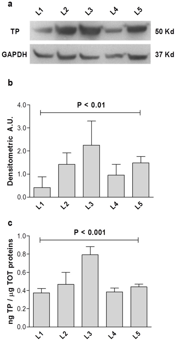
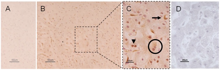
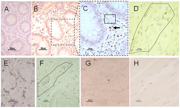
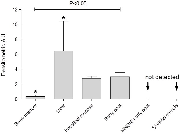
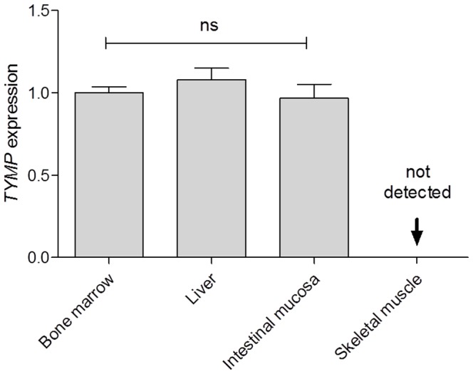
Mitochondrial neurogastrointestinal encephalomyopathy (MNGIE) is a rare autosomal recessive mitochondrial disease associated with mutations in the nuclear TYMP gene. As a result, the thymidine phosphorylase (TP) enzyme activity is markedly reduced leading to toxic accumulation of thymidine and therefore altered mitochondrial DNA. MNGIE is characterized by severe gastrointestinal dysmotility, neurological impairment, reduced life expectancy and poor quality of life. There are limited therapeutic options for MNGIE. In the attempt to restore TP activity, allogenic hematopoietic stem cell transplantation has been used as cellular source of TP. The results of this approach on ∼ 20 MNGIE patients showed gastrointestinal and neurological improvement, although the 5-year mortality rate is about 70%. In this study we tested whether the liver may serve as an alternative source of TP. We investigated 11 patients (7M; 35-55 years) who underwent hepatic resection for focal disorders. Margins of normal liver tissue were processed to identify, quantify and localize the TP protein by Western Blot, ELISA, and immunohistochemistry, and to evaluate TYMP mRNA expression by qPCR. Western Blot identified TP in liver with a TP/GAPDH ratio of 0.9 ± 0.5. ELISA estimated TP content as 0.5 ± 0.07 ng/μg of total protein. TP was identified in both nuclei and cytoplasm of hepatocytes and sinusoidal lining cells. Finally, TYMP mRNA was expressed in the liver. Overall, our study demonstrates that the liver is an important source of TP. Orthotopic liver transplantation may be considered as a therapeutic alternative for MNGIE patients.


Conflict of interest statement

Competing Interests: The authors have declared that no competing interests exist.

Figures

Figure 1
Figure 1. TP occurrence and concentration in control liver.
Figure 1A shows an example of a WB separation of 50 ng total protein from healthy liver. TP and reference protein GAPDH chemioluminescence are reported from liver L1 to L5. Figure 1B illustrates the densitometric arbitrary units (A.U.) calculated normalizing TP chemioluminescent signal on the internal reference protein GAPDH ± SD and showing a significant variability among non-MNGIE subjects (P< 0.01, one way non parametric test ANOVA). The graph in Figure 1C reports the TP concentration measured by ELISA and expressed as ng TP/μg total proteins for the 5 liver tissue samples ± SD and showing a significant variability among non-MNGIE subjects (P< 0.001, one way non parametric test ANOVA).
Figure 2
Figure 2. Representative photomicrographs showing TP immunoreactivity in control and MNGIE liver.
Figure 2A demonstrates the lack of TP immunolabeling in a normal liver section in which the primary antibody was omitted (negative control). Figure 2B (low magnification) and 2C (high magnification) illustrate TP immunoreactivity in a normal liver section. In Figure 2C, the arrowhead and circle, (Figure 2C) point to TP immunostained nuclei and cytoplasm of hepatocytes, while the arrow indicates non-hepatocytic cells with features of bile duct elements. Also, note the lack of any TP immunolabeling in a MNGIE liver section (Figure 2D). Calibration bars  = 20 µm and 50 µm in 2C, 2D and 2A, 2B, respectively.
Figure 3
Figure 3. TP immunoreactivity in the duodenum and skeletal muscle of control and in a MNGIE patient.
TP immunolabeling is lacking in a tissue section of normal duodenal mucosa in which primary antibody was omitted (negative control) (Figure 3A). Figure 3B (low magnification) and 3C (high magnification) show TP immunolabeling in the control mucosa. The black arrow indicates TP immunolabeled cells with features of immunocytes distributed throughout the lamina propria; the rectangle shows a less intense TP immunostaining in the cytoplasm of cells with features of fibroblasts. The dotted line area in figure 3D indicates a myenteric plexus displaying TP immunolabelling in non-neuronal cells (likely glial cells). Note the lack of TP immunolabeling in the mucosal lamina propria (Figure 3E) and myenteric plexus (dotted line) of a MNGIE patient (Figure 3F). The TP immunolabeling was negative also in normal (Figure 3G) and MNGIE (Figure 3H) skeletal muscle. Calibration bars  = 20 µm and 50 µm in 3C, 3D, 3E, 3F, 3H and 3A, 3B, 3G respectively.
Figure 4
Figure 4. TP concentration and distribution in a panel of selected human tissues.
The graph reports the A.U. calculated normalizing TP chemioluminescent signal on total proteins. Data are reported as mean ± SD using bone marrow as calibrator tissue. The TP concentrations vary significantly among different tissues (P< 0.05 one way ANOVA non-parametric test; *P< 0.05 Tukey's post-test).
Figure 5
Figure 5. TYMP mRNA transcription in a panel of healthy human tissues.
The amount of TYMP mRNA has been reported ± SD. Bone marrow was used as a calibrator tissue. TYMP mRNA expression did not vary significantly in bone marrow, liver, and duodenal mucosa. No TYMP transcript was found in skeletal muscle.

References

    1. DiMauro S, Schon EA (2003) Mitochondrial respiratory-chain diseases. N Engl J Med 348: 2656–2668. - PubMed
    1. Nishino I, Spinazzola A, Hirano M (1999) Thymidine phosphorylase gene mutations in MNGIE, a human mitochondrial disorder. Science 283: 689–692. - PubMed
    1. Spinazzola A, Marti R, Nishino I, Andreu AL, Naini A, et al. (2002) Altered thymidine metabolism due to defects of thymidine phosphorylase. J Biol Chem 277: 4128–4133. - PubMed
    1. Marti R, Nishigaki Y, Hirano M (2003) Elevated plasma deoxyuridine in patients with thymidine phosphorylase deficiency. Biochem Biophys Res Commun 303: 14–18. - PubMed
    1. Ferraro P, Pontarin G, Crocco L, Fabris S, Reichard P, et al. (2005) Mitochondrial deoxynucleotide pools in quiescent fibroblasts: a possible model for mitochondrial neurogastrointestinal encephalomyopathy (MNGIE). J Biol Chem 280: 24472–24480. - PubMed

Publication types

MeSH terms