. 2014 Sep;32(9):2397-406.
doi: 10.1002/stem.1736.

Importance of cell-cell contact in the therapeutic benefits of cardiosphere-derived cells

Affiliations

Importance of cell-cell contact in the therapeutic benefits of cardiosphere-derived cells

Yucai Xie et al. Stem Cells. 2014 Sep.

Abstract

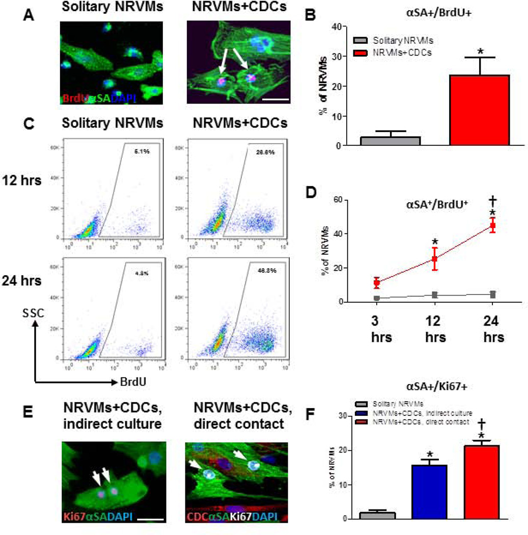
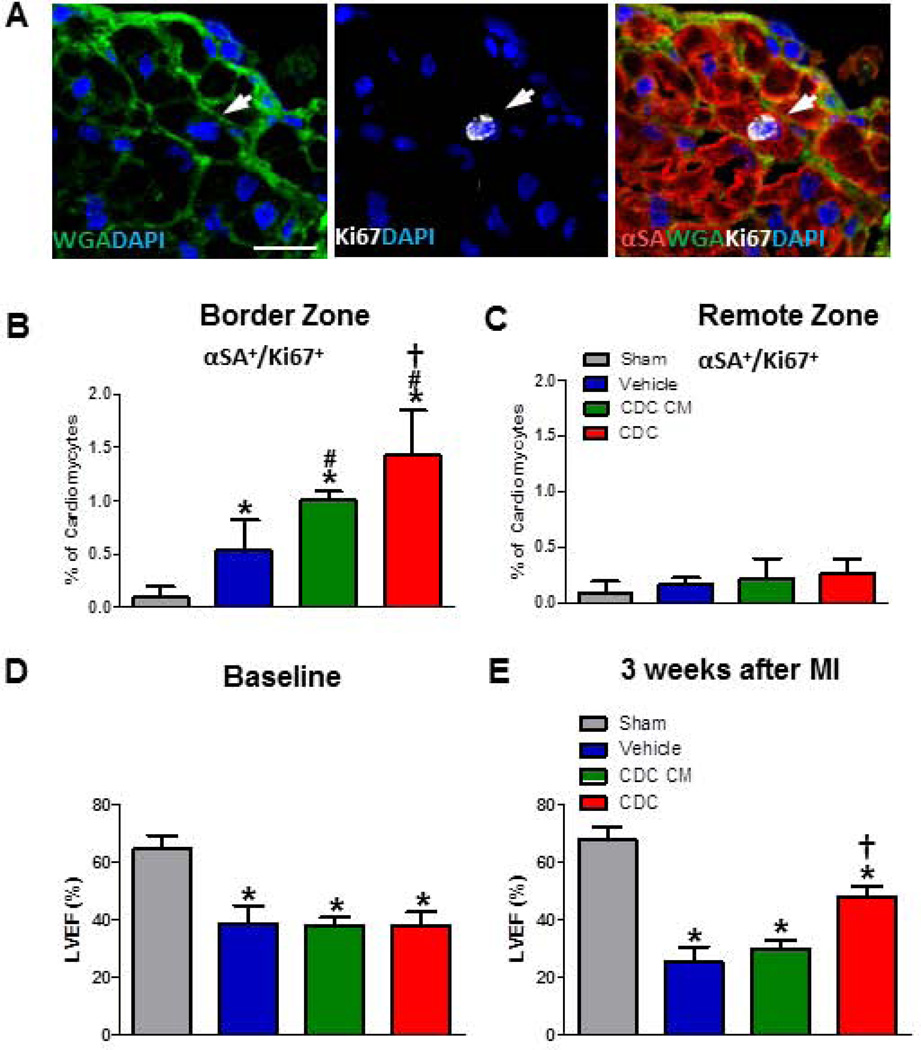
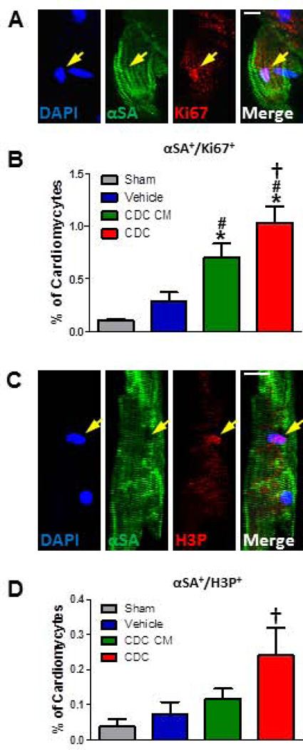
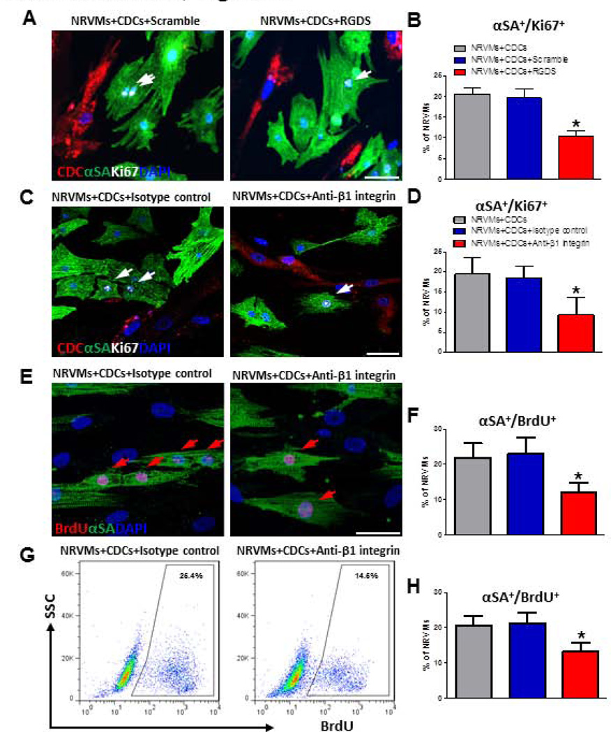
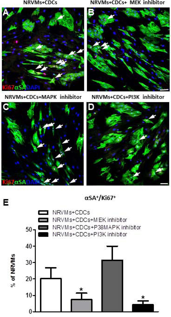
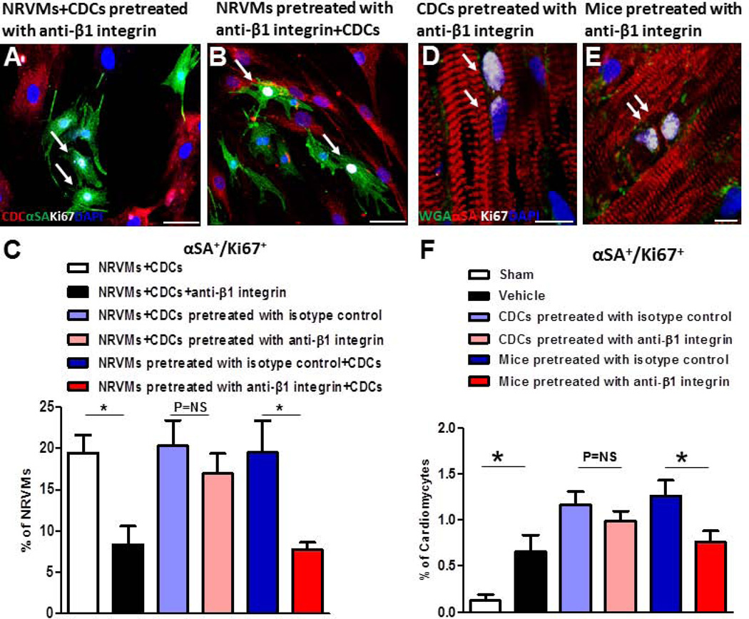
Cardiosphere-derived cells (CDCs) effect therapeutic regeneration after myocardial infarction (MI) both in animal models and in humans. Here, we test the hypothesis that cell-cell contact plays a role in mediating the observed therapeutic benefits of CDCs, above and beyond conventional paracrine effects. Human CDCs or vehicle were injected into immunodeficient (SCID) mouse hearts during acute MI. CDC transplantation augmented the proportion of cycling (Ki67(+) ) cardiomyocytes and improved ventricular function. CDC-conditioned media only modestly augmented the percentage of Ki67(+) cardiomyocytes (>control but <CDCs), but did not improve pump function. When neonatal rat ventricular myocytes (NRVMs) were cocultured with human CDCs in vitro, the percentage of cycling NRVMs (Ki67(+) or BrdU(+) nuclei) increased relative to solitary NRVM culture. To further dissect the relative contributions of soluble factors versus contact-dependent mechanisms, we compared CDCs grown with NRVMs in a transwell contact-free system versus admixed coculture. The percentage of cycling NRVMs was higher in admixed coculture than in the contact-free system. Pretreatment with inhibitors of MEK and PI3K, or with β1 integrin neutralizing antibody, blocked the ability of CDCs to promote myocyte cycling. While conditioned media are not inert, direct apposition of CDCs to cardiomyocytes produces greater enhancement of cardiomyocyte proliferation in vitro and in vivo, and improves function post-MI. Intact cardiomyocyte β1 integrin signaling is necessary for the contact-dependent cardioproliferative effects of CDCs.

Keywords: Cardiomyocyte; Cardiosphere-derived cells; Cell-cell communication; Paracrine factors; Proliferation; β1 integrin.


Conflict of interest statement

Disclosure of potential conflicts of interest

Eduardo Marbán holds equity in Capricor Therapeutics, Inc. K. Malliaras receives a consulting fee from Capricor Therapeutics, Inc. Capricor Therapeutics provided no funding for the present study. The remaining authors report no conflicts.

Figures

Figure 1
Figure 1. Admixed co-culture CDCs and CDC-conditioned media stimulate NRVM cycling in vitro
A, Representative confocal images of BrdU+ NRVMs, with αSA shown as green, BrdU as red, and DAPI as blue. B, Quantification of BrdU+ NRVMs admixed co-cultured with CDCs, or cultured alone (solitary NRVMs, n=4). C, Representative flow cytometry analysis of BrdU+ NRVMs at 3, 12 and 24 hours after incubation with BrdU. D, Percentage, as reported by flow cytometry, of BrdU+ NRVMs admixed co-cultured with CDCs, or cultured alone (solitary NRVMs) (n=3 per group). E, Representative confocal images of NRVMs cultured indirectly with CDCs (left panel) or cultured with direct contact with CDCs (right panel). F, The quantification of Ki67+ NRVM co-cultured with CDCs or cultured alone (solitary NRVMs, n=5 to 6). Arrows point to BrdU+/Ki67+ NRVMs. * indicates p<0.05 when compared to solitary NRVM culture group or 3-hour group; † indicates p<0.05 compared to all other groups. Scale bars indicate 50 µm.
Figure 2
Figure 2. CDC transplantation increases the percentage of cycling cardiomyocytes and improves cardiac function compared to conditioned media after MI in vivo
A, Representative confocal images of Ki67+ cardiomyocyte from CDC group. (αSA: red; WGA: green; Ki67: white; DAPI: blue) in the heart injected with CDCs. B and C, Quantification of cycling (Ki67+) cardiomyocytes from the infarct border (B) or remote zone (C) at days 7 after MI (n=6–7 per group). D and E, Left ventricular ejection fraction (LVEF) in baseline (D) and three weeks after MI (E) with different treatment. Arrows point to Ki67+ cardiomyocytes. * indicates p<0.05 when compared to sham operation groups; # indicates p<0.05 compared to vehicle groups; † indicates p<0.05 compared to vehicle groups or CDC condition media (CDC CM) groups. Scale bars indicate 10 µm.
Figure 3
Figure 3. CDCs promote more resident cycling cardiomyocyte turnover in the adult MI mouse heart than conditioned media assessed by immunocytochemistry
A and B, Representative confocal images (A) and quantification (B) of Ki67+ single enzymatically-isolated cardiomyocyte from the sham or MI heart injected with vehicle, CDC CM or CDCs. C and D, The representative single H3P+ cardiomyocyte (C) and the quantitative data (D) from four groups after MI (n=3–4 per group). Arrows point to Ki67+/H3P+ cardiomyocytes. * indicates p<0.05 when compared to sham operation groups; # indicates p<0.05 compared to vehicle groups; † indicates p<0.05 compared to vehicle groups or CDC CM groups. Scale bars indicate 10 µm.
Figure 4
Figure 4. Integrin blockade abrogates the effect of CDCs on NRVM proliferation
A and B, Ki67+ NRVMs co-cultured with CDCs in the presence of a pan-integrin blocker, RGDS or a control peptide (GRGDTP, “scramble”). n=4 for the average values. C and D, Ki67+ NRVMs co-cultured with CDCs in the presence of an anti-β1 integrin antibody or an isotype control. n=5 for each group. E and F, Same experiment as C and D, but immunostained for BrdU rather than Ki67. G and H, BrdU+ NRVMs detected by flow cytometry in CDC co-culture with or without anti-β1 integrin antibody. Representative flow cytometry plots (G) and the quantitative data (H) of BrdU+ NRVMs (n=4 to 5 per group). Arrows point to Ki67+ or BrdU+ cardiomyocytes. * indicates p<0.05 when compared to control groups. Scale bars indicate 50 µm.
Figure 5
Figure 5. Inhibition of MEK or PI3K signaling abolishes pro-proliferative effects of CDCs on NRVMs
A–D, Representative confocal images of NRVMs co-cultured with human CDC (A), with a MEK inhibitor (B), with a P38MAPK inhibitor (C), or a PI3K inhibitor (D). E, Averaged percentages of Ki67+ NRVMs with different inhibitors (n=4 per group). Arrows point to Ki67+ cardiomyocytes. * indicates p<0.05 compared to no inhibitor control. Scale bars indicate 50 µm.
Figure 6
Figure 6. Blockage of β1 integrin on cardiomyocytes, but not on CDCs, eliminates the pro-proliferative effects of CDCs
A and B, Representative confocal images of Ki67+ NRVMs co-cultured with CDCs which were pretreated with anti-β1 integrin antibody (A) and CDCs co-cultured with NRVMs which were pretreated with anti-β1 integrin antibody (B). C. Average percentages of cycling NRVMs (n=3 to 5). D and E, Representative confocal images of Ki67+ cardiomyocytes, αSA (red), WGA (green), Ki67 (white) and DAPI (blue), from mouse hearts transplanted with CDCs that were pretreated with anti-β1 integrin antibody (D) or mouse hearts injected with anti-β1 integrin antibody before CDC transplantation (E). F, Quantification of Ki67+ cardiomyocytes from border zone 7 days after MI (n=3 to 5 per group). *p<0.05. Scale bar indicates 50 µm in A and B and 10 µm in D and E. Arrows point to Ki67+ cardiomyocytes.

References

    1. Bergmann O, Bhardwaj RD, Bernard S, et al. Evidence for cardiomyocyte renewal in humans. Science. 2009;324:98–102. - PMC - PubMed
    1. Malliaras K, Zhang Y, Seinfeld J, et al. Cardiomyocyte proliferation and progenitor cell recruitment underlie therapeutic regeneration after myocardial infarction in the adult mouse heart. EMBO Mol Med. 2013;5:191–209. - PMC - PubMed
    1. Senyo SE, Steinhauser ML, Pizzimenti CL, et al. Mammalian heart renewal by pre-existing cardiomyocytes. Nature. 2013;493:433–436. - PMC - PubMed
    1. Robey TE, Saiget MK, Reinecke H, Murry CE. Systems approaches to preventing transplanted cell death in cardiac repair. J Mol Cell Cardiol. 2008;45:567–581. - PMC - PubMed
    1. Loffredo FS, Steinhauser ML, Gannon J, Lee RT. Bone marrow-derived cell therapy stimulates endogenous cardiomyocyte progenitors and promotes cardiac repair. Cell Stem Cell. 2011;8:389–398. - PMC - PubMed

Publication types