

Meta-analysis of molecular imaging of serotonin transporters in major depression
- PMID: 24802331
- PMCID: PMC4083395
- DOI: 10.1038/jcbfm.2014.82
Meta-analysis of molecular imaging of serotonin transporters in major depression
Abstract
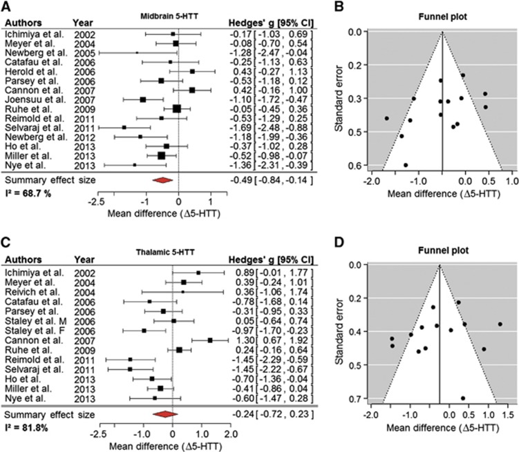
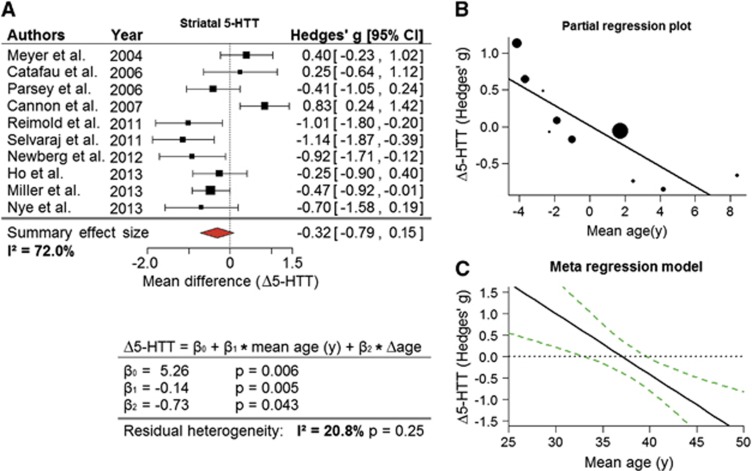
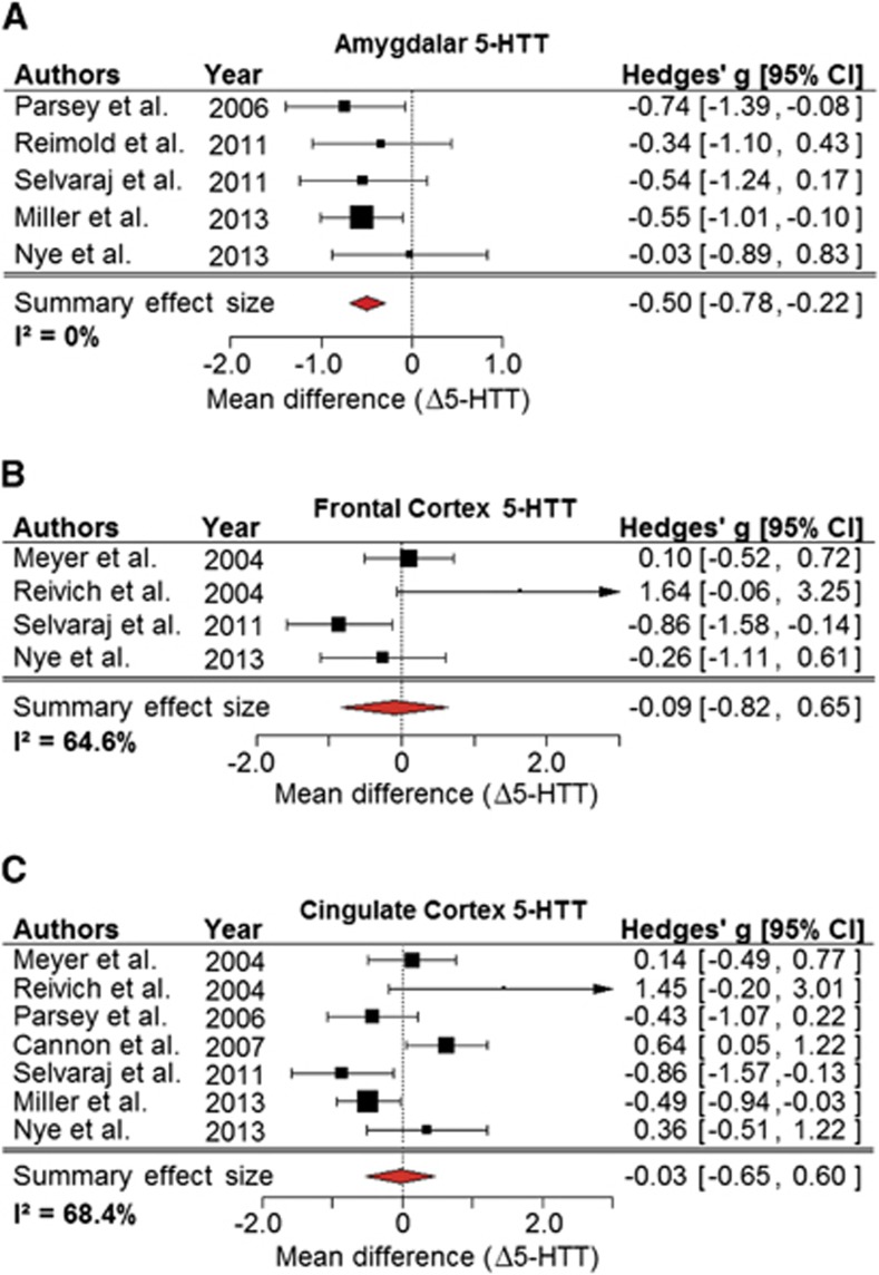
The success of serotonin-selective reuptake inhibitors has lent support to the monoamine theory of major depressive disorder (MDD). This issue has been addressed in a number of molecular imaging studies by positron emission tomography or single-photon emission computed tomography of serotonin reuptake sites (5-HTT) in the brain of patients with MDD, with strikingly disparate conclusions. Our meta-analysis of the 18 such studies, totaling 364 MDD patients free from significant comorbidities or medication and 372 control subjects, revealed reductions in midbrain 5-HTT (Hedges' g=-0.49; 95% CI: (-0.84, -0.14)) and amygdala (Hedges' g=-0.50; 95% CI: (-0.78, -0.22)), which no individual study possessed sufficient power to detect. Only small effect sizes were found in other regions with high binding (thalamus: g=-0.24, striatum: g=-0.32, and brainstem g=-0.22), and no difference in the frontal or cingulate cortex. Age emerged as an important moderator of 5-HTT availability in MDD, with more severe reductions in striatal 5-HTT evident with greater age of the study populations (P<0.01). There was a strong relationship between severity of depression and 5-HTT reductions in the amygdala (P=0.01). Thus, molecular imaging findings indeed reveal widespread reductions of ∼10% in 5-HTT availability in MDD, which may predict altered spatial-temporal dynamics of serotonergic neurotransmission.
Figures
Similar articles
-
5-HTT binding in recovered depressed patients and healthy volunteers: a positron emission tomography study with [11C]DASB.Am J Psychiatry. 2007 Dec;164(12):1858-65. doi: 10.1176/appi.ajp.2007.06111933. Am J Psychiatry. 2007. PMID: 18056241
-
Imaging the serotonin transporter during major depressive disorder and antidepressant treatment.J Psychiatry Neurosci. 2007 Mar;32(2):86-102. J Psychiatry Neurosci. 2007. PMID: 17353938 Free PMC article. Review.
-
Brain serotonin transporter binding potential measured with carbon 11-labeled DASB positron emission tomography: effects of major depressive episodes and severity of dysfunctional attitudes.Arch Gen Psychiatry. 2004 Dec;61(12):1271-9. doi: 10.1001/archpsyc.61.12.1271. Arch Gen Psychiatry. 2004. PMID: 15583118
-
Elevated serotonin transporter binding in major depressive disorder assessed using positron emission tomography and [11C]DASB; comparison with bipolar disorder.Biol Psychiatry. 2007 Oct 15;62(8):870-7. doi: 10.1016/j.biopsych.2007.03.016. Epub 2007 Aug 2. Biol Psychiatry. 2007. PMID: 17678634
-
Receptor and transporter imaging studies in schizophrenia, depression, bulimia and Tourette's disorder--implications for psychopharmacology.World J Biol Psychiatry. 2002 Jul;3(3):133-46. doi: 10.3109/15622970209150614. World J Biol Psychiatry. 2002. PMID: 12478878 Review.
Cited by
-
No association between peripheral serotonin-gene-related DNA methylation and brain serotonin neurotransmission in the healthy and depressed state.Clin Epigenetics. 2024 May 27;16(1):71. doi: 10.1186/s13148-024-01678-y. Clin Epigenetics. 2024. PMID: 38802956 Free PMC article.
-
Changes in RNA expression levels during antidepressant treatment: a systematic review.J Neural Transm (Vienna). 2021 Sep;128(9):1461-1477. doi: 10.1007/s00702-021-02394-0. Epub 2021 Aug 20. J Neural Transm (Vienna). 2021. PMID: 34415438
-
In Vivo Cerebral Translocator Protein (TSPO) Binding and Its Relationship with Blood Adiponectin Levels in Treatment-Naïve Young Adults with Major Depression: A [11C]PK11195 PET Study.Biomedicines. 2021 Dec 24;10(1):34. doi: 10.3390/biomedicines10010034. Biomedicines. 2021. PMID: 35052718 Free PMC article.
-
Monoamine Oxidase Inhibition by Plant-Derived β-Carbolines; Implications for the Psychopharmacology of Tobacco and Ayahuasca.Front Pharmacol. 2022 May 2;13:886408. doi: 10.3389/fphar.2022.886408. eCollection 2022. Front Pharmacol. 2022. PMID: 35600851 Free PMC article. Review.
-
Modulation of serotonin transporter expression by escitalopram under inflammation.Commun Biol. 2024 Jun 8;7(1):710. doi: 10.1038/s42003-024-06240-3. Commun Biol. 2024. PMID: 38851804 Free PMC article.
References
-
- Brown GL, Goodwin FK, Ballenger JC, Goyer PF, Major LF. Aggression in humans correlates with cerebrospinal fluid amine metabolites. Psychiatry Res. 1979;1:131–139. - PubMed
-
- Traskman L, Asberg M, Bertilsson L, Sjostrand L. Monoamine metabolites in CSF and suicidal behavior. Arch Gen Psychiatry. 1981;38:631–636. - PubMed
-
- Roy A, De Jong J, Linnoila M. Cerebrospinal fluid monoamine metabolites and suicidal behavior in depressed patients. A 5-year follow-up study. Arch Gen Psychiatry. 1989;46:609–612. - PubMed
-
- Dewar KM, Reader TA, Grondin L, Descarries L. [3H]paroxetine binding and serotonin content of rat and rabbit cortical areas, hippocampus, neostriatum, ventral mesencephalic tegmentum, and midbrain raphe nuclei region. Synapse. 1991;9:14–26. - PubMed
-
- Andersson A, Eriksson A, Marcusson J. Unaltered number of brain serotonin uptake sites in suicide victims. J Psychopharmacol. 1992;6:509–513. - PubMed
Publication types
MeSH terms
Substances
Grants and funding
LinkOut - more resources
Full Text Sources
Other Literature Sources
