. 2015 Jul;220(4):2171-90.
doi: 10.1007/s00429-014-0778-9. Epub 2014 May 7.

Increased hippocampal excitability and impaired spatial memory function in mice lacking VGLUT2 selectively in neurons defined by tyrosine hydroxylase promoter activity

Affiliations

Increased hippocampal excitability and impaired spatial memory function in mice lacking VGLUT2 selectively in neurons defined by tyrosine hydroxylase promoter activity

Karin Nordenankar et al. Brain Struct Funct. 2015 Jul.

Abstract

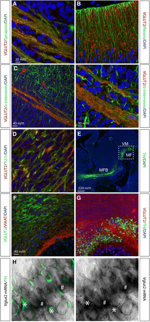
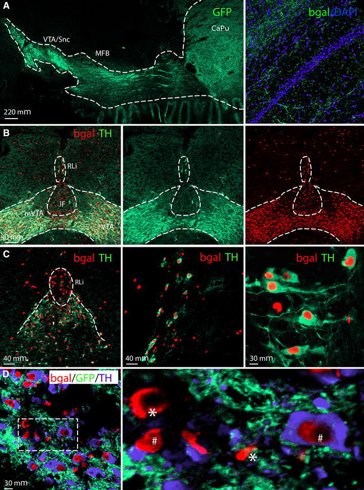
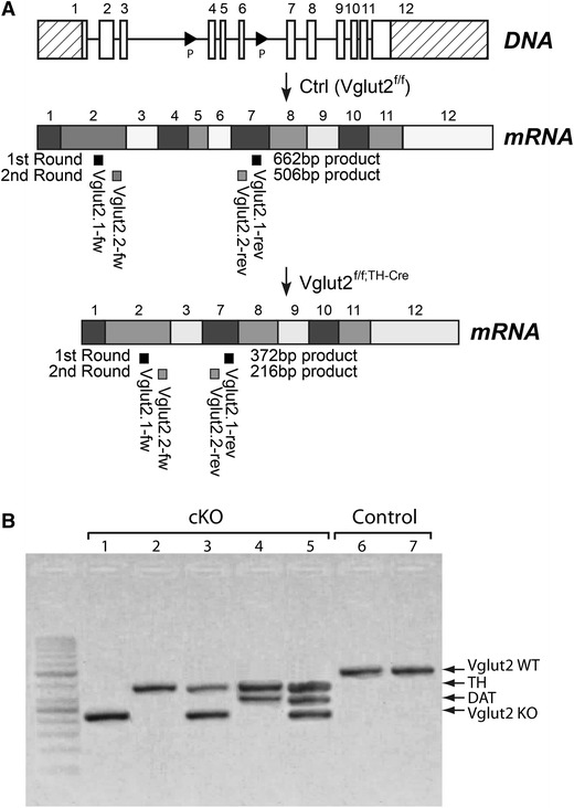
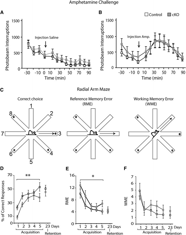
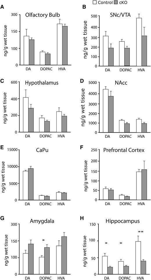
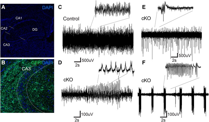
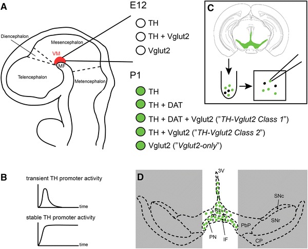
Three populations of neurons expressing the vesicular glutamate transporter 2 (Vglut2) were recently described in the A10 area of the mouse midbrain, of which two populations were shown to express the gene encoding, the rate-limiting enzyme for catecholamine synthesis, tyrosine hydroxylase (TH).One of these populations ("TH-Vglut2 Class1") also expressed the dopamine transporter (DAT) gene while one did not ("TH-Vglut2 Class2"), and the remaining population did not express TH at all ("Vglut2-only"). TH is known to be expressed by a promoter which shows two phases of activation, a transient one early during embryonal development, and a later one which gives rise to stable endogenous expression of the TH gene. The transient phase is, however, not specific to catecholaminergic neurons, a feature taken to advantage here as it enabled Vglut2 gene targeting within all three A10 populations expressing this gene, thus creating a new conditional knockout. These knockout mice showed impairment in spatial memory function. Electrophysiological analyses revealed a profound alteration of oscillatory activity in the CA3 region of the hippocampus. In addition to identifying a novel role for Vglut2 in hippocampus function, this study points to the need for improved genetic tools for targeting of the diversity of subpopulations of the A10 area.


Figures

Fig. 1
Fig. 1
Immunohistochemical and in situ hybridization analysis on sagittal sections of the embryonal mouse brain. Immunofluorescent analysis of VGLUT2 (red) with cell nuclei labelled with DAPI (blue) at embryonic (E) day 12. Synaptic colocalization of VGLUT2 (red) and Synapsin (green) (a); Nestin-expressing neuronal progenitors (green) interspersed with VGLUT2 (red) (b); localization of VGLUT2 (red) within and parallel to fibrous structures immunoreactive for neuronal marker alpha-Internexin (a-Internexin; green) (c left and right); localization of vesicular VGLUT2 (red dots) within neuronal fibre bundles labelled by β-III-Tubulin (Tuj1; green) (d); overview (E15) of ventral midbrain (VM) to visualize the area above mesencephalic flexure (MF) and TH-positive (green) neurons with projections in median forebrain bundle (MFB) to striatum (e), dotted square marks the area of sections shown in f, g; VGLUT2-positive fibres (note, green) in the VM above the MF, no colocalization with the marker for inhibitory neurons, VIAAT (red) (f); VGLUT2-positive fibres (red) in VM area of TH-positive neurons (green) (g); (h) in situ hybridization combined with immunohistochemistry of Vglut2 mRNA (black) and TH protein (green) in the VM.Cells positive for both Vglut2 (black) and TH (green) marked with single asterisk cells positive for Vglut2 but not TH marked with hash
Fig. 2
Fig. 2
TH-Cre transgene analysis in the adult mouse brain via the tau-mGFP double Cre-reporter. Cell nuclei marked with DAPI (blue) ( a). Projections from the ventral tegmental area and substantia nigra pars compacta (VTA/SNc) to the entire striatal area (left) via the median forebrain bundle (MFB) visualized by immunofluorescence for the green fluorescent protein (GFP), close-up of GFP fibres in the hippocampus (right) (a). Nuclear β-galactosidase (β-gal; red) shows the position of cells in which TH-Cre activity has enabled β-gal expression; endogenous TH protein expression is indicated by TH immunoreactivity (green); co-localization (β-gal (red) nuclei in centre of TH (green) cytoplasm) is mainly seen in medial (m) and lateral (l) VTA, less so in the medially located rostral linear nucleus (RLi) and Interfascicular nucleus (IF) where many cells show β-gal (red) only (b, c). Triple β-gal (red)/TH (blue)/GFP (green) immunofluorescence in RLi shows some β-gal/GFP-positive TH-Cre-reporting cells that immunopositive for TH (hash) and some that do not have TH (single asterisk) (d left and close-up to the right)
Fig. 3
Fig. 3
Localization of TH protein and Vglut2 mRNA by combined immunohistochemistry and in situ hybridization in coronal section of the adult mouse midbrain. Representative section at Bregma −3.16. Illustration of anatomically distinct A10 areas, including the rostral linear nucleus of the raphé nuclei (RLi); paranigral nucleus (PN); interfascicular nucleus (IF); parabrachial pigmented nucleus (PbP); also marked is the substantia nigra pars compacta (SNc); substantia nigra pars reticulata (SNr). The cerebral peduncle (CP) and third ventricle (3 V) were added as landmarks a; Overview of ventral midbrain showing Vglut2 mRNA broadly dispersed (grey) and TH-immunoreactivity (brown) in A10 and SNc areas, with the above outlined structures as overlay (black dotted outline) and indicating the area of the close-ups below (orange dotted line) (b). Close-up images of the RLi and IF as indicated in B (orange outline) showing TH-immunoreactivity (brown) and Vglut2 mRNA (light green) (c) and Vglut2 mRNA detected as silver grains (d). Arrows denote the same neurons in both panels; the upper arrow indicating a neuron positive for both TH and Vglut2, the lower arrow indicating a neuron positive for TH only
Fig. 4
Fig. 4
Targeting construct and single-cell RT-PCR analysis. a Illustration of the genomic DNA sequence showing the Vglut2 locus in a wildtype mouse (uppermost panel), followed by the mRNA generated from the wildtype (WT) floxed allele that has not seen TH-Cre activity (middle), and the mRNA of the conditional knockout allele generated after TH-Cre-activity has excised the DNA sequence between the LoxP (P) sites (lower panel). The Vglut2 primers for RT-PCR analysis on single cell material were designed around exons 4 and 6 to detect both the wildtype and knockout (KO) allele, the primer annealing sites for nested RT-PCR are indicated below the transcript as follows: first round in middle panel, second round in lower panel. b Representative example of a gel picture showing final RT-PCR products in single cell material (derived from GFP fluorescent, dissected VTA/SNc P1 conditional knockout (cKO) and control brains, resp.);Vglut2 (WT allele 506 bp, uppermost band; KO allele 372 bp, lowermost band), TH and DAT, all as indicated to the right of the picture
Fig. 5
Fig. 5
Analysis of basal and amphetamine-induced locomotion and of spatial memory. Horizontal locomotion (recorded as at least two consecutive photobeam interruptions) before and after saline- (a) and amphetamine- (b) injections; shown 30 minutes prior to injection (−30 to −10), injection indicated by time point 0, and 90 min post-injection (10–90) in 10 min intervals. Spatial memory testing in the baited radial arm maze. Schematic drawing of the maze depicting a correct choice (left), a reference memory error (RME) (middle) and a working memory error (WME) (right) (see materials and methods for details) (c). Performance of the mice during the acquisition days (1–5) and retention testing (day 23) as indicated by the number of correct choices (left), RMEs (middle) and WMEs (right) (d). Single asterisk p < 0.05; double asterisk p < 0.001(2-way ANOVA)
Fig. 6
Fig. 6
HPLC analysis of DA and its metabolites in dissected brain material. Tissue content of dopamine (DA), 3,4-Dihydroxyphenyl-acetic acid (DOPAC) and homovanillic acid (HVA) of control (white bars) and cKO (grey bars) mice, all in ng/g wet tissue. Olfactory bulb (a); substantia nigra pars compacta/Ventral tegmental area (SNc/VTA) (b); hypothalamus (c); nucleus accumbens (NAcc) (d); caudate putamen (CAPu) (e); prefrontal cortex (f); amygdala (g); hippocampus (h). Single asterisk p < 0.05; double asterisk p < 0.001 (Mann–Whitney U test)
Fig. 7
Fig. 7
Local field potential (LFP) recordings in the CA3 of the hippocampal slice preparation. Immunofluorescent analysis of coronal hippocampal sections. Overview of hippocampus regions [CA1, CA2, CA3 and dentate gyrus (DG)] with cell bodies visualized by DAPI (blue) (a); higher magnification close-up image of the CA3 region revealing projections labelled with green fluorescent protein (GFP; green) from TH-Cre-reporter activity, cell bodies marked with DAPI (blue) (b). LFP recordings of the CA3 region of hippocampal slices taken from Control and cKO mice; representative example of a control slice showing stable gamma oscillations (c); three examples of different forms of epileptiform activity observed in CA3 slices of cKO mice (df)
Fig. 8
Fig. 8
Illustration of the analysis of subpopulations in the ventral midbrain. a Schematic drawing of an E12 mouse embryo (seen in sagittal view) in which the ventral midbrain (VM) area, where the dopamine (DA) neurons develop, is highlighted in red. In our analysis (Fig. 1), we looked at the colocalization of Vglut2 mRNA with TH immunoreactivity and found cells expressing both and either of these genes, i.e. TH only, Vglut2 only and TH–Vglut2 together. At this stage, DAT is not expressed, and was therefore not analysed. b Representation of the two known phases of TH promoter activity, a transient phase which is not specific to cells that will develop into catecholaminergic neurons, and a stable phase which gives rise to the stable TH expression in catecholaminergic neurons. c Schematic drawing of the single cell selection we performed at P1 (Fig. 3) to analyse gene expression in TH-Cre cells by RT-PCR. Coronal midbrain slices of Ctrl-Cre-GFP and cKO-Cre-GFP mice, in which TH-Cre activity has led to expression of the Cre-double reporter TaumGFP thus enabling localization of these cells by GFP in fluorescent microscope, were prepared on vibratome and the entire GFP-positive ventral area [encompassing the A10 area (the rostral linear nucleus of the raphé nuclei (RLi); paranigral nucleus (PN); interfascicular nucleus (IF); medial parabrachial pigmented nucleus (PbP)] together with the lateral VTA and substantia nigra pars compacta (SNc), but not substantia nigra pars compacta (SNr) was dissected and triturated into single cell solution from which single GFP-positive cells were picked. d The subsequent RT-PCR analysis using Vglut2 primers (designed to recognize mRNA both from the wildtype and knockout in the same reaction), DAT primers and TH primers led us to conclude that TH-Cre is active in several populations of cells in the GFP-positive dissected area of the ventral midbrain. Based on ours (Fig. 2) and previous studies that have shown that, in the adult, Vglut2 is most highly expressed in the A10 nuclei and not in the lateral VTA and SNc, and our observation of TH-Cre activity (detected as β-galacosidase in Fig. 3) suggest that the Vglut2 mRNA we detect is derived from the A10 nuclei. We cannot rule out that the Vglut2 we observe is derived from the lateral structures (shadowed in grey) that were also dissected, although this seems less likely. In summary, we found single GFP-positive cells expressing the combination of TH, Vglut2 and DAT as illustrated by the green dots in the figure: TH only; TH and DAT; TH and DAT and Vglut2; TH and Vglut2 but no DAT; Vglut2 only. Cells showing TH and DAT and Vglut2 have previously been termed “THVglut2 Class 1” and cells showing TH and Vglut2 but no DAT have been termed “THVglut2 Class 2”, a terminology we adapted and refer to in the current study. All literature referred to is listed in text

Similar articles

Cited by

References

    1. Alsiö J, Nordenankar K, Arvidsson E, Birgner C, Mahmoudi S, Halbout B, Smith C, Fortin GM, Olson L, Descarries L, Trudeau LE, Kullander K, Levesque D, Wallén-Mackenzie Å. Enhanced sucrose and cocaine self-administration and cue-induced drug seeking after loss of VGLUT2 in midbrain dopamine neurons in mice. J Neurosci. 2011;31(35):12593–12603. doi: 10.1523/JNEUROSCI.2397-11.2011. - DOI - PMC - PubMed
    1. Birgner C, Nordenankar K, Lundblad M, Mendez JA, Smith C, le Greves M, Galter D, Olson L, Fredriksson A, Trudeau LE, Kullander K, Wallén-Mackenzie Å. VGLUT2 in dopamine neurons is required for psychostimulant-induced behavioral activation. Proc Natl Acad Sci U S A. 2010;107(1):389–394. doi: 10.1073/pnas.0910986107. - DOI - PMC - PubMed
    1. Crusio WE. Flanking gene and genetic background problems in genetically manipulated mice. Biol Psychiatry. 2004;56(6):381–385. doi: 10.1016/j.biopsych.2003.12.026. - DOI - PubMed
    1. El Mestikawy S, Wallén-Mackenzie Å, Fortin GM, Descarries L, Trudeau LE. From glutamate co-release to vesicular synergy: vesicular glutamate transporters. Nat Rev Neurosci. 2011;12(4):204–216. doi: 10.1038/nrn2969. - DOI - PubMed
    1. Fauchey V, Jaber M, Caron MG, Bloch B, Le Moine C. Differential regulation of the dopamine D1, D2 and D3 receptor gene expression and changes in the phenotype of the striatal neurons in mice lacking the dopamine transporter. Eur J Neurosci. 2000;12(1):19–26. doi: 10.1046/j.1460-9568.2000.00876.x. - DOI - PubMed

Publication types

MeSH terms

Substances

LinkOut - more resources