. 2014 May 20;111(20):7343-8.
doi: 10.1073/pnas.1311254111. Epub 2014 May 6.

Leucophores are similar to xanthophores in their specification and differentiation processes in medaka

Affiliations

Leucophores are similar to xanthophores in their specification and differentiation processes in medaka

Tetsuaki Kimura et al. Proc Natl Acad Sci U S A. .

Abstract

Animal body color is generated primarily by neural crest-derived pigment cells in the skin. Mammals and birds have only melanocytes on the surface of their bodies; however, fish have a variety of pigment cell types or chromatophores, including melanophores, xanthophores, and iridophores. The medaka has a unique chromatophore type called the leucophore. The genetic basis of chromatophore diversity remains poorly understood. Here, we report that three loci in medaka, namely, leucophore free (lf), lf-2, and white leucophore (wl), which affect leucophore and xanthophore differentiation, encode solute carrier family 2, member 15b (slc2a15b), paired box gene 7a (pax7a), and solute carrier family 2 facilitated glucose transporter, member 11b (slc2a11b), respectively. Because lf-2, a loss-of-function mutant for pax7a, causes defects in the formation of xanthophore and leucophore precursor cells, pax7a is critical for the development of the chromatophores. This genetic evidence implies that leucophores are similar to xanthophores, although it was previously thought that leucophores were related to iridophores, as these chromatophores have purine-dependent light reflection. Our identification of slc2a15b and slc2a11b as genes critical for the differentiation of leucophores and xanthophores in medaka led to a further finding that the existence of these two genes in the genome coincides with the presence of xanthophores in nonmammalian vertebrates: birds have yellow-pigmented irises with xanthophore-like intracellular organelles. Our findings provide clues for revealing diverse evolutionary mechanisms of pigment cell formation in animals.

Keywords: genome evolution; neural crest differentiation; pigment cell variation; vertebrate body color.


Conflict of interest statement

The authors declare no conflict of interest.

Figures

Fig. 1.
Fig. 1.
Phenotypes of the mutants. (A) The wild-type larva had orange leucophores (triangles) and a yellow-colored body surface, although the outline of the xanthophores was not visible. (B) This lf mutant had no visible leucophores. (C) This lf-2 mutant had no visible leucophores. (D) The wl mutant had white instead of orange leucophores (triangles). The lf, lf-2, and wl mutants all appeared pale due to the loss of pigmented xanthophores. (EI) Rescued phenotypes. lf (E, stage 26) and lf-2 (G, stage 40) mutants displayed no fluorescent leucophores under blue light (leucophores usually emit yellow fluorescence under blue light). Microinjection of BACs GOLWFno17_n04 and ola1-008A15 partially rescued leucophore formation in the lf (F) and lf-2 (G) mutants, respectively, as evidenced by fluorescence (triangles). (I) Microinjection of BAC GOLWFno599_n14 partially rescued the orange pigmentation of leucophores (triangles) in the wl mutant (stage 40).
Fig. 2.
Fig. 2.
Expression patterns. (AD, M, and N) pax7a, (EH, Q, and R) slc2a15b, and (IL, O, P, S, and T) slc2a11b. In wild-type fish, pax7a was expressed in the tectum, hindbrain, and dorsal neural tube (A), which was not altered in the lf-2 mutant (B). Cells expressing pax7a were detected in dots laterally on the surface of the head and trunk (C), whereas they were lost in the lf-2 mutant (D). The dotted expression signal of slc2a15b was not affected in the head region of the lf mutant (E and F), whereas it was lost in the trunk region (G and H). Compared with wild-type fish (I and K), slc2a11b expression was not affected in either the head (J) or trunk (K) region in the wl mutant. (MP) In the lf mutant, pax7a (M and N) and slc2a11b (O and P) mRNA were normally expressed. (QT) In the lf-2 mutant, whereas expression of both slc2a15b and slc2a11b was unaffected in the head region (Q and S), their dotted signals were lost in the trunk (R and T). (A, B, E, F, I, J, M, O, Q, and S) Stage 21. (C, D, G, H, K, L, N, P, R, and T) Stage 26. (A, B, E, F, I, J, M, O, Q, and S) Dorsal view, and (C, D, G, H, K, L, N, P, R, and T) lateral view; anterior to the Left. Triangles indicate signals on body surface.
Fig. 3.
Fig. 3.
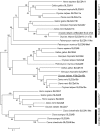
Phylogenetic tree of amino acid sequence of SLC2A class II. The phylogenetic tree was constructed using the neighbor-joining method and displayed using MEGA5. Numbers indicate the percentage of replicate trees in which the associated clade clustered together in the bootstrap test (1,000 replicates). The tree is drawn to scale, with branch lengths in the same units as those of the evolutionary distances used to infer the phylogenetic tree. Reptilia were excluded from this analysis due to short amino acid sequences with no information on expressed sequence tags (ESTs) of SLC2A genes.
Fig. 4.
Fig. 4.
Model for pigment cell development. Chromatoblast cells were generated from neural crest cells through fate restriction. Leucophore and xanthophore development was driven by pax7a. Both leucophore and xanthophore precursor cells became positive for slc2a15b and slc2a11b. Yellow pigmentation was promoted by both slc2a15b and slc2a11b, and slc2a15b promoted leucophore differentiation.

Comment in

Similar articles

Cited by

References

    1. Fujii R. Cytophysiology of fish chromatophores. Int Rev Cytol. 1993;143:191–255.
    1. Fujii R. The regulation of motile activity in fish chromatophores. Pigment Cell Res. 2000;13(5):300–319. - PubMed
    1. Hama T. Chromatophores and iridocytes. In: Yamamoto T, editor. MEDAKA (KILLIFISH) Biology and Strains. Tokyo: Keigaku; 1975. pp. 138–153.
    1. Matsumoto J. Studies on fine structure and cytochemical properties of erythrophores in swordtail, Xiphophorus helleri, with special reference to their pigment granules (Pterinosomes) J Cell Biol. 1965;27(3):493–504. - PMC - PubMed
    1. Odiorne JM. The occurrence of guanophores in Fundulus. Proc Natl Acad Sci USA. 1933;19(7):750–754. - PMC - PubMed

MeSH terms

Substances

Associated data

LinkOut - more resources