. 2014 May 6;5(3):e00880-14.
doi: 10.1128/mBio.00880-14.

Differences in lactococcal cell wall polysaccharide structure are major determining factors in bacteriophage sensitivity

Differences in lactococcal cell wall polysaccharide structure are major determining factors in bacteriophage sensitivity

Stuart Ainsworth et al. mBio. .

Abstract

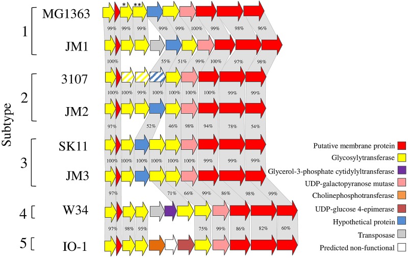
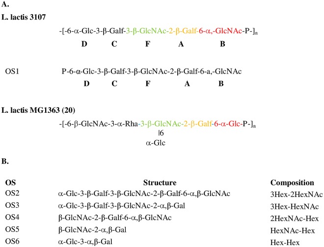
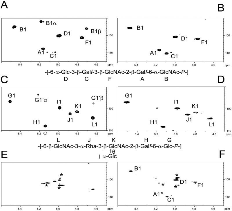
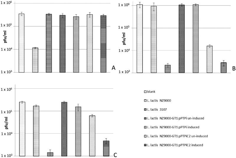
ABSTRACT Analysis of the genetic locus encompassing a cell wall polysaccharide (CWPS) biosynthesis operon of eight strains of Lactococcus lactis, identified as belonging to the same CWPS type C genotype, revealed the presence of a variable region among the strains examined. The results allowed the identification of five subgroups of the C type named subtypes C1 to C5. This variable region contains genes encoding glycosyltransferases that display low or no sequence homology between the subgroups. In this study, we purified an acidic polysaccharide from the cell wall of L. lactis 3107 (subtype C2) and confirmed that it is structurally different from the previously established CWPS of subtype C1 L. lactis MG1363. The CWPS of L. lactis 3107 is composed of pentasaccharide repeating units linked by phosphodiester bonds with the structure 6-α-Glc-3-β-Galf-3-β-GlcNAc-2-β-Galf-6-α-GlcNAc-1-P. Combinations of genes from the variable region of subtype C2 were introduced into a mutant of subtype C1 L. lactis NZ9000 deficient in CWPS biosynthesis. The resulting recombinant mutant synthesized a polysaccharide with a composition characteristic of that of subtype C2 L. lactis 3107 and not wild-type C1 L. lactis NZ9000. By challenging the recombinant mutant with various lactococcal phages, we demonstrated that CWPS is the host cell surface receptor of tested bacteriophages of both the P335 and 936 groups and that differences between the CWPS structures play a crucial role in determining phage host range. IMPORTANCE Despite the efforts of nearly 80 years of lactococcal phage research, the precise nature of the cell surface receptors of the P335 and 936 phage group receptors has remained elusive. This work demonstrates the molecular nature of a P335 group receptor while bolstering the evidence of its role in host recognition by phages of the 936 group and at least partially explains why such phages have a very narrow host range. The information generated will be instrumental in understanding the molecular mechanisms of how phages recognize specific saccharidic receptors located on the surface of their bacterial host.


Figures

FIG 1
FIG 1
Comparison of the variable regions in the type C CWPS biosynthesis cluster of lactococcal strains MG1363, JM1, 3107, JM2, SK11, JM3, W34, and IO-1. Genes with a high level of identity (indicated as percent nucleotide sequence identity) are joined by gray blocks compared to the adjoining strain. Five subtypes (C1 to C5) of the C genotype are highlighted. Genes marked with * and ** are interrupted in MG1363 derivatives NZ9000-GT1 (LLNZ_01145) and NZ9000-GT2 (LLNZ_01150), respectively. Genes with diagonal lines represent strain 3107 subtype C2 genes cloned into pPTPiC2.
FIG 2
FIG 2
(A) Structure of L. lactis 3107 CWPS and OS1. The structure of the MG1363 CWPS (20) is shown as a comparison. Similar component constituents of L. lactis 3107 and MG1363 CWPS are colored to highlight similarity. Bold letters D, C, F, A, and B represent individual detected monosaccharide residues. (B) Representation of the oligosaccharide fractions (OS2 to OS6) resulting from partial hydrolysis of L. lactis 3107 CWPS. Fractions were purified by RP-HPLC and analyzed by MALDI-TOF MS, confirming the L. lactis 3107 CWPS structure in panel A. The nature of the linkages present in the L. lactis 3107 CWPS, determined by methylation analysis, is displayed adjacent to each fraction.
FIG 3
FIG 3
NMR analysis of intact cell surface polysaccharides. 1H-13C HSQC NMR liquid spectra of purified polysaccharides (A and C) were compared with HR-MAS spectra of intact cells (B, D to F). Comparison of the NMR spectra of purified L. lactis 3107 CWPS (A) and intact L. lactis 3107 cells (B) confirms that the L. lactis 3107 CWPS is clearly expressed at the cell surface. Intact L. lactis NZ9000 cells clearly express a CWPS at their surface (D) that is, as expected, identical to the previously described (20) purified CWPS of L. lactis MG1363 (C). HR-MAS NMR analysis of uninduced NZ9000-GT1/pPTPiC2 cells (E) does not reveal the presence of the L. lactis NZ9000 CWPS (compare panel E with panels C and D), whereas the analysis of nisin-induced cells shows the presence of L. lactis 3107 CWPS (compare panel F with panels A and B). Asterisks indicate anomer signals associated with an undefined underlying polysaccharide. B1α/β and G1α/β signals originate from free reducing ends of both polysaccharides.
FIG 4
FIG 4
Irreversible-adsorption assay results displaying various free-phage titers (A, sk1; B, ViridusJM2; C, φLC3) postadsorption to various strains. Induction of pPTPi and pPTPiC2 was done with a 1/2,000 dilution of L. lactis NZ9700 cell-free supernatant containing nisin.

References

    1. Garneau JE, Moineau S. 2011. Bacteriophages of lactic acid bacteria and their impact on milk fermentations. Microb. Cell Fact. 10(Suppl 1):S20. 10.1186/1475-2859-10-20 - DOI - PMC - PubMed
    1. Mahony J, Murphy J, van Sinderen D. 2012. Lactococcal 936-type phages and dairy fermentation problems: from detection to evolution and prevention. Front. Microbiol. 3:335. 10.3389/fmicb.2012.00335 - DOI - PMC - PubMed
    1. Dupont K, Janzen T, Vogensen FK, Josephsen J, Stuer-Lauridsen B. 2004. Identification of Lactococcus lactis genes required for bacteriophage adsorption. Appl. Environ. Microbiol. 70:5825–5832. 10.1128/AEM.70.10.5825-5832.2004 - DOI - PMC - PubMed
    1. Deveau H, Labrie SJ, Chopin MC, Moineau S. 2006. Biodiversity and classification of lactococcal phages. Appl. Environ. Microbiol. 72:4338–4346. 10.1128/AEM.02517-05 - DOI - PMC - PubMed
    1. Chandry PS, Moore SC, Boyce JD, Davidson BE, Hillier AJ. 1997. Analysis of the DNA sequence, gene expression, origin of replication and modular structure of the Lactococcus lactis lytic bacteriophage sk1. Mol. Microbiol. 26:49–64. 10.1046/j.1365-2958.1997.5491926.x - DOI - PubMed

Publication types

Substances