. 2014 May;141(10):2139-49.
doi: 10.1242/dev.107029.

Sertoli cells control peritubular myoid cell fate and support adult Leydig cell development in the prepubertal testis

Affiliations

Sertoli cells control peritubular myoid cell fate and support adult Leydig cell development in the prepubertal testis

Diane Rebourcet et al. Development. 2014 May.

Abstract

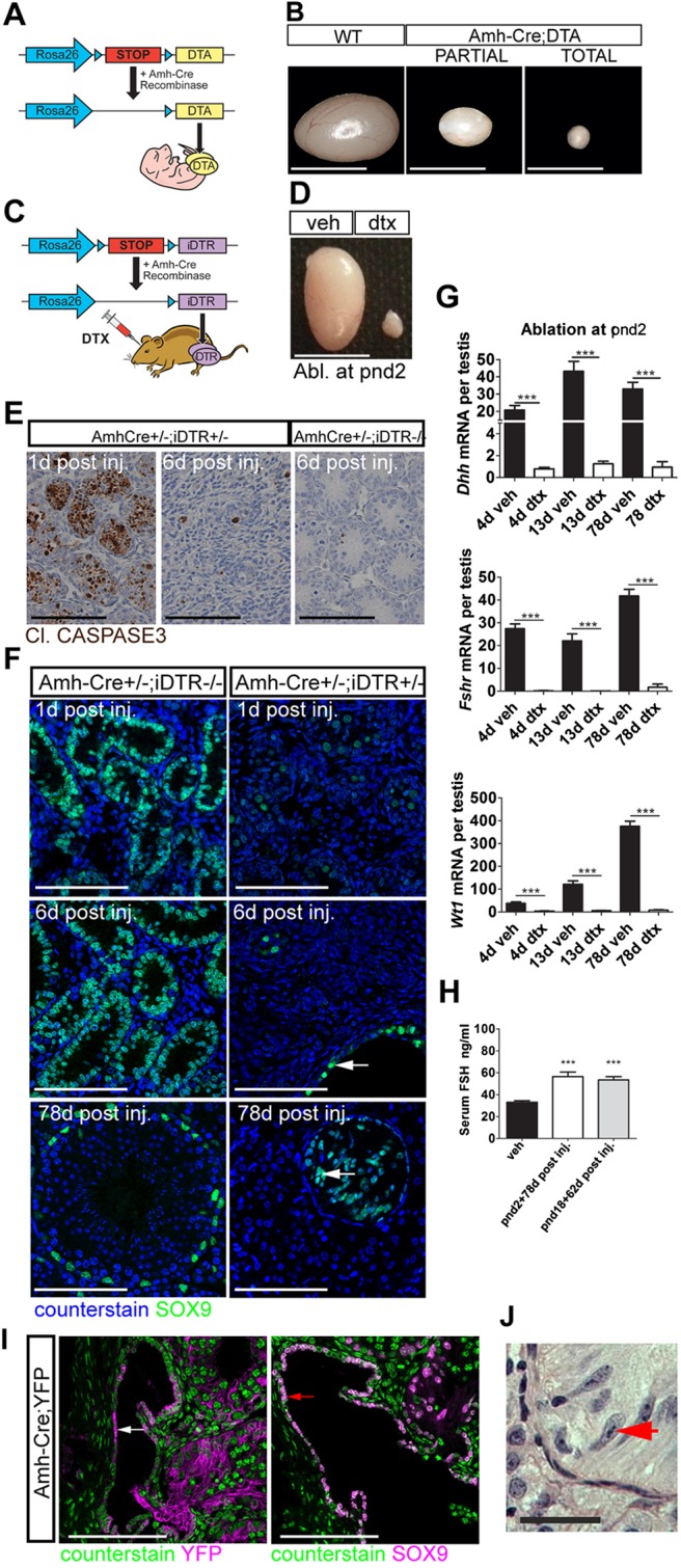
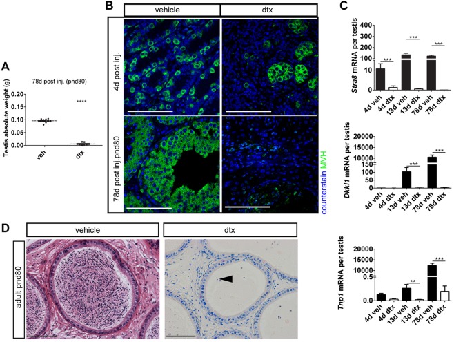
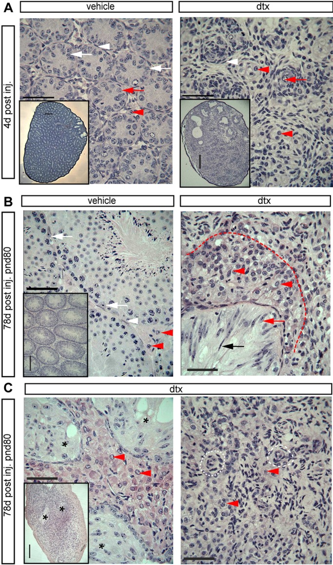
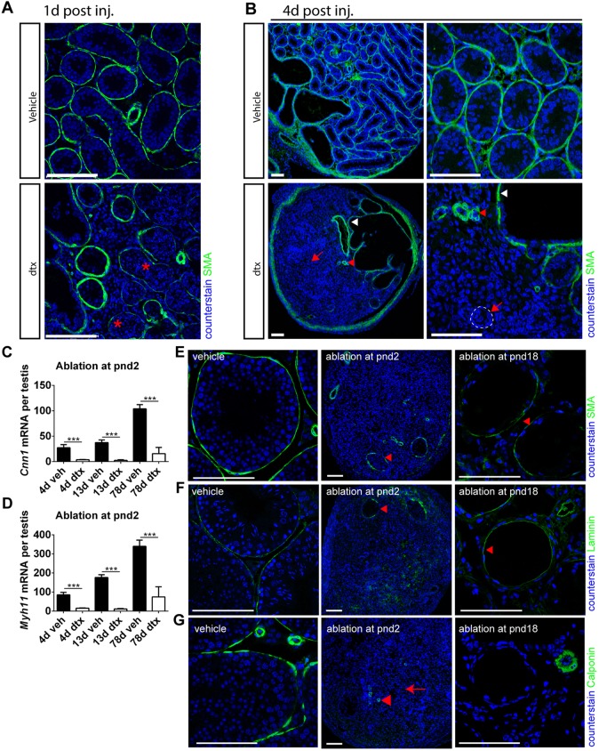
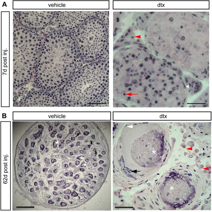
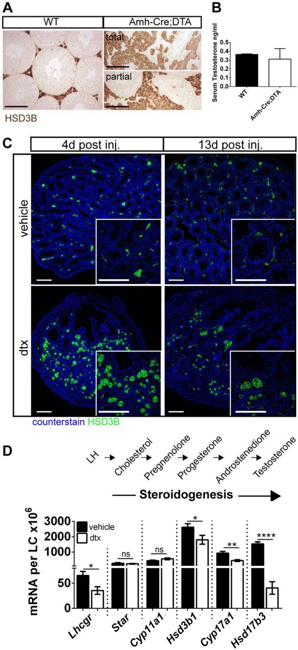
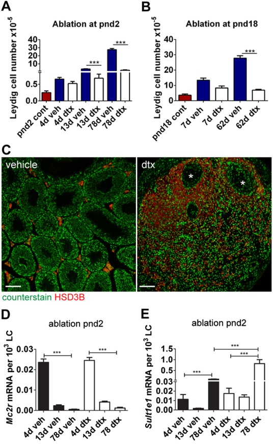
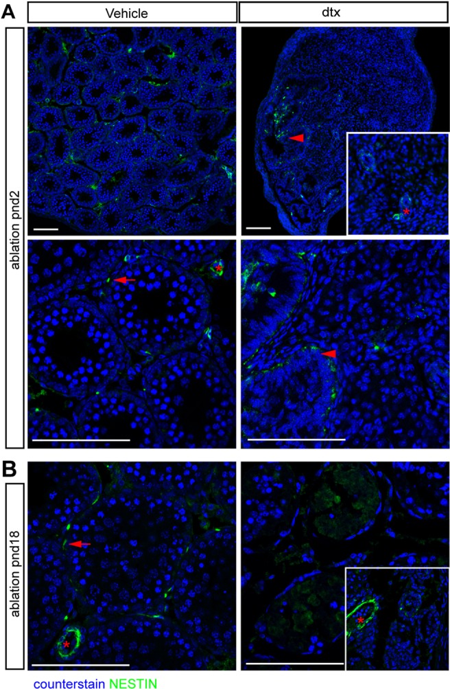
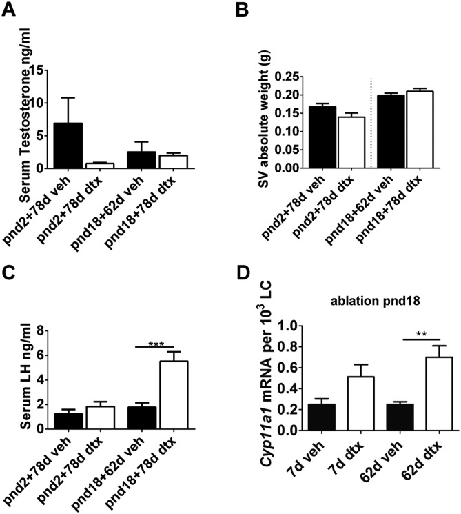
Sertoli cells (SCs) regulate testicular fate in the differentiating gonad and are the main regulators of spermatogenesis in the adult testis; however, their role during the intervening period of testis development, in particular during adult Leydig cell (ALC) differentiation and function, remains largely unknown. To examine SC function during fetal and prepubertal development we generated two transgenic mouse models that permit controlled, cell-specific ablation of SCs in pre- and postnatal life. Results show that SCs are required: (1) to maintain the differentiated phenotype of peritubular myoid cells (PTMCs) in prepubertal life; (2) to maintain the ALC progenitor population in the postnatal testis; and (3) for development of normal ALC numbers. Furthermore, our data show that fetal LCs function independently from SC, germ cell or PTMC support in the prepubertal testis. Together, these findings reveal that SCs remain essential regulators of testis development long after the period of sex determination. These findings have significant implications for our understanding of male reproductive disorders and wider androgen-related conditions affecting male health.

Keywords: Diphtheria; Leydig; Male fertility; Mouse; Peritubular myoid; Sertoli; Testis.


Figures

Fig. 1.
Fig. 1.
SC-specific ablation. (A) Mouse model one: Amh-Cre:DTA. (B) DTA induced testicular atrophy with different degrees of severity. (C) Mouse model two: Amh-Cre:iDTR. (D) DTX induced testicular atrophy in adulthood following injection at pnd2. (E) Apoptosis is restricted to SCs and is resolved 6 d post DTX injection. (F) SC ablation is mirrored by decreased SC SOX9 expression. Note that the expression of SOX9 is retained in the rete testis epithelium (arrows). Scale bar: 100 µm. (G) Relative expression of the SC-specific markers Dhh, Fshr and Wt1 (one-way ANOVA, n=7-9; ***P<0.001). (H) Circulating FSH concentrations at pnd80 (one-way ANOVA, n=7-9; ***P<0.001). (I) Immunolocalisation of YFP (white arrow), and SOX9 (red arrow) in Amh-Cre:YFP testis (d17). (J) Rete testis epithelium with tripartite nucleolus consistent with an SC origin for these cells (red arrow). WT, wild type; Veh, vehicle control. Scale bars: 500 µm in B,D; 100 µm in E,I; 50 µm in F,J.
Fig. 2.
Fig. 2.
Ontogeny of GC loss. (A) Testis weight was significantly reduced in adulthood following SC ablation at pnd2 (t-test, n=9-13; ****P<0.0001). (B) Immunolocalisation of MVH (DDX4) protein (green) identified GC loss following SC ablation. (C) Expression levels of the GC-specific markers Stra8 (spermatogonia), Dkkl1 (spermatocytes) and Tnp1 (spermatids) were all significantly reduced following SC ablation at pnd2 (one-way ANOVA, n=7-9; **P<0.01, ***P<0.001). (D) Consistent with GC loss, spermatozoa were absent and cellular debris remained (arrowhead) in cauda epididymides when examined in adulthood. Scale bars: 100 µm.
Fig. 3.
Fig. 3.
Testicular histology following SC ablation at pnd2. (A) Testicular histology of testes 4 d after ablation at pnd2. In DTX-treated animals no intact seminiferous tubules are present, although the rete testis remains present. In control testes, primordial germ cells (red arrows) and SCs (white arrows) are present inside the tubules and are surrounded by an intact PTMC layer (white arrowheads). FLCs (red arrowheads) are apparent in the interstitial tissue. In DTX-treated animals the tubules appear to have collapsed with the PTMCs forming concentric rings (white arrowheads), occasionally surrounding a surviving spermatogonial stem cell (red arrow). LCs remain present in the tissue between the collapsed tubules (red arrowheads). The insets, which are at lower magnification, show the overall structure of the seminiferous tubules in vehicle- or DTX-treated testes. (B) Testicular histology of adult testes (80 d) after ablation at pnd2. Representative SCs (white arrows), PTMCs (white arrowheads) and LCs (red arrowheads) are highlighted in vehicle-treated testis. In DTX-treated animals there was no tubular structure, although the rete testis remained intact (black arrow). The epithelium of the rete testis either had an SC-like appearance or had a highly elongated, pseudostratified appearance (black and red arrows). Abundant LCs were present in the vicinity of the rete testis (red arrowheads and delineated by the red dashed line), but there was a sharp reduction in LC numbers further from the rete testis. (C) Some variation between animals was seen in the size of the LC population surrounding the rete testis (black asterisks). In the parenchyma of the testis, concentric circles of cells were seen that might represent collapsed tubules seen at pnd6 (white dashed line). LCs were present but scarce in this region (red arrowheads). Scale bars: 50 µm in A-C; 250 µm in insets.
Fig. 4.
Fig. 4.
SCs maintain the PTMC differentiated phenotype in prepubertal life. (A) Disruption of PTMCs (as indicated by loss of SMA, asterisks) 1 d post DTX injection at pnd2. (B) Four days after injection, loss of SMA expression is consistent with a collapse of the tubular architecture (arrows and dashed line). Note that SMA expression is also restricted to blood vessels (red arrowhead) and rete testis (white arrowhead). (C,D) Expression of myoid cell markers (C) Cnn1 and (D) Myh11 following SC ablation at pnd2 (one-way ANOVA, n=7-9, ***P<0.001). (E) SMA expression is retained if SC ablation occurs at pnd18 (arrowhead). (F) Disruption to tubular basement membrane (BM) (laminin) at pnd2, whereas rete testis BM remains intact (arrowhead), whereas there is retention of gross tubule morphology from pnd18 (arrowhead). (G) Loss of calponin expression, a functional marker of PTMCs, at both pnd2 [arrow; whereas blood vessels retain it (arrowhead)], consistent with a collapse of the tubular architecture, and at pnd18 albeit with retention of gross tubule morphology. Scale bars: 100 µm.
Fig. 5.
Fig. 5.
Testicular histology following SC ablation at pnd18. (A) Testicular histology of mice 7 d after ablation at pnd18. Testes retained tubular architecture with representative PTMCs (white arrowhead) surrounding tubules and LCs in the interstitium (red arrowhead). Seminiferous tubules exhibited altered spermatogenesis and apoptotic GCs were clearly visible (red arrow). Scale bars: 100 µm (left), 50 µm (right). (B) In adult mice (pnd80) injected at pnd18, the tubular structure of the testis remained intact, although the tubules were marked by calcium salt deposits in the lumen (asterisks). PTMCs were present around the tubules (white arrowhead), forming multilayers in places. The lumen of some of the tubules contained unidentified cells (black arrow). LCs (B, red arrowheads) were present between the tubules. Scale bars: 400 µm (left), 50 µm (right).
Fig. 6.
Fig. 6.
FLCs are retained and function independently in perinatal life. (A) Immunolocalisation of LCs (HSD3B) and (B) circulating testosterone in adult (pnd60) wild-type and Amh-Cre:DTA testes (t-test, n=3-6). (C) FLCs (HSD3B) in Amh-Cre:iDTR animals 4 d and 13 d following SC ablation at pnd2. Note retention of the FLC population despite SC, GC and PTMC loss. (D) Comparative testicular expression of steroidogenic transcripts 4 d following SC ablation at pnd2, when only FLCs are present (t-tests, n=7-9; *P<0.05, **P<0.01, ****P<0.0001; ns, not significant). Scale bars: 100 µm.
Fig. 7.
Fig. 7.
SC ablation reduces final ALC number in adulthood. (A,B) Total LC number per testis at key time points following SC ablation at pnd2 (A) or pnd18 (B) (one-way ANOVA, n=4-12). (C) LCs (HSD3B) are localised around the rete testis (asterisks) in adulthood following SC ablation at pnd2. (D,E) Markers of FLC (Mc2r) (D) and ALC (Sult1e1) (E) show similar gene expression profiles in vehicle-treated and pnd2 SC ablation groups (one-way ANOVA, n=7-9; ***P<0.001).
Fig. 8.
Fig. 8.
ALC progenitor cells are restricted to the rete testis following SC ablation. LC progenitor cells (as marked by nestin) localise to peritubular (arrows) and perivascular (asterisk) regions in vehicle-treated testis (A,B). By contrast, LC progenitor cells are absent from the testicular parenchyma 13 d after SC ablation at pnd2 (A) and 7 d after SC ablation at pnd18 (B), but remain adjacent to the rete testis (A, arrowheads). Scale bars: 100 µm.
Fig. 9.
Fig. 9.
Functional compensation of ALCs after SC ablation. (A) Serum testosterone (one-way ANOVA, n=9-12), (B) seminal vesicle weight (t-test, n=9-12) and (C) LH concentrations in adulthood following SC ablation at pnd2 and pnd18 (ANOVA, n=9-12). (D) Expression of Cyp11a1 following SC ablation at pnd18 (ANOVA, n=7-9; **P<0.01, ***P<0.001).

References

    1. Ailenberg M., Tung P. S., Fritz I. B. (1990). Transforming growth factor-beta elicits shape changes and increases contractility of testicular peritubular cells. Biol. Reprod. 42, 499-509 10.1095/biolreprod42.3.499 - DOI - PubMed
    1. Ariyaratne H. B. S., Mendis-Handagama S. M. L. C., Hales D. B., Mason J. I. (2000). Studies on the onset of Leydig precursor cell differentiation in the prepubertal rat testis. Biol. Reprod. 63, 165-171 10.1095/biolreprod63.1.165 - DOI - PubMed
    1. Auharek S. A., Lara N. L. M., Avelar G. F., Sharpe R. M., Franca L. R. (2012). Effects of inducible nitric oxide synthase (iNOS) deficiency in mice on Sertoli cell proliferation and perinatal testis development. Int. J. Androl. 35, 741-751 10.1111/j.1365-2605.2012.01264.x - DOI - PubMed
    1. Baker P. J., O'Shaughnessy P. J. (2001). Role of gonadotrophins in regulating numbers of Leydig and Sertoli cells during fetal and postnatal development in mice. Reproduction 122, 227-234 10.1530/rep.0.1220227 - DOI - PubMed
    1. Baker P. J., Sha J. A., McBride M. W., Peng L., Payne A. H., O'Shaughnessy P. J. (1999). Expression of 3beta-hydroxysteroid dehydrogenase type I and VI isoforms in the mouse testis during development. Eur. J. Biochem. 260, 911-917 10.1046/j.1432-1327.1999.00245.x - DOI - PubMed

Publication types